Research Article
Austin J Obstet Gynecol.2017; 4(2): 1073.
Ultrasound Diagnosis of Polycystic Ovarian Syndrome: Current Guidelines, Criticism and Possible Update
Fulghesu AM, Canu E*, Porru C and Cappai A
Department of Scienze Chirurgiche, University of Cagliari, Cagliari, Italy
*Corresponding author: Elena Canu, Department of Scienze Chirurgiche, University of Cagliari, Cagliari, Italy
Received: August 29, 2017; Accepted: October 25, 2017; Published: November 01, 2017
Abstract
The US diagnosis of PCOS is still an open problem. The frequency of over diagnosis in particular phase of reproductive age, the contrasting guidelines and the technological advances lead to be a difficult charge for the sonographer. Follicle number and disposition and ovarian volume represents most used criteria, but other aspects as stromal study and ovarian vascularization could help. On the other hand, recently, the introduction of Anti-mullerian hormone assay introduced a support for US diagnosis.
Keywords: PCOS; US; FNPS; FNPO; S/A ratio
Abbreviations
AFC: Antral follicular count; AMH: Anti-mullerian hormone; FNPO: Follicle number per ovary; FNPS: Follicle number per section; LH: Luteinizing hormone; PCO: Polycystic Ovary; PCOM: Polycystic Ovarian Morphology PCOS: Polycystic Ovary Syndrome; S/A: Stroma/Area (Ovarian stromal area to total ovarian area); TA: Transabdominal; TV: Transvaginal; US: Ultrasonography, Ultrasonographic
Introduction
Body text
Definition: Polycystic ovarian syndrome is the most common female endocrine disorder, affecting 5-10% of women in the reproductive age [1]. PCOS has a wide variety of clinical manifestations: from alterations of the menstrual cycle secondary to ovulatory dysfunction, to dermatological manifestations, such acne and hirsutism, to metabolic alterations, often accompanied by ultrasound features of polycystic ovaries [2].
From the introduction in the current clinical practice of the first clinical screening of gynecological health in young women and the use of the pelvic ultrasound as routine exam, the diagnosis of PCOS are increased to the point that it is calculated that every four healthy girls, at least one was diagnosed of PCOS [3].
The definition of polycystic ovarian syndrome represents a clinical debate between specialists interested to pelvic ultrasound and/or gynecological endocrinology and constitutes a discussion topic in the literature.
Stein and Leventhal [4] discovered in 1935 the existence of a precise association between some clinical elements (infertility, amenorrhea, hirsutism and obesity) and the morphology of the ovaries: increased volume and pearly appearance and texture. From 1935 many other definitions have been proposed and the two main used by authors in the last twenty years are those of the NIH/NICHD (National Institute of Health/National Institute of Child Health and Human Development) of 1990 [5] and that of the Consensus of Rotterdam (2003) [6].
The definition proposed by NIH/NICHD envisages the presence of clinical hyperandrogenism or hyperandrogenemia, oligo or an ovulation and the exclusion of other disorders that cause hyperandrogenism, as congenital adrenal hyperplasia, androgen secreting neoplasms, hyperprolactinemia and thyroid dysfunction. This definition would exclude the use of ultrasound in diagnosis and this is not supported by many authors.
Current Guidelines
Recent diagnostic criteria proposed by the American Society of Reproductive Medicine (ASRM) and European Society of Human Reproduction and Embryology (ESHRE) in 2003 [7], as well as by the Androgen Excess and PCOS Society in 2006 [8], have reasserted the importance of ovarian morphology to the diagnosis of PCOS.
The consensus establishes, that the diagnosis of PCOS, may be done in the presence of at least two of the following symptoms (excluding other causes of hyperandrogenism):
- Oligoanovulation (oligoamenorrhea)
- Clinical hyperandrogenism and/or biological hyperandrogenemia
- Polycystic ovary morphology at ultrasound
About the phenotype, with the adoption of the Rotterdam criteria, two new subtypes have been introduced (not included in the criteria of the NIH, 1990): in addition to patients with ovulatory problems and hyperandrogenism (both with and without ovary PCO to US examination), fall within the definition of PCOS two other groups of patients, i.e. patients with anovulation and PCO ovary but without signs of hyperandrogenism, and patients with PCO ovary and hyperandrogenism but with normal ovulatory cycles.
Ultrasonographic features of polycystic ovaries and clinical correlates
From Consensus, ovarian ultrasound evaluation becomes a crucial stage in the diagnostic work-up of PCOS. The introduction of endovaginal probes with excellent sharpness of the image made possible to study more precisely both the size and the ovarian morphology, also in obese patients, in which the transabdominal scan was not sufficiently reliable.
For more than 15 years sonographic criteria to define the polycystic ovary was heavily influenced by the definition of Adams et al [9]: multiple follicles (n> 10) of small size (average diameter 2-8 mm) arranged in the subcortical seat around a hyperechogenic stroma and with enlarged volume ovaries (> 8ml).
In 2003, the Consensus Conference of Rotterdam [6], considering the literature, redefined the sonographic criteria of polycystic ovaries:
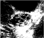
- Presence of at least 12 follicles in each ovary: the calculation must take account of all the follicles present, from the inner edge to the outer one, regardless of their arrangement and, for a more exhaustive study, different sections produced on different planes must be evaluated (Figure 1);

Figure 1: Presence of multiple follicles of small size on TA ovarian scan.
- Follicular diameter between 2 and 9mm: the follicular diameter is the average of the diameters in the three sections or the diameter of the follicle in the scan in which appears circular;
- Increased ovarian volume (>10mm3): for the volume calculation, several different formulas, through the preliminary identification of the three diameters have been proposed. All the methods demonstrated almost the same validity, as well as the calculations made by the software of modern ultrasound devices. It is recommended the use of the ellipsoid formula 0,52 × (D1×D2×D3).
The ultrasound examination should be performed in accordance with certain specific rules:
The operator must have performed a training to ensure the careful evaluation of the clinical image and the correlation with the endocrinological data. In particular, the experience helps in the differential diagnosis with multifollicular ovaries. Multifollicolar ovaries encoded, as provided by the same Consensus on studies conducted by Adams et al [9], are diagnosed by a number of follicles > 6, follicles with greater diameter (4-10 mm) and by a normal echogenicity of stromal component.
When possible transvaginal ultrasonography should be preferred especially in obese patients.
In women with regular menstrual cycles the exam is carried out in the early follicular phase (days 3-5). In oligo-amenorrheic women it can choose a random day, or rather, the first 3-5 days following a bleeding induced by progesterone. This type of timing ensures the optimal approach for the quantitative assessment of ovarian volume and area.
If there is evidence of a dominant follicle (10mm3) or of a corpus luteum, the ultrasound should be repeated in the next menstrual cycle.
The presence in a single ovary of one of the features described above constitutes a sufficient element for ultrasound diagnosis. Conversely are excluded from the diagnosis ovaries with cysts presenting pathological aspect.
All the above criteria do not apply to women with estrogenprogestin therapy. The study in these patients should be postponed to a later time to the suspension of treatment when it has the resumption of a normal functional activity.
The number of follicles, to perform an ultrasound diagnosis, has been variously reported as more than 10, or 12, or 15, etc; currently all authors are agree to consider > 12 the number of follicles needed for the diagnosis of PCOS (identified on a single median ovarian scan FNPS) [10,11] (Table 1).
    Author
    N.    Follicles


    Fox    (1991)
    15


    Adams    (1985)
    10


    Jonaard    (2003)
    12
Table 1: Follicles number in PCOS.
With the advance of US technology, recent guidelines [12,13], based on two studies performed in adults, recommended that the US features diagnostic for PCOM will be modified increasing the threshold of Follicle Number to 25 Per Ovary (FNPO), scanning by TV transducer frequency >8MHz, and counting by a specific software [14], but because the low availability of such probes and software, this cut-off is disregarded in the clinical practice.
About to the size of the follicles, recent studies point out that the diameter of follicles between 2 and 5mm are more characteristic of the syndrome and more related to the presence of clinical symptoms.
About the ovarian volume, 10cm3 threshold value is general agreement, but Jonard, in 2006 [10], proposed to reduce the threshold value to 7cm3, and Allemand, in 2007 [11], showed that 7cm3 is the average value calculated by 3D in the control populations. In the other hand, the definition of polycystic ovaries in adolescence is still uncertain. The symptoms as oligomenorrhea or amenorrhea and clinical or biochemical signs of hyperandrogenism, as acne and/or hirsutism are often present also in healthy adolescents [15].
Concerning the US PCOM pattern, both follicle number and ovarian size in this age are under discussion: the follicular count may be difficult in TA ultrasound and the follicle number may be apparently high [16], because, if it use the threshold value of normal follicular count in adults, could be misleading when applied to young girls [17]. Also, the ovarian size is larger during adolescence than in adulthood, and progressively decreases with age [18], therefore the ovarian size, suggested to be a workable parameter in the TA diagnosis of PCOS, could not be reliable in very young subjects.
On the other hand, several authors reported presence of PCOM in young girls, not regarding it as pathological something [19], but others indicate this morphology as a predictor of PCOS development [20].
Also stroma represents an acknowledged US marker for polycystic ovary syndrome. The proportion between the stroma and the ovary area in the middle section (S/A ratio) had been indicated as a reliable marker for hyperandrogenism [21]. The S/A ratio measure had been suggested to differentiate, by US, normoandrogenic PCOM from PCOS.
This examination should be performed in TV mode but unfortunately it is not always possible at a young age. It is necessary in these cases a transabdominal exam. This method is reliable about the size of the ovary and the number of follicles, less valid in the evaluation of the stromal component that is not easily distinguished within the ovarian parenchyma, if the subject is overweight.
All that increased prevalence of PCOS compared to that found using the criteria of the NIH. The real belonging to the large category of patients with PCOS of the last two phenotype, patients with anovulation and PCO ovary but without signs of hyperandrogenism, and patients with PCO ovary and hyperandrogenism but with normal ovulatory cycles, has done and is doing much to discuss.
A scientific work published by Belosi, Fulghesu, Lanzone et al [22] reclassified the population of patients with diagnosis of PCOS by applying separately the two diagnostic models. Has been studied 375 patients: 345 complied with the Rotterdam criteria and, of these, 273 also meet the criteria of NIH in 1990. Thus, there are 72 patients who have a diagnosis based only on the Rotterdam criteria. Clinical and hormonal features of these patients show that the 72 not-NIH show BMI, androgens and insulin much lower that patients positive for NIH criteria.
Another study carried out by Welt et al [23], analyzed the hormonal features of PCOS patients and showed that patients not- NIH, who had menstrual irregularities and PCO ovary, showed testosterone and androstenedione levels similar to those of control population and significantly lower than two groups of patients with hyperandrogenism and menstrual irregularities or hyperandrogenism and PCO ovary to US examination [23]. Therefore it is estimated that the set of sonographic criteria proposed by the Consensus ESHRE/ ASRM of Rotterdam in 2003 appears to be sufficiently sensitive and specific for its good correlation with circulating androgens, but overestimate probably cases of PCOS. For this reason, the presence of more than 10 follicles does not appear to be the most important ultrasound feature of PCOS.
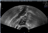
In particular the main difficulties are found in differential diagnosis of the multifollicular ovary, as described by Adams in 1985: this feature is described in subject with weight near the lower limits or less than the standard, and notes a relative hypogonadism. In these cases, there are reduction of the pulsatility of gonadotropins, low levels of androgens, US features similar to the PCO ovary in size and number of follicles and with slightly larger follicles (6- 10 mm) occupying the entire ovarian parenchyma (Figure 2), but without the component of stromal hyperplasia.

Figure 2: Multifollicular Ovary.
This feature is frequently associated with secondary amenorrhea or oligomenorrhea due to the alterations of gonadotropin pulsatility and frequently found in subjects with alterations in eating behavior, athletes or under psychophysical stress subjects. Current studies, not yet published, have shown that this feature is present physiologically in 20-30% of subjects in the first years from menarche: our study group in 2008, conducted a work, currently in publication, with 302 young women, with regular menstrual cycles and without clinical signs of hyperandrogenism, aged between 11 and 20 years. This study showed that 56% had a normal ultrasound patterns, 30% multifollicular pattern, 13% PCO pattern and the remaining 1% presence of dysfunctional ovarian cysts. Furthermore, it showed that patients with multifollicular ovaries had age and years from menarche significantly less to those of other groups, and with the increase of years from menarche occurred an increase in the prevalence of normal ovarian pattern and a significant rate reduction of multifollicular pattern (7% after the fifth year from menarche).
This suggests that the multifollicular ovary is probably the physiological expression of an ovarian functional immaturity during the late pre-pubescent and puberty and not a pathological finding. These aspects both morphological that endocrine are important to avoid placing a diagnosis of PCOS to subjects that are crossing a physiological step of the hypotalamic-pituitary axis and are suffering probably from slight dysfunctions.
In these cases, the correct evaluation of the stroma can represent a valuable tool.
The stroma in the polycystic ovary
In 1985 Adams et al. reported the peripheral arrangement of follicles in the PCO ovary around a core of hyperechogenic stromal tissue. Dewailly in 1994 [24] observed that the choice of studying the ovarian hypertrophy appeared to be justified because this parameter is not only easy to measure, but also is directly related with the hypertrophy of the stroma and constitutes an indirect indicator.
For these reasons, recently, several studies undertook to improve the diagnostic specificity of ultrasound by focusing on the evaluation of the stromal hypertrophy. Various systems have been proposed in order to define the increase of the stromal presence and the echogenicity (the normal stromal echogenicity is slightly lower than that of the myometrium) but none of these proposals has been a great result because strongly subjective and depending on the operator and on equipement.
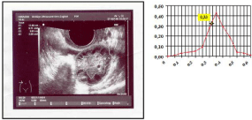
In 2001, Fulghesu et al [21], proposed quantification by the stroma rate compared to the total ovarian parenchyma (S/A ratio). This measure is obtained by the assessment of the ratio between the area of the stromal central thickening and the total area of the ovary. Stromal area is obtained by defining the peripheral profile of the stroma with the calliper, total area is obtained by defining, always with the calliper, the outer limit of the organ image sampled to the larger section of the ovary (Figure 3). With this assessment the diagnosis of ovarian PCO corresponds to values of S/A > 0.3, i.e. the diagnosis of PCOS is possible when the stroma represents more than one-third of the ovary in the middle section.

    Figure 3:  Quantitative evaluation of the stroma area/total area ratio of the ovary obtained by transvaginal track. The increase of the stromal area, more than onethird
of the total, is a parameter of diagnostic specificity of 96%. Youden index identify in 0,33 the S/A cut-off for identifying hyperandrogenized subjects.
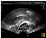
The stromal area/ovary total area ratio may be evaluated without additional technologies using the bidimensional ultrasound and can reduces subjectivity problems in the stroma evaluation; a scientific work published in 2006 by Belosi et al indicated the ratio S/A as a measure which little affected by variations inter-operator and that guarantees great diagnostic sensitivity and specificity, strictly correlated with the values of the plasma androgens. S/A ratio, in fact, is increased (greater than 0.33) in subjects with higher levels of androgens and could represent a further criterion of ultrasonographic diagnosis simplifying the identification of the hyperandrogenic subjects (Figure 4).

    Figure 4:  In patients with PCOS, stromal area/total area ratio has a value >
0.34 differently from patients with multifollicular ovaries and by controls.
The introduction of such method allowed Belosi group to identify within the group of NIH negative patients, subjects that have the most remarkable hyperandrogenism, menstrual and/or metabolic disorders [22].
In 2007 Fulghesu et al published a multicenter work about the stroma evaluation in PCOS [25]. The data confirmed with great precision that when the stroma exceeds 1/3 of the ovarian surface is very high the possibility to find high androgen levels and that eliminate the risk of overlap diagnosis with the multifollicular ovaries. Study carried out in over 300 subjects in 5 Italian Centers, verified that the ratio S/A can be measured in a homogeneous way by all operators (also not trained) and with equipements that had very simple software. The ROC curve showed that this method is the best predictor of hyperandrogenism of all US parameters. Battaglia et al obtained same results in a 3D study, confirming the presence that stromal thickness of more of one-third of ovarian tissue is related to hyperandrogenism and represents a valid US criterion for PCOS diagnosis [26].
Moreover, the S/A ratio demonstrated the best US PCOS diagnostic performance when associated with Total Ovarian Follicular count (FNPO).
Recently, nevertheless it was excluded from diagnostic criteria for a supposed difficulty in clinical practice [27].
Additional Diagnostic Prospects
Recently, to try to improve the ultrasound diagnosis of PCO, was studied the morphology of the PCO ovary by the use of color Doppler in the transvaginal US for the evaluation of the ovarian and uterine vascular flows. It initially paid attention to the large vessels (uterine and ovarian artery) that showing an increase of pulsatile index of the uterine artery for effect of high levels of androgens on reduction of uterine vessels perfusion [27].
Afterwards the focus shifted on the ovarian stroma vessels because there is an association between high levels of LH and increased stromal vascularization. Consequently a decrease of the intraovarian resistances causes stromal hyperplasia in patients with PCOS pattern.
The significance of higher stromal blood flow is accepted as predictor of hyperandrogenism, but actually the use is not recommended in the diagnosis of PCOS [30].
Recently, in the context of reproductive medicine, became increasingly widespread study of the PCOS ovary with 3D transvaginal US that appears more reliable for the assessment of ovarian volumes and blood flow compared to the bidimensional.
A recent work of Sun et al, showed that patients with elevated androgens levels, had S/A ratio about double compared to the group with ovarian feature of PCO, but with normal levels of circulating androgens.
On the other hand, PCOS is characterized by an increased number of follicles at all growing stages [31,32]. This increase is especially shown in the pre-antral and small antral follicles, which primarily produce AMH [33,34]. Elevated serum AMH level, may be considered as an effect of the stock of pre antral and small antral follicles [2,4] and appears higher in women with PCOS ovary in all populations [37,38] than in healthy women [35,36]. Due to its strong connection with the pathophysiology of PCOS, serum AMH could be considered the “gold standard” in the diagnosis of PCOS. Although serum AMH would be theoretically more accurate than AFC, as it reflects also the excess of small follicles unseen on US [38,39], it is not still considered a diagnostic criterion.
The robust association between AMH and AFC induced some authors to include anyway these values in the PCOS diagnosis.
Conclusion
The ultrasound diagnosis of the PCOS may need an update, which takes in consideration the unresolved problems about the over diagnosis, and which would be easily and quickly achievable with a common US equipement.
Rotterdam criteria (2003) are actually the official criteria for the PCOS diagnosis; however it appears possible to refine diagnostic ultrasound performance using the method, validated by international scientific literature, of the stroma evaluation with S/A ratio, which would appear more linked with the endocrine findings of the syndrome. The use of the quantification of the stromal area and the evaluation of the arrangement of the follicles is thus very helpful to perform a correct diagnosis of PCOS. This will give the possibility to better characterize the different phenotypes of PCOS for optimize the management of patients and the possiblities of medical therapy.
References
- Zawadzki, JADA. Diagnostic criteria for polycystic ovary syndrome: towards a rational approach. Dunaif A, Givens JR, Haseltine FP, Merriam GR, editors. In: Polycystic Ovary Syndrome. Boston: Blackwell Scientific. 1992: 377-384.
- Diamanti-Kandarakis E, Panidis D. Unravelling the phenotypic map of polycystic ovary syndrome (PCOS): a prospective study of 634 women with PCOS. Clin Endocrinol. 2007; 67: 735-742.
- Duijkers IjM, Klipping C. Polycystic ovaries, as defined by the 2003 Rotterdam consensus criteria, are found to be very common in young healthy women. Gynecol Endocrinol. 2010; 26: 152-160.
- Stein IF, Leventhal ML. Amenorrhea associated with bilateral polycystic ovaries. Am J Obstet Gynecol. 1935; 29: 181-191.
- Zawadzki JK, Dunaif A. Diagnostic criteria for polycystic ovary syndrome: towards a rational approach. Boston: Blackwell Scientific Publications. 1992.
- Group REA-SPCW. Revised 2003 consensus on diagnostic criteria and longterm health risks related to polycystic ovary syndrome (PCOS). Hum Reprod. 2004; 19: 41-47.
- Rotterdam ESHRE/ASRM-Sponsored PCOS Consensus Workshop Group. Revised 2003 consensus on diagnostic criteria and long-term health risks related to polycystic ovary syndrome. Fertil Steril. 2004; 8: 34-38.
- Azziz R, Carmina E, Dewailly D, Diamanti-Kandarakis E, Escobar-Morreale HF, Futterwiet W. et al. The Androgen Excess and PCOS Society criteria for the polycystic ovary syndrome: the complete task force report. Fertil Steril. 2009; 91: 456-488.
- Adams J, Franks S, Polson DW, Mason HD, Abdulwahid N, Tucker M, et al. Multifollicular ovaries: clinical and endocrine features and response to pulsatile gonadotropin releasing hormone. Lancet. 1985; 2: 8469-8470.
- Jonard S, Robert Y, Cortet-Rudelli C, Pigny P, Decanter C, Dewailly D. Ultrasound examination of polycystic ovaries: is it worth counting the follicles? Hum Reprod. 2003; 18: 598-603.
- Allemand MC, Tummon IS, Phy JL, Foong SC, Dumesic DA, Session DR. et al. Diagnosis of polycystic ovaries by three dimensional transvaginal ultrasound. Fertil Steril. 2006; 85: 214-229.
- Lujan ME, Jarrett BY, Brooks ED, Reines JK, Peppin AK, Muhn N, et al. Updated ultrasound criteria for polycystic ovary syndrome: reliable thresholds for elevated follicle population and ovarian volume. Hum Reprod. 2013; 28: 1361-1368.
- Dewailly D, Gronier H, Poncelet E, Robin G, Leroy M, Pigny P, et al. Diagnosis of polycystic ovary syndrome (PCOS): revisiting the threshold values of follicle count on ultrasound and of the serum AMH level for the definition of polycystic ovaries. Hum Reprod. 2011; 26: 3123-3129.
- Carmina E, Oberfield SE, Lobo RA. The diagnosis of polycistic ovary syndrome in adolescents AM J Obstet Gynecol. 2010; 203: 201-205.
- Venturoli S, Porcu E, Fabbri R, Pluchinotta V, Ruggeri S, Macrelli S, et al. Longitudinal change of sonographical ovarian aspects and endocrine parameters in irregular cycles of adolescence. Pediatr Res. 1995; 38: 974- 980.
- Mortensen M, Rosenfield RL, Littlejohn E. Functional significance of polycystic size ovaries in healthy adolescence. J Clin Endocrinol Metab. 2006; 911: 3786-3790.
- Fruzzetti F, Campagna AM, Perini D, Carmina E. Ovarian volume in normal and hyperandrogenic adolescent women. Fertil Ster. 2015; 104: 196-199.
- Ersen A, Onal H, Yildirim D, Adal E. Ovarian and uterine ultrasonography and relation to puberty in healthy girls between 6 and 16 years in the Turkish population: a cross-sectional study. J PediatrEndocrinol Metab. 2012; 25: 447-451.
- Zhu RY, Wong YC, Yong EL. Sonographic evaluation of polycystic ovaries. Best Pract Res Clin Obstet Gynaecol. 2016; 37: 25-37.
- Fulghesu AM, Ciampelli M, Belosi C, Apa R, Pavone V, Lanzone A, et al. A new ultrasound criterion for the diagnosis of polycystic ovary syndrome: the ovarian stroma/total area ratio. Fertil Steril. 2001; 76: 326-331.
- Belosi C, Selvaggi L, Apa R, Guido M, Romualdi D, Fulghesu AM, et al. Is the PCOS diagnosis solved by ESHRE/ASRM 2003 consensus or could it include ultrasound examination of the ovarian stroma? Hum Reprod. 2006; 21: 3108-3115.
- Welt CK, Gudmundsson JA, Arason G, Adams J, Palsdottir H, Gudlaugsdottir G, et al. Characterizing discrete subsets of polycystic ovary syndrome as defined by the Rotterdam criteria: the impact of weight on phenotype and metabolic features. JCEM. 2006; 91: 4842-4848.
- Dewailly D, Robert Y, Helin I, Ardaens Y, Thomas-Desrousseaux P, Lemaitre L, et al. Ovarian stromal hypertrophy in hyperandrogenic women. Clin endocrinol oxford. 1994; 41: 557-562.
- Fulghesu AM, Angioni S, Frau E, Belosi C, Apa R, Mioni R, et al. Ultrasound in polycystic ovary syndrome--the measuring of ovarian stroma and relationship with circulating androgens: results of a multicentric study. Hum reprod. 2007; 22: 2501-2508.
- Battaglia C, Mancini F, Persico N, Zaccaria V, De Aloysio D. Ultrasound evaluation of PCO, PCOS and OHSS. Biol med online. 2004; 9: 614-619.
- Christ JP, Willis AD, Brooks ED, Vanden Brink H, Jarrett BY, Pierson RA, et al. Follicle number, not assessments of the ovarian stroma, represents the best ultrasonographic marker of polycystic ovary syndrome. Fertil Steril. 2014; 101: 280-287.
- Leerasiri P, Wongwananuruk T, Rattanachaiyanont M, Indhavivadhana S, Techatraisak K, Angsuwathana S, et al. Ratio of ovarian stroma and total ovarian area by ultrasound in prediction of hyperandrogenemia in reproductive-aged Thai women with polycystic ovary syndrome: a diagnostic test. J Obstet gynecol research. 2015; 41: 248-253.
- Sun L, Fu Q. Three-dimensional transrectal ultrasonography in adolescent patients with polycystic ovarian syndrome. Int Journal Gynecol Obstet. 2007; 98: 34-38.
- Maciel GA, Baracat EC, Benda JA, Markham SM, Hensinger K, Chang RJ, et al. Stockpiling of transitional and classic primary follicles in ovaries of women with polycystic ovary syndrome. J Clin Endocrinol Metab. 2004; 89: 5321- 5327.
- Weenen C, Laven JS, Von Bergh AR, Cranfield M, Groome NP, Visser JA, et al. Anti-Mullerian hormone expression pattern in the human ovary; potential implications for initial and cyclic follicle recruitment. Mol Hum Reprod. 2004; 10: 77-83.
- Bhide P, Dilgil M, Gudi A, Shah A, Akwaa C, Homburg R, et al. Each small antral follicle in ovaries of women with polycystic ovary syndrome produces more antimullerian hormone than its counterpart in a normal ovary: an observational cross-sectional study. Fertil Steril. 2014.
- Lie Fong S, Schipper I, de Jong FH, Themmen AP, Visser JA, Laven JS, et al. Serum Anti-Mullerian hormone and inhibinB concentrations are not useful predictors of ovarian response during ovulation induction treatment with recombinant folliclke-stimulatin hormone in women with polycystic ovary syndrome. Fertil Steril. 2011; 96: 459-463.
- Villarroel C, Merino PM, Lopez P, Eyzaguirre FC, Van Velzen A, Iniguez G, et al. Polycystic ovarian morphology in adolescents with regular menstrual cycles is associated with elevated anti-Mullerian hormone. Hum Reprod. 2011; 26: 2861-2868.
- Laven JS, Mulders AG, Visser JA, Themmen AP, De Jong FH, Fauser BC. Anti-Mullerian hormone serum concentrations in normoovulatory and anovulatory women of reproductive age. J Clin Endocrinol Metab. 2004; 89: 318-323.
- Pigny P, Merlen E, Robert Y, Cortet Rudelli C, Decanter C, Jonard S, et al. Elevated serum level of anti-mullerian hormone in patient with polycystic ovary syndrome: relationship to the ovarian follicleexcessand to the follicular arrest. J Clin Endocrinol Metab. 2003; 88: 5957-5962.
- Jeppesen JV, Anderson RA, Kelsey TW, Christiansen SL, KristensenSG, JayaprakasanK, et al. Which follicles make the most anti-Mullerian hormone in humans? Evidence for an adrupt decline in AMH production at the time of follicle selection. Mol Hum Reprod. 2013; 19: 519-527.
- Dewailly D, Andersen CY, Balen A, Breekmans F, Dilaver N, Fanchin R, et al. The physiology and clinical utility of anti-Mullerian hormone in women. Hum Reprod Update. 2014; 20: 370-385.
- Dewailly D. Rationale for the use of serum AMH assay as a probe for PCOM. Hum Reprod Update. 2014.
- Dumont A, Robin G, Catteau-Jonard S, Dewailly D. Role of Anti-Müllerian Hormone in pathophysiology, diagnosis and treatment of Polycystic Ovary Syndrome: a review. reprod biol endocrinol. 2015; 13: 137.