
Editorial
Austin J Obstet Gynecol. 2018; 5(3): 1103.
Ectopic Molar Pregnancy in the Broad Ligament: A Case Report
Viviani V¹*, Lecointre L², Host A¹ and Garbin O¹
¹Department Gynecology, Cmco Unit, Gynecology- Obstetrics Hub, University Hospitals, Strasbourg, France
²Department Gynecology, Hautepierre Hospital, Gynecology-Obstetrics Hub, University Hospitals, Strasbourg, France
*Corresponding author: Viviani Victor, Department Gynecology, Cmco Unit, Gynecology-Obstetrics Hub, University Hospitals, Strasbourg, France
Received: February 08, 2018; Accepted: March 12, 2018; Published: March 26, 2018
Abstract
We report below the first case of complete molar pregnancy situated in the broad ligament in a nulliparous 23-year-oldwoman. Ectopic molar pregnancies are extremely rare forms of gestational trophoblastic disease. Reports in the literature indicate that they can occur in any part of the pelvic cavity. Their clinical presentation mimics that of an extra-uterine pregnancy. Although their diagnosis may be problematic, they are invariably managed by surgical treatment.
Keywords: Ectopic Molar Pregnancy; Abdominal Pregnancy; Broad Ligament
Introduction
Trophoblastic diseases are a group of placental lesions characterized by proliferation and abnormal maturation of the trophoblast including cancers derived from the trophoblast [1,2]. The incidence of complete hydatidiform mole is one per thousand pregnancies and that of partial mole three per thousand pregnancies in Western countries [3]. Ectopic molar pregnancy and pregnancy in the abdominal cavity are both extremely rare entities. Complete ectopic molar pregnancy, i.e. outside the uterine cavity, tends to be under diagnosed and its true incidence is not known [4,5]. Abdominal pregnancy is a situation described in very rare cases in the literature and concerns approximately 1% of extrauterine pregnancies [6]. The case which we report below is the first case identified in the literature of complete molar pregnancy situated in the broad ligament of the uterus.
Case Presentation
The patient was a nulliparous primigravida aged 23. She had a history of hyper prolactinemia complicating a pituitary microadenoma and obesity, with a BMI at 34kg/m². The patient initially presented owing to metrorrhagiaat 8 Weeks’ Gestation (WG) on January 23, 2016. Endovaginal echography revealed what then appeared to be two gestational sacs although no embryo was visualized. In the absence of echographic development, a diagnosis of failed pregnancy was made. The patient was treatedmedically on February 26, 2016 with oral mifepristone and misoprostol. Three months later, she consulted on account of metrorrhagia; Beta Fraction-Hcg (beta-hCG) was assayed at 2500IU/mL. Subsequently the beta-hCG level displayed a slow falloff and then leveled off until June 2016. In view of the suspicion of a right-sided extrauterine pregnancy with an echographic image suggestive of a right hematosalpinx, the patient received an injection of methotrexate in July 2016: this was complicated by moderate hepatic cytolysis. BetahCG levels decreased thereafter, and finally leveled off at between 30 and 100. She underwent hysteroscopy in January 2017, which showed a T-shaped uterine cavity within normal limits. Pelvic MRI was performed but did not reveal any apparent anomaly; however hysterosal pingography detected a minute circled left ampullary subtraction defect image, consistent with a persistent small left-sided extra-uterine pregnancy. A second injection of methotrexate was administered in January 2017 after review of her hepatic enzymes: these showed minimal hepatic cytolysis and echography suggested steatosis. A viral screen was negative.
Following the injection, beta-hCG levels gradually decreased: they were 404 on January 25, then 437 on D4 post-injection, and159 on D7 post-injection. A trough value of 19 was reached on February 20, 2017. Subsequent lab tests indicated that the beta-hCG level had climbed again to 106. Changes in the beta-hCG level over time are shown in Figure 1. In view of this renewed rise, and with the patient’s consent, we decided exploratory laparoscopy was indicated; this was performed on March 22, 2017. On laparoscopy, the abdominal cavity, uterus, Fallopian tubes and ovaries appeared normal. A bluish subperitoneal swelling measuring 7 mm was visualized at the base of the right broad ligament between the right utero-ovarian ligament and the right Fallopian tube interiorly to the ovarian vein. We proceeded with the dissection and excision of this structure (Figure 2). The operating specimen was removed in a retrieval bag. Histological examination revealed syncytio-trophoblastic cells and intermediate trophoblastic cells with a moderate degree of nuclear atypia. After the slides were reviewed at the French expert center for trophoblastic diseases in Lyon, the definitive histological conclusion was of a complete hydatidiform mole. This was therefore a case of ectopic complete hydatidi form mole in the right broad ligament. The post-operative course was uncomplicated, and weekly monitoring of the beta-hCG level was instituted along with oral contraception. Beta-hCG levels were negative in April 2017 and did not rise again subsequently (Figure 2), at any of the monthly monitoring checks for 6 months. After this period, the patient stopped her contraception because she was eager to become pregnant again.
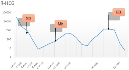
    Figure 1:  Changes in beta-hCG levels on a semi-logarithmic scale. Mtx:
methotrexate injection. Chir: surgical treatment. Key: Mois = Months.
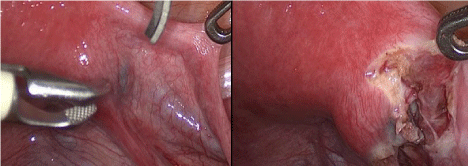
    Figure 2:  Laparoscopy: Bluish subperitoneal swelling at the base of the
broad ligament between the Fallopian tube (top right), ovarian vein and uteroovarian
ligament corresponding to the ectopic molar pregnancy. On the righthand
image, dissection and excision.
Discussion
Gestational trophoblastic diseases consist of benign entities, complete and partial hydatidiform moles, as well as clinically malignant entities known as gestational trophoblastic tumors: these include some invasive moles, choriocarcinomas, placental site trophoblastic tumors and epithelioid trophoblastic tumors [7]. The majority of gestational trophoblastic diseases develop in the uterus. Ectopic molar pregnanciesare therefore extremely rare.
In a review of the literature conducted by Yamada et al between 1960 and 2014, the authors found 31 cases of ectopic molar pregnancy [8]. Their incidence is estimated at 1.5 pregnancies per million [8]. Most of them were found in the Fallopian tubes (61%), but 16% were located in the ovaries, and 3% in the uterine horns. Two cases were described where ectopic molar pregnancy occurred in the peritoneum, one in the uterine cervix and another in a Cesarean scar [9]. No case of molar pregnancy affecting the broad ligament has been described until now. Pregnancy occurring in the broad ligament is a form of abdominal pregnancy. It accounts for approximately 1 %of all extra-uterine pregnancies [6]. Its incidence is thought to be 1 per 10,000 live births. Two pathophysiological theories have been advanced to explain this phenomenon. Broad ligament pregnancies may be primary through implantation of the ovum in the abdominal cavity or secondary to tubo-abdominal miscarriage or tubal rupture [10]. The principal risk factors are those of extra-uterine pregnancy: sterility, intra-uterine device, pregnancy post-IVF, and history of uterine injury, termination of pregnancy by aspiration, uterine scarring, genital infections and insufficient follow-up. The etiology and pathogenesis of broad ligament pregnancies is however not fully known. They could result from a tubal pregnancy which has ruptured into the mesosalpinx and continued its development, or from an ovarian pregnancy that has migrated to the broad ligament [11,12]. The case described in this article would appear to be the first case of ectopic complete molar pregnancy situated in the broad ligament which has been reported in the literature. The main difficulty with this case was diagnostic. The initial clinical picture was very similar to that of a relatively asymptomatic extra-uterine pregnancy. In view of a persisting positive beta-hCG level in spite of two injections of methotrexate additional investigations were requested. None of them, not even MRI, was able to substantiate the diagnosis. The final diagnosis, as with any molar pregnancy, was made by histological analysis. Treatment is the same as for other hydatidiform moles, namely surgical resection of the pathological trophoblast; this is usually accomplished by aspiration although our case was treated by laparoscopic surgery. Follow-up is based on monitoring of the decreasing beta-hCG level every week until it becomes negative, and thereafter every month for 6 months. Contraception is recommended in order to avoid the advent of a new pregnancy which would hamper beta-hCG monitoring, crucial for the diagnosis of persistent trophoblastic disease.
Conclusion
Ectopic molar pregnancy is an extremely rare disorder which can occur at any site in the pelvic cavity. Ours is the first case report of a molar pregnancy occurring in the broad ligament: it was successfully treated by laparoscopic surgery.
References
- Vassilakos P. Riotton G, Kajii T. “Hydatidiform Mole: Two Entities. A Morphologic and Cytogenetic Study with Some Clinical Consideration.” American Journal of Obstetrics and Gynecology. 1977; 127: 167-170.
- Szulman AE, Surti U. “The Clinicopathologic Profile of the Partial Hydatidiform Mole.” Obstetrics and Gynecology. 1982; 59: 597-602.
- Jacobs PA, Hunt PA, Matsuura JS, Wilson C, Szulman AE. Complete and partial hydatidiform mole in Hawaii: cytogenetics, morphology and epidemiology. Br J Obstet Gynaecol. 1982; 89: 258-266.
- Burton JL, Lidbury EA, Gillespie AM, Tidy JA, Smith O. Lawry J, Hancock BW, Wells M. “Over-Diagnosis of Hydatidiform Mole in Early Tubal Ectopic Pregnancy.” Histopathology. 2001; 38: 409-417.
- Sebire NJ, Lindsay I, Fisher RA, Savage P, Seckl MJ. “Overdiagnosis of Complete and Partial Hydatidiform Mole in Tubal Ectopic Pregnancies.” International Journal of Gynecological Pathology: Official Journal of the International Society of Gynecological Pathologists. 2005; 24: 260-264.
- Astrash HK, Friede A, Hogue CJR. Abdominal pregnancy in the United States: frequency and maternal morbidity. Obstet Gynecol. 1987; 69: 333-337.
- “Gestational Trophoblastic Diseases. Report of a WHO Scientific Group.” World Health Organization Technical Report Series. 1983; 692: 7-81.
- Gillespie AM, Lidbury EA, Tidy JA, Hancock BW. The clinical presentation, treatment, and outcome of patients diagnosed with possible ectopic molar gestation. International Journal of Gynecological Cancer. 2004; 14: 366-369.
- Yamada, Yasushi, Satoshi Ohira, Teruyuki Yamazaki, and Tanri Shiozawa. “Ectopic Molar Pregnancy: Diagnostic Efficacy of Magnetic Resonance Imaging and Review of the Literature.” Case Reports in Obstetrics and Gynecology. 2016: 1-7.
- Alto WA. Abdominal pregnancy. Am Fam Physician. 1990; 41: 209-214.
- Faller E, Kauffmann E, Chevrière S, Heisert M, Ranjatoelina H, Boumahni B, Sitty-Amina AA, and Barau G, et al. “Grossesse abdominale menéée à terme Full term abdominal pregnancy.” Journal de Gynécologie, Obstétrique et Biologie de la Reproduction. 2006; 35: 732-735.
- Rakotomahenina H, Andrianampy HA, Ramamonjinirina P, Solofomalala GD, and Brun JL. “Grossesse à terme dans le ligament large.” Gynécologie Obstétrique & Fertilité. 2014; 42: 537-539.