
Research Article
Austin J Obstet Gynecol. 2019; 6(2): 1138.
The Value Fetal Umbilical Artery and Middle Cerebral Artery Doppler Indices in Women with Pregnancy Induced Hypertension in the Prediction of Adverse Perinatal Outcome
Mahmoud Alalfy1*, E.Eltaieb2
¹Reproductive Health and Family Planning Department, National Research Centre, Consultant Obstetrics and Gynecology, Algezeera Hospital, Egypt
²Lecturer in Obstetrics and Gynecology, Ainshams University, Cairo, Egypt
*Corresponding author: Mahmoud Alalfy, Reproductive Health and Family Planning Department, National Research Centre, Consultant Obstetrics and Gynecology, Algezeera Hospital, Egypt
Received: April 01, 2019; Accepted: April 18, 2019; Published: April 25, 2019
Abstract
Background: During normal gestation, vascularization of placenta bed happened that helps blood flow in between the pregnant woman and the fetus. Whereas the placenta of ladies who have preeclampsia is abnormal and has reduced trophoblastic invasion which cause improper placental perfusion.
Patients and Methods: A prospective observational study was conducted at Ain Shams University Maternity Hospital, in Obstetrics and Gynecology Department, Faculty of Medicine and at Algezera Hospital over a period from January 2017 to June 2018 on pregnant women from 32-40 weeks of gestation with PIH who attended outpatient clinic and emergency room.
Results: Cases of abnormal CPR had significant higher severe PE, CS, fetal distress, SGA, NICU and death and significantly lower elective CS, GA at delivery, BW and APGAR scores.
Conclusion: Middle cerebral artery pulsatility index is more sensitive than umblical artery pulsatility index and middle cerebral artery/umblical artery pulsitility index ratio in prediction of adverse perinatal outcomes. However, middle cerebral artery/umblical artery pulsitility index ratio showed more specificity in predection of perinatal outcome in patients with pregnancy induced hypertension.
Introduction
Pregnancy is associated with some hypertensive complications and might induce maternal and neonatal morbidity and mortality. Hypertension that occurs with pregnancy accounts for five to fifteen percent [1].
To reach a successful uncomplicated pregnancy, we should have a normally formed uteroplacental and placentofetal circulation all through pregnancy. Whereas changes in the development and formation of placenta is linked to hypertensive disorders with pregnancy that result in altered circulation that in turn leads to intrauterine growth restriction ,premature babies and fetal demise sequeles [1].
Gestational hypertension points to increased blood pressure firstly diagnosed after twenty weeks of pregnancy without proteinuria or other diagnostic manifestations of preeclampsia. With the advance of gestation, some women with gestational hypertension might develop proteinuria, edema that denotes preeclampsia development [2].
During normal gestation, vascularization of placenta bed happened that helps blood flow in between the pregnant woman and the fetus. Whereas the placenta of ladies who have preeclampsia is abnormal and has reduced trophoblastic invasion which cause improper placental perfusion [3].
Vasodilatation of the cerebral vessels as a reaction to maintain enough oxygen needs to the brain is denoted by reduced pulsatility index in the cerebral vessels [4].
Aim of the Work
The aim of this work is to evaluate the accurateness of middle cerebral artery and umbilical artery Doppler in expecting perinatal outcome in women with PIH in the third trimester.
Patients and Methods
A prospective observational study was conducted at Ain Shams University Maternity Hospital, in Obstetrics and Gynecology Department, Faculty of Medicine and at Algezeera Hospital, Egypt over a period from January 2017 to June 2018 on pregnant women from 32-40 weeks of gestation with PIH who attended outpatient clinic and emergency room.
Sample size justification
Depending on (Rozeta et al., 2010) who found that frequency of Abnormal MCA/UA ratio 42.5% and NICU admission among normal and abnormal MCA/UA ratio 77.6% & 47.4% respectively, and assuming the power=0.80 and a=0.05, and by using PASS 11th release the minimal sample size for a single group prospective observational study to detect the difference between normal and abnormal MCA/ UA ratio regarding NICU admission is 100 women [5].
Intervention: All the patients will undergo accurate color Doppler velocimetry examination. The ultrasound machine that was used is Medison Sonoace R5, with a Doppler unit and a 3.5MHz convex linear probe. The output power of 50m/cm2 will be used, and the high-pass filter will set to 100Hz. The study population was divided into two groups depending on the normal or abnormal values of MCA/UA pulsatility index ratio. All the patients were followed up till delivery, delivery was attended and the neonates were assessed immediately postnatal.
Inclusion criteria:
1. All adult pregnant women with pregnancy induced hypertension;
Gestational Hypertension defined as:
• BP =140/90 mmHg for the first time during pregnancy after 20 weeks gestation.
• No Proteinuria.
• BP return to normal < 12 weeks postpartum.
Preeclampsia:
Minimum criteria:
• BP =140/90 mmHg for the first time during pregnancy after 20 weeks gestation.
• Proteinuria = 300mg/24hr or =+1 dipstick.
Criteria of severity:
• BP =160/110 mmHg.
• Proteinuria = 5gm/24hr or =+2 dipstick.
• Serum creatinine > 1.2mg/dl unless known to be previously elevated.
• Increased LDH.
• Persistent epigastric pain.
• Persistent headache or other cerebral or visual disturbances.
2. Gestational age: 32-40 weeks of gestation.
Singleton pregnancy.
3. Maternal age :< 40 years old.
Exclusion criteria:
Chronic hypertension, Diabetes mellitus or gestational diabetes.
Multiple pregnancies, Fetal congenital anomalies, polyhydramnios.
Pregnancy complicated with other medical disorders (cardiac disorders, renal disorders, .. etc)
Outcome criteria
Primary outcome: Neonatal outcome will be detected by APGAR score at 1 & 5 minutes.
Secondary outcome: Variables were; admission to the NICU and the duration of treatment, Mode of delivery, Gestational age at delivery, Neonatal birth weight Early neonatal death, The outcome for each pregnancy will be obtained by examining the labour ward records and NICU records.
Intervention: An informed written consent was taken from all patients and was approved by local ethical committee.
All patients were subjected to careful and detailed history including History of the present pregnancy: Medical or surgical condition to define high risk pregnancy.
Symptoms of severity e.g. epigastric pain, blurring of vision, headache.
Examination of the patients: General examination including Vital signs, lower limb edema,..etc and abdominal examination. Then Ultrasound examination to assess viability of pregnancy.
Determine gestational age and to exclude major abnormalities and Assess fetal growth.
Doppler velocimetry examination
The MCA/UA PI ratio (Cerebroplacental ratio) is usually constant during the last 10 weeks of gestation [1].
(Arbeille et al.,1996) also found the cerebroplacental ratio constant during the third trimester of pregnancy and suggested 1 as the cut off value; all values below 1 were considered abnormal; Gramellini et al. (1992) also used a single cut off value of 1.08 [6,7].
Therefore, in our study a single cut off value (1.08) was used:
• Group A: with middle cerebral artery and umbilical artery pulsatility index ratio>1.08
• Group B: with middle cerebral artery and umbilical artery pulsatility index ratio<1.08.
These both groups were followed up and their delivery and neonatal records were studied. Group A and Group B will be compared for perinatal outcomes.
Results
Table 1 and Figure 1 show that: More than half of the studied cases had severe PE.
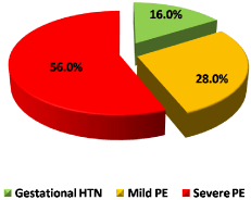
Figure 1: PIH among the studied cases.
![]()
    Characteristics
    Mean±SD
    Range


    Age (years)
    30.0±5.9
    18.0–38.0


    BMI (Kg/m2)
    29.6±3.6
    21.0–37.0


    GA at enrollment (weeks)
    36.6±1.4
    33.0–39.0


    Parity
    1.5±1.3
    0.0–4.0


    N
    %


    PIH
    Gestational HTN
    16
    16


    Mild PE
    28
    28


    Severe PE
    56
    56


    Total=100.
Table 1: Demographic characteristics of the studied cases.
Table 2 and Figure 2 show that: More than two thirds of cases had abnormal MCA-PI abnormalities, less than half of the studied cases UA-PI abnormalities, less than half of the studied cases MCA/UA PI ratio abnormalities.
Figure 2: Doppler abnormalities among the studied cases.
![]()
    Mean±SD
    Range
    Abnormalities


    MCA-PI
    1.28±0.26
    0.95–1.95
    67 (67.0%)


    UA-PI
    1.00±0.14
    0.77–1.23
    42 (42.0%)


    MCA/UA-PI
    1.33±0.41
    0.81–2.21
    44 (44.0%)


    Total=100.
Table 2: Doppler findings among the studied cases.
Table 3 and Figure 3 show that: More than two thirds of the studied cases underwent CS, more than half of the studied cases admitted to NICU, small for GA and death were in few cases.
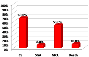
Figure 3: Outcomes among the studied cases.
![]()
    Outcomes
    N
    %


    MOD
    NVD
    31
    31


    CS
    69
    69


    Indication of CS^
    Elective
    29
    42


    Fetal distress
    24
    34.8


    Accidental hemorrhage
    10
    14.5


    Oligohydamnios
    4
    5.8


    CPD
    2
    2.9


    Small for GA
    8
    8


    NICU
    53
    53


    Death
    10
    10


    Mean±SD
    Range


    GA at delivery (weeks)
    37.3±1.3
    33.0–39.0


    Birth weight (kg)
    2.84±.41
    1.65–3.4


    APGAR1
    5.6±1.8
    0.0–8.0


    APGAR5
    7.5±2.0
    0.0–10.0


    #NICU duration (days)
    9.7±2.3
    6.0–14.0


    Total=100, ^Among CS cases (N=69), #Among    cases of NICU (N=53).
Table 3: Outcomes among the studied cases.
Table 4 show that: Cases abnormal MCA-PI had significant higher severe PE, CS, SGA, NICU admission and death and significantly lower elective CS, GA at delivery, BW and APGAR scores.
![]()
    Characteristics
    Abnormal
    Normal
    P


    (N=67)
    (N=33)


    Age (years)
    28.7±5.7
    32.6±5.3
    0.101


    BMI (Kg/m2)
    29.1±3.3
    30.4±4.0
    ^0.096


    GA at enrollment (weeks)
    36.4±1.4
    36.9±1.5
    ^0.091


    Parity
    1.2±1.1
    1.9±1.5
    0.107


    PIH
    Gestational HTN
    1 (1.5%)
    15 (45.5%)
    #<0.001*


    Mild PE
    13 (19.4%)
    15 (45.5%)


    Severe PE
    53 (79.1%)
    3 (9.1%)


    MOD
    NVD
    9 (13.4%)
    22 (66.7%)
    #<0.001*


    CS
    58 (86.6%)
    11 (33.3%)


    Indication of CS
    Elective
    20 (29.9%)
    9 (81.8%)
    &0.006*


    Fetal distress
    23 (34.3%)
    1 (9.1%)
    &0.082


    Accidental Hge
    10 (14.9%)
    0 (0.0%)
    &0.345


    Oligohydamnios
    4 (6.0%)
    0 (0.0%)
    &1.000


    CPD
    1 (1.5%)
    1 (9.1%)
    &0.295


    Small for GA
    8 (11.9%)
    0 (0.0%)
    &0.050*


    NICU
    46 (68.7%)
    7 (21.2%)
    #<0.001*


    Death
    10 (14.9%)
    0 (0.0%)
    &0.028*


    GA at delivery (weeks)
    36.8±1.3
    38.2±0.8
    ^<0.001*


    Birth weight (kg)
    2.65±.37
    3.23±.12
    ^<0.001*


    APGAR1
    5.0±1.9
    6.7±1.0
    ^<0.001*


    APGAR5
    6.9±2.1
    8.6±1.1
    ^<0.001*


    NICU duration (days)
    9.9±2.3
    8.1±2.3
    0.064


    ^Independent t-test, #Chi square    test, &Fisher's Exact test, *Significant.
Table 4: Comparison according to modes of delivery regarding Doppler
abnormalities.
Table 5 show that: Cases abnormal UA-PI had significant higher severe PE, CS, fetal distress, SGA, NICU and death and significantly lower elective CS, GA at delivery, BW and APGAR scores.
![]()
    Characteristics
    Abnormal
    Normal
    P


    (N=42)
    (N=58)


    Age (years)
    28.9±6.1
    30.8±5.6
    ^0.105


    BMI (Kg/m2)
    29.2±3.6
    29.8±3.6
    ^0.403


    GA at enrollment (weeks)
    36.9±1.2
    36.4±1.5
    ^0.058


    Parity
    1.2±1.1
    1.6±1.4
    ^0.105


    PIH
    Gestational HTN
    2 (4.8%)
    14 (24.1%)
    #<0.001*


    Mild PE
    6 (14.3%)
    22 (37.9%)


    Severe PE
    34 (81.0%)
    22 (37.9%)


    MOD
    NVD
    6 (14.3%)
    25 (43.1%)
    #0.002*


    CS
    36 (85.7%)
    33 (56.9%)


    Indication of CS
    Elective
    8 (19.0%)
    21 (36.2%)
    #<0.001*


    Fetal distress
    21 (50.0%)
    3 (5.2%)
    #<0.001*


    Accidental Hge
    5 (11.9%)
    5 (8.6%)
    &1.000


    Oligohydamnios
    1 (2.4%)
    3 (5.2%)
    &0.343


    CPD
    1 (2.4%)
    1 (1.7%)
    &1.000


    Small for GA
    6 (14.3%)
    2 (3.4%)
    &0.066


    NICU
    32 (76.2%)
    21 (36.2%)
    #<0.001*


    Death
    7 (16.7%)
    3 (5.2%)
    &0.090


    GA at delivery (weeks)
    37.1±1.1
    37.3±1.5
    ^0.431


    Birth weight (kg)
    2.57±.33
    3.04±.35
    ^<0.001*


    APGAR1
    4.5±1.8
    6.4±1.4
    ^<0.001*


    APGAR5
    6.4±2.0
    8.2±1.7
    ^<0.001*


    NICU duration (days)
    10.9±1.7
    7.7±1.7
    ^<0.001*


    ^Independent    t-test, #Chi square test, &Fisher's Exact test, *Significant.
Table 5: Comparison according to UA-PI.
Table 6 show that: Cases of abnormal CPR had significant higher severe PE, CS, fetal distress, SGA, NICU and death and significantly lower elective CS, GA at delivery, BW and APGAR scores.
![]()
    Characteristics
    Abnormal
    Normal
    P


    (N=44)
    (N=56)


    Age (years)
    29.2±6.1
    30.7±5.6
    ^0.205


    BMI (Kg/m2)
    29.4±3.7
    29.7±3.6
    ^0.694


    GA at enrollment (weeks)
    36.9±1.2
    36.4±1.5
    ^0.061


    Parity
    1.3±1.1
    1.6±1.4
    ^0.155


    PIH
    Gestational HTN
    1 (2.3%)
    15 (26.8%)
    #<0.001*


    Mild PE
    7 (15.9%)
    21 (37.5%)


    Severe PE
    36 (81.8%)
    20 (35.7%)


    MOD
    NVD
    5 (11.4%)
    26 (46.4%)
    #<0.001*


    CS
    39 (88.6%)
    30 (53.6%)


    Indication of CS
    Elective
    10 (22.7%)
    19 (33.9%)
    #0.002*


    Fetal distress
    21 (47.7%)
    3 (5.4%)
    #<0.001*


    Accidental Hge
    5 (11.4%)
    5 (8.9%)
    &0.737


    Oligohydamnios
    2 (4.5%)
    2 (3.6%)
    &1.000


    CPD
    1 (2.3%)
    1 (1.8%)
    &1.000


    Small for GA
    6 (13.6%)
    2 (3.6%)
    &0.133


    NICU
    32 (72.7%)
    21 (37.5%)
    #<0.001*


    Death
    7 (15.9%)
    3 (5.4%)
    &0.101


    GA at delivery (weeks)
    37.1±1.1
    37.4±1.5
    ^0.238


    Birth weight (kg)
    2.55±.30
    3.07±.34
    ^<0.001*


    APGAR1
    30 (68.2%)
    16 (28.6%)
    ^<0.001*


    APGAR5
    24 (54.5%)
    12 (21.4%)
    ^<0.001*


    NICU duration (days)
    11.0±1.6
    7.6±1.6
    ^<0.001*


    ^Independent    t-test, #Chi square test, &Fisher's Exact test, *Significant.
Table 6: Comparison according to CPR.
Table 7,8 and Figure 4 show that: Cases underwent CS had significant more frequent Doppler PI abnormalities.

    Figure 4: Comparison according to modes of delivery regarding Doppler
abnormalities.
![]()
    Characteristics
    NVD
    CS
    P


    (N=31)
    (N=69)


    MCA-PI
    Mean±SD
    1.47±0.27
    1.20±0.21
    ^<0.001*


    Abnormality
    9 (29.0%)
    58 (84.1%)
    #<0.001*


    UmA-PI
    Mean±SD
    0.92±0.11
    1.04±0.14
    ^<0.001*


    Abnormality
    6 (19.4%)
    36 (52.2%)
    #0.002*


    CPR
    Mean±SD
    1.63±0.40
    1.19±0.34
    ^<0.001*


    Abnormality
    5 (16.1%)
    39 (56.5%)
    #<0.001*


    ^Independent    t-test, #Chi square test.
Table 7: Comparison according to modes of delivery regarding Doppler indices.
![]()
    Characters
    Value
    95% CI
    Value
    95% CI
    Value
    95% CI


    MCA-PI
    UA-PI
    MCA/UA PI Ratio


    Sensitivity
    84.10%
    73.3%–91.8%
    52.20%
    39.8%–64.4%
    56.50%
    44.0%–68.4%


    Specificity
    71.00%
    52.0%–85.8%
    80.60%
    62.5%–92.5%
    83.90%
    66.3%–94.5%


    DA
    80.00%
    70.8%–87.3%
    61.00%
    50.7%–70.6%
    65.00%
    54.8%–74.3%


    YI
    55.00%
    36.9%–73.2%
    32.80%
    14.6%–51.0%
    40.40%
    22.9%–57.8%


    PPV
    86.60%
    76.0%–93.7%
    85.70%
    71.5%–94.6%
    88.60%
    75.4%–96.2%


    NPV
    66.70%
    48.2%–82.0%
    43.10%
    30.2%–56.8%
    46.40%
    33.0%–60.3%


    LR+
    2.9
    1.65–5.07
    2.7
    1.27–5.73
    3.5
    1.53–8.03


    LR-
    0.22
    0.12–0.40
    0.59
    0.44–0.80
    0.52
    0.38–0.71


    LR
    12.89
    4.70–35.33
    4.55
    1.66–12.46
    6.76
    2.32–19.69


    Kappa
    0.541
    0.364–0.717
    0.265
    0.107–0.423
    0.331
    0.172–0.489


    CI: Confidence interval; DA: Diagnostic accuracy; PPV: Positive Predictive value; NPV: Negative Predictive    value; LR+: Positive likelihood ratio; LR-: Negative likelihood ratio; LR:    Diagnostic odd ratio.
      MCA-PI had highest sensitivity &    NPV; while CPR    had highest specificity and PPV in prediction of CS.
Table 8: Diagnostic characteristics of Doppler abnormalities in predicting CS.
Table 9,10 and Figure 5 show that: Cases with SGA had nonsignificant more frequent doppler PI abnormalities
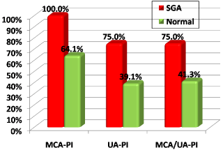
    Figure 5: Comparison according to weight for GA regarding doppler
abnormalities.
![]()
    Characteristics
    Small
    Normal
    P


    (N=8)
    (N=92)


    MCA-PI
    Mean±SD
    1.13±0.05
    1.30±0.27
    ^<0.001*


    Abnormality
    8 (100.0%)
    59 (64.1%)
    #0.051


    UmA-PI
    Mean±SD
    1.12±0.09
    0.99±0.14
    ^<0.001*


    Abnormality
    6 (75.0%)
    36 (39.1%)
    #0.066


    CPR
    Mean±SD
    1.01±0.11
    1.35±0.42
    ^<0.001*


    Abnormality
    6 (75.0%)
    38 (41.3%)
    #0.166


    ^Independent    t-test, #Fisher's Exact test.
Table 9: Comparison according to weight for GA regarding Doppler indices.
![]()
    Characters
    Value
    95% CI
    Value
    95% CI
    Value
    95% CI


    MCA-PA
    UmA-PI
    CPR


    Sensitivity
    100%
    63.1%–100%
    75.00%
    34.9%–96.8%
    75.00%
    34.9%–96.8%


    Specificity
    35.90%
    26.1%–46.5%
    58.70%
    47.9%–68.9%
    60.90%
    50.1%–70.9%


    DA
    41.00%
    31.3%–51.3%
    62.00%
    51.7%–71.5%
    60.00%
    49.7%–69.7%


    YI
    35.90%
    26.1%–45.7%
    35.90%
    4.3%–67.5%
    33.70%
    2.0%–65.3%


    PPV
    11.90%
    5.3%–22.2%
    13.60%
    5.2%–27.4%
    14.30%
    5.4%–28.5%


    NPV
    100%
    89.4%–100%
    96.60%
    88.1%–99.6%
    96.40%
    87.7%–99.6%


    LR+
    1.56
    1.34–1.82
    1.92
    1.19–3.08
    1.82
    1.14–2.90


    LR-
    0
    0.00–0.00
    0.41
    0.12–1.38
    0.43
    0.13–1.43


    LR
    Infinity
    Infinity–Infinity
    4.67
    0.89–24.40
    4.26
    0.82–22.27


    Kappa
    0.082
    0.023–0.142
    0.122
    -0.008–0.252
    0.11
    -0.013–0.234


    CI: Confidence interval; DA: Diagnostic accuracy; PPV: Positive Predictive value; NPV: Negative Predictive    value; LR+: Positive likelihood ratio; LR-: Negative likelihood ratio; LR:    Diagnostic odd ratio.
      MCA-PI had highest sensitivity&    NPV; while MCA/UA-PIratio had highest specificity and PPV in prediction of GA.
Table 10: Diagnostic characteristics of doppler abnormalities in predicting SGA.
Table 11,12 and Figure 6 show that: Cases admitted to NICU had significant more frequent doppler PI abnormalities.
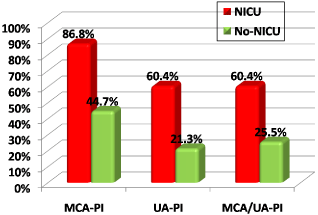
    Figure 6: Comparison according to NICU admission regarding doppler
abnormalities.
![]()
    Characteristics
    NICU
    No-NICU
    P


    (N=53)
    (N=47)


    MCA-PI
    Mean±SD
    1.15±0.15
    1.43±0.28
    ^<0.001*


    Abnormality
    46 (86.8%)
    21 (44.7%)
    #<0.001*


    UA-PI
    Mean±SD
    1.06±0.13
    0.94±0.13
    ^<0.001*


    Abnormality
    32 (60.4%)
    10 (21.3%)
    #<0.001*


    CPR
    Mean±SD
    1.12±0.25
    1.56±0.43
    ^<0.001*


    Abnormality
    32 (60.4%)
    12 (25.5%)
    #<0.001*


    ^Independent    t-test, #Chi square test.
Table 11: Comparison according to NICU admission regarding doppler indices.
![]()
    Charac-ters
    Value
    95% CI
    Value
    95% CI
    Value
    95% CI


    MCA-PI
    UA-PI
    CPR


    Sensitivity
    86.80%
    74.7%––94.5%
    60.40%
    46.0%–73.5%
    60.40%
    46.0%–73.5%


    Specificity
    55.30%
    40.1%–69.8%
    73.70%
    61.3%–86.3%
    74.50%
    59.7%–86.1%


    DA
    72.00%
    62.1%–80.5%
    69.00%
    59.0%–77.9%
    67.00%
    56.9%–76.1%


    YI
    42.10%
    25.2%–59.0%
    39.10%
    21.5%–56.7%
    34.80%
    16.7%–53.0%


    PPV
    68.70%
    56.2%–79.4%
    72.20%
    56.5%–83.9%
    72.70%
    57.2%–85.0%


    NPV
    78.80%
    61.1%–91.0%
    61.80%
    48.1%–74.0%
    62.50%
    48.5%–75.1%


    LR+
    1.94
    1.39–2.72
    2.84
    1.57–5.13
    2.36
    1.39–4.04


    LR-
    0.24
    0.11–0.50
    0.5
    0.35–0.72
    0.53
    0.37–0.77


    LR
    8.14
    3.05–21.71
    5.64
    2.32–13.72
    4.44
    1.89–10.46


    Kappa
    0.428
    0.257–0.600
    0.386
    0.210–0.562
    0.345
    0.164–0.525


    CI: Confidence interval; DA: Diagnostic accuracy; PPV: Positive Predictive value; NPV: Negative Predictive    value; LR+: Positive likelihood ratio; LR-: Negative likelihood ratio; LR:    Diagnostic odd ratio.
      MCA-PI had highest sensitivity&    NPV; while MCA/UA-PI had highest specificity and PPV in prediction of NICU admission.
Table 12: Diagnostic characteristics of doppler abnormalities in predicting NICU admission.
Table 13 and Figure 7 show that: Cases with neonatal mortality had significant more frequent MCA-PI abnormalities non-significant more frequent UA-PI&CPR abnormalities.
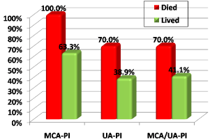
    Figure 7: Comparison according to neonatal mortality regarding doppler
abnormalities.
![]()
    Characteristics
    Died
    Lived
    P


    (N=10)
    (N=90)


    MCA-PI
    Mean±SD
    1.09±0.05
    1.30±0.27
    ^<0.001*


    Abnormality
    10 (100.0%)
    57 (63.3%)
    #0.028*


    UmA-PI
    Mean±SD
    1.11±0.10
    0.99±0.14
    ^0.003*


    Abnormality
    7 (70.0%)
    35 (38.9%)
    #0.090


    CPR
    Mean±SD
    0.99±0.13
    1.36±0.41
    ^<0.001*


    Abnormality
    7 (70.0%)
    37 (41.1%)
    #0.101


    ^Independent    t-test, #Chi square test.
Table 13: Comparison according to neonatal mortality regarding doppler indices.
![]()
    Charac-ters
    Value
    95% CI
    Value
    95% CI
    Value
    95% CI


    MCA-PI
    UA-PI
    CPR


    Sensitivity
    100%
    69.2%–100.0%
    70.00%
    34.8%–93.3%
    70.00%
    34.8%–93.3%


    Specificity
    36.70%
    26.8%–47.5%
    58.90%
    48.0%–69.2%
    61.10%
    50.3%–71.2%


    DA
    43.00%
    33.1%–53.3%
    62.00%
    51.7%–71.5%
    60.00%
    49.7%–69.7%


    YI
    36.70%
    26.7%–46.6%
    31.10%
    1.0%–61.2%
    28.90%
    -1.3%–59.1%


    PPV
    14.90%
    7.4%–25.7%
    15.90%
    6.6%–30.1%
    16.70%
    7.0%–31.4%


    NPV
    100%
    89.4%–100.0%
    94.60%
    85.1%–98.9%
    94.80%
    85.6%–98.9%


    LR+
    1.58
    1.35–1.85
    1.8
    1.11–2.91
    1.7
    1.06–2.74


    LR-
    0
    0.00–0.00
    0.49
    0.19–1.28
    0.51
    0.19–1.33


    LR
    Infinity
    Infinity–Infinity
    3.67
    0.89–15.13
    3.34
    0.81–13.77


    Kappa
    0.104
    0.036–0.171
    0.128
    -0.012–0.269
    0.115
    -0.019–0.249


    CI: Confidence interval; DA: Diagnostic accuracy; PPV: Positive Predictive value; NPV: Negative Predictive    value; LR+: Positive likelihood ratio; LR-: Negative likelihood ratio; LR:    Diagnostic odd ratio.
      MCA-PI had highest sensitivity&    NPV; while MCA/UA-PI had highest specificity and PPV in prediction of neonatal mortality.
Table 14: Diagnostic characteristics of doppler abnormalities in predicting neonatal mortality.
Discussion
Several studies revealed that middle cerebral artery and umbilical artery pulsatility index (PI) ratio have a higher sensitivity and specificity when compared with umbilical artery alone for the prediction of fetal prognosis.
Middle cerebral artery and umbilical artery PI ratio denotes not only the circulatory deficiency of the umbilical velocimetry of the placenta but also the adaptive events resulting in the adjustment of the middle cerebral artery and umbilical artery pulsatility index ratio [4].
Our study was a prospective observational study that was held at Ain Shams university maternity hospital. The aim was to assess the accuracy of middle cerebral artery and umbilical artery Doppler in predicting perinatal outcome in women with PIH in the third trimester.
The study included 100 women with pregnancy-induced hypertension. 16 (16%) women had gestational hypertension, 28 (28%) women had mild PE and 56 (56%) women had severe PE.
The mean (SD) of age was 30.0±5.9 years and the mean (SD) of gestational age was 36.6±1.4 weeks. We found that 69% of women were delivered by C.S, 53% of neonates were admitted to NICU with median duration of admission was 10 (6–14) days, as regards APGAR score at 5min 36% neonates were less than 7, the mean (SD) of birth weight was 2840±410 gm., 8% neonates were SGA, perinatal mortality was 10%.
Our study showed that the group of abnormal MCA/UA PI ratio ‹1.08 (which included 44 women) was significantly associated with higher rates of C.S 39 (88.6%), NICU admission 32 (72.7%), the median duration of NICU admission was longer (11 days), 5 minutes APGAR score < 7 24(54.5%), while abnormal MCA/UA PI ratio < 1.08 was non significantly associated with the rate of perinatal mortality 7 (15.9%) and SGA 6 (13.6%).
The current study also showed a positive correlation between hypertensive disorders in pregnancy and MCA/UA PI ratio less than 1.8, where the rate of severe pre-eclampsia was significantly higher 36 (81.8%) in women who had an MCA/UA PI ratio ‹1.08.
As regarding prediction of NICU admission, we found that MCA/ UA PI ratio had 60.4% sensitivity and 74.5% specificity as predictors of NICU admission, with diagnostic accuracy 67%, positive predictive value 72.7% and negative predictive value 62.5% while our study showed that MCA PI had the highest sensitivity 86.8% and negative predictive value 78.8% in prediction of NICU admission.
Shahinaj et al. (2010) in their prospective observational study that included738 women with preeclampsia and gestational hypertension revealed that in the group of abnormal MCA/UA PI there were a significant higher rates of NICU admission (77.6%), Apgar scores less than 7 at 5 minute (61.9%). MCA /UA PI had 50.1% sensitivity and 79.3% specificity to detect need for NICU admission, with 77.6% positive and 52.5% negative predictive values [8].
Moreover a previous research made by Shahinaj et al. (2010) uncovered that in the group of abnormal MCA/UA PI ratio there was a significant association between women who had abnormal MCA/ UA PI ratio and women who did not as regard the median duration of NICU admission, whereas longer duration of receiving treatment in NICU was needed and this agreed with the results of our study [8].
A previous study made by El-Sokarry et al. in (2011) agreed with the current study in their prospective case-control study that included 100 women. They concluded that the rate of NICU admission and Apgar score ‹7 were higher with abnormal MCA/UA PI ratio (P ‹ 0.05) [9].
In the present study regarding the relation between Doppler indices and small for gestational age we found that MCA PI had the heightest secetivity of (100%) and heightest negative predective value (100%) compared to umblical artery PI and CPR PI but regarding to specificity and positive predictive value CPR PI showed the highest specificity of (60.9%) and highest positive predictive value of (14.3%) in prediction of small of gestational age.
Mishra et al. (2013) announced that all fetuses with absent or reversed diastolic flow in UA had poor perinatal outcome. Revealing that ratio of PI of MCA/UA is more sensitive & specific index in expecting unfavorable perinatal outcome than UA PI or MCA PI used alone with sensitivity & specificity as high as 86% and 92% respectively [10].
The current study disagreed with a previous research made by Ozerena et al. (2009) in their cross sectional prospective study that conducted on 125 normal pregnancy and 62 post term pregnant patients to determine the best index for predicting adverse perinatal outcome or IUGR. As the study demonstrated that UA PI was the best indicator for predection of small of gestational age and admission to NICU. In such study the diagnostic accuracy of cerebro/ umbilical ratio was lower than the S/D ratio of UA denoting a strong relation between UA Doppler indices and adverse perinatal outcome regardless MCA Doppler indices. the UA S/D ratio showed a higher sensitivity and diagnostic accuracy 88% and 94% in predicting adverse perinatal outcome when compared with cerebro/umbilical ratio 81% and 85%, the UA PI (69% and 85%) and the MCA PI (42% and 58%), these findings were against our study, where cerebro/umbilbical ratio showed a better prediction of adverse neonatal outcome than each index separately [11].
In a previous research made by Gramellini et al. (1992), they used a single cut-off value (1.08) to illustrate that the cerebral-umbilical ratio has a higher sensitivity and diagnostic accuracy (68% and 90%) when compared with the UA PI (64% and 83%) and the MCA PI (24% and79%) respectively, the results of the present study seem to be similar, except that in the present study the cutoff value was (1.21) and also higher diagnostic accuracy in the MCA/UA (RI), a ratio which Gramellini did not consider [7].
In the current study, regarding prediction of perinatal mortality MCA PI showed the heightest sensitivity of (100%) and heightest negative perfective value (100%) when compared to umblival artery PI or MCA/ UA PI ratio whereas the MCA/UA PI ratio showed the heighest specificity (61.1%) and positive predective value (16.7%).
In disagreement to our stud, a study made by Abderazek et al. (2016) found that the MCA/UA pulsatility index ratio has low sensitivity and positive predictive value, 58.06 % and 72% respectively, in predicting adverse perinatal mortality.
In our study MCA PI had the heightest sensitivity of (84.1%) and heightest negative predictive value of (66.7%) to predict c.s as mode of delivery, while the CPR PI had less sensitivity of (56.5%) and less negative predective value of (46.4%). On the other hand CPR PI had the best spesificity (83.9%) and positive predective value (88.6%) [12].
In a previous study made by Mahmoud Alalfy et al. (2017) showed that they made a study on estimation of gestational age by US with Doppler indices of umbilical artery and MCA in different growth patterns of fetuses and in women with hypertension and found uteroplacental insufficiency in clinical situations of pregnancy induced hypertension, preeclampsia and IUGR that showed high PI and RI values of umbilical artery Doppler above the 95th percentile for gestational age and revealed pathologically decreased PI and RI values of MCA Doppler below the 5th centile for GA abnormally inverted CPR [13].
Conclusion
Middle cerebral artery pulsatility index is more sensitive than umblical artery pulsatility index and middle cerebral artery/umblical artery pulsitility index ratio in prediction of adverse perinatal outcomes. However middle cerebral artery/umblical artery pulsitility index ratio showed more specificity in predection of perinatal outcome in patients with pregnancy induced hypertension.
Doppler data combining both umbilical and cerebral velocimetery provide additional information on fetal consequences of the placental abnormality; hence, Doppler can be a useful tool in the management of patients with preeclampsia and can help in deciding the time of delivery so that fetuses can be saved.
Recommendations
The use of Doppler ultrasound in high-risk pregnancies appears to improve a number of obstetric outcomes and promising in reducing perinatal morbidity and mortality.
Acknowledgement
We would like to thank the staff members who helped us in this study in, Ainshams university hospital and Algezeerah hospital.
Informed Consent
Informed consent was obtained from all the individual participants included in the study.
Author Contributions
The team of authors participated in this study in its aspects with:
• Substantial contributions to the conception or design of the work; or the acquisition, analysis, or interpretation of data for the work.
• Drafting the work or revising it critically for important intellectual content.
• Final approval of the version to be published.
• Agreement to be accountable for all aspects of the work in ensuring that questions related to the accuracy or integrity of any part of the work are appropriately investigated and resolved.
References
- Smitha K, Sowmya K and Malathi T. Study of Doppler waveforms in pregnancy induced hypertension and its correlation with perinatal outcome. Int J Reprod Contracept Obstet Gynecol. 2014; 3: 428-433.
- American College of Obstetricians and Gynecologists, Report of the American College of Obstetricians and Gynecologists’ Task Force on Hypertension in Pregnancy. Obstet Gynecol. 2013; 122: 1122.
- Venuto and Rocco C. “A Comprehensive Review of Hypertension in Pregnancy”. Journal of Pregnancy. 2012: 1–19.
- Varsha D, Yelikas KA and Priyanka D. Cerebral-umbilical doppler ratio as predictor of perinatal outcome in pregnancies with hypertension disorders. Journal of Evolution of Medical and Dental Sciences. 2013; 2: 7366-7372.
- Rozela Shahinaj, Nikita Manoku, Enriketa Kroi and Ilir Tasha. The value of the middle cerebral to umbilical artery Doppler ratio in the prediction of neonatal outcome in patient with preeclampsia and gestational hypertension. J Prenat Med. 2010; 4: 17-21.
- Arbeille PH, Trauquant F and Body G. Evolution de la circulation arterielle ombilicale et cerebrale du fetus au cours de la grossesse. In: Arbeille PH, Trauquant F, Body G, eds. Progres en Neonatologie.1st ed. Basel: Karger Editions. 1996; 30-37.
- Gramellini D, Folli MC, Raboni S, Vadora E and Merialdi A. Cerebral umbilical Doppler ratio as a predictor of adverse perinatal outcome. Obstet Gynecol. 1992; 79: 416-420.
- Shahinaj R, Manoku N, Kroi E, Tasha I. The value of the middle cerebral to umbilical artery Doppler ratio in the prediction of neontatal outcome in patient with preeclampsia and gestational hypertension. Journal of Prenatal Medicine. 2010; 4: 17-21.
- El-Sokkary M, Omran M and Ahmed H. Ratio of Middle Cerebral Artery / Umbilical Artery Doppler Velocimetry and Status of Newborn in Postterm Pregnancy. 2011.
- Mishra D, Sakhi P, Saraf A, Gupta A, Soni K and Dhruw S. Role of obstetric Doppler in prediction of adverse perinatal outcome in intrauterine growth retardation and pregnancy induced hypertension. Sch J App Med Sci. 2013; 1016-1020.
- Ozerena M, Dinc H and Ekmena U. Umbilical and middle cerebral artery Doppler indices in patient with postterm. Eur J Obstet Gynecol Reprod Biol. 1999; 82: 11-16.
- Abd El-Razek M, Hassan N, Elghareeb N. Middle cerebral artery and umbilical artery pulsatility index ratio in patients with pregnancy-induced hypertension and its correlation with perinatal outcome. 2016.
- Alalfy, Idris, Gaafar, Saad, Nagy, et al. The value of fetal trans cerebellar diameter in detecting GA in different fetal growth patterns in Egyptian fetuses Imaging Med. 2017; 9.