Research Article
Austin J Obstet Gynecol. 2019; 6(4): 1148.
Comparison between Unilateral and Bilateral Sacrospinous Ligament Fixation for Treatment of Uterine Prolapse using Miya Hook
Amin MOA, Azab HI* and Abdellatif HA
Department of Obstetrics and Gynecology, Faculty of Medicine, Alexandria University, Egypt
*Corresponding author: Hossam Ibrahim Azab, Department of Obstetrics and Gynecology, Faculty of Medicine, Alexandria University, Egypt
Received: July 23, 2019; Accepted: August 28, 2019; Published: September 04, 2019
Abstract
Purpose: The aim of the work was the surgical assessment of vaginal sacrospinous ligament fixation for the treatment of second, third and fourth stages of uterine prolapse by the use of Miya hook.
Methods: The study was a prospective randomized clinical study carried out on 20 women randomly selected with uterine prolapse from outpatient clinic of El-shatby maternity university hospital. The degree of prolapse was classified according to the POP-Q system. All 20 patients were operated upon by performing vaginal sacrospinous ligament fixation which were randomly divided into 2 groups (10 patients right Unilateral and 10 patients Bilateral) by the use of the Miya Hook, after exposing the sacrospinous ligament facilitated by the Breisky-Navritil retractors. Patients were evaluated immediately post operatively (after 24 hours) and six months later.
Results: All 20 patients had matching ages, BMI, parity; with mean symptoms, duration of 2.5 years. Both groups had comparable results regarding immediate and late postoperative complications. In the bilateral group, both operative duration and immediate postoperative gluteal pain were significantly higher than in the unilateral group. Sacrospinous ligament fixation in general is an effective and safe procedure for treatment of uterine prolapse with a 90% and 85% for both subjective and objective cure rates.
Conclusions: In conclusion, sacrospinous ligament fixation is an effective and safe procedure for treatment of uterine prolapse with a relatively higher incidence of cystocele recurrence than other procedures due to alteration of the vaginal axis.
Keywords: Sacrospinous; Unilateral; Bilateral; Uterine Prolapse; Miya Hook
Introduction
Pelvic organ prolapse is a common condition in women and its treatment is one of the most common surgical procedures in women. The prevalence of pelvic organ prolapse varies between studies, but has been reported in larger observational studies of menopausal women in the range of 31-41.1% [1,2].
Repair of pelvic support defects can be performed either abdominally or vaginally [3]. These range from partial or total colpocleisis [4] and colpectomy to the newer laparoscopic techniques [5,6] and, more recently, infracoccygealsacropexy (posterior intravaginalslingplasty) [7,8].
One of the more popular procedures is the Sacrospinous Ligament Fixation (SSLF), a simple procedure in which the highest point of the vagina is sutured to the sacrospinous ligament. The procedure was first described by Richter in 1968 as a transvaginal procedure for treatment of vaginal vault prolapse [9], which can be performed both abdominally, and more commonly through the vaginal route. The Miya Hook ligature carrier is an instrument invented by Miyazaki in 1987 making the sacrospinous ligament suspension of the prolapsed vagina or uterus easier [10].
The aim of the work was surgical assessment of vaginal sacrospinous ligament fixation for the treatment of second, third and fourth stages of uterine prolapse by the use of Miya hook.
Patients
The study was conducted on 20 women randomly selected with uterine prolapse from outpatient clinic of El-shatby maternity university hospital.
All patients’ age ranged between 30 to 65 years old, they had normal pelvic scan, had second and third degrees uterine prolapse, were postmenstrual at the time of the procedure for the menstruating patients, medically fit for surgery, and have all signed a consent to be included in the study.
All patients who were pregnant, with pelvic inflammatory diseases, severe medical conditions, with multiple fibroids, ascites, pelvic tumours, present or suspected pelvic malignancy were excluded from the study.
Methods
All 20 unhysterectomized patients included in the study were randomly divided into 2 equal groups, 10 patients who had right unilateral sacrospinous fixation, and 10 patients who had bilateral sacrospinous fixation done to them. All 20 patients were subjected to the following:

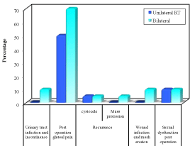
    Figure 1: Comparison between unilateral and bilateral sacrospinous fixation
with postoperative complications.
Full history taking, and examination, both general and local, to make sure that the patient is fit for the procedure according to the defined inclusion criteria and that none of the exclusion criteria is present. The degree of prolapse was classified according to the Pelvic Organ Prolapse Quantification System (POP-Q system) [11].
The procedure: Spinal analgesia was used, and broad spectrum antibiotic had been used. The patient is positioned in the Lithotomy position. Vaginal septal space entered through a longitudinal incision of the posterior vaginal wall, with midline dissection extended to the vaginal apex. Sharp and blunt dissection to the vaginal mucosa was done to be separated from the rectovaginal septum, and lateral dissection to expose either the right lateral space (Right unilateral procedure), or both lateral spaces (Bilateral procedure) by dissecting the vaginal mucosa at the lateral sulci down to the ischial spines. Exposure of the sacrospinous ligaments was facilitated by using Breisky-Navritil retractors. After palpating the ischial spine with 1 finger vaginally, a needle is passed on the same side through a perianal stab incision into the ischiorectal fossa. The finger in the vagina follows the needle tip to help protect the rectum. The needle was passed along the lateral pelvic sidewall toward the ischial spine to penetrate the levator muscle 1cm distal to the ischial spine. The same procedure was repeated on the contralateral side in bilateral cases. No other hysteropexy procedures were done either on a previous setting nor on the present one. No polypropylene mesh was used during the procedure, and all patients kept their uteri intact as they were chosen to have normal uterine anatomy.
Women were evaluated immediately post operatively (24 hours postoperative) and six months later. Subjective assessment of common symptoms of pelvic floor dysfunction was done, as well as objective assessment by vaginal examination.
Results
This trial was conducted at El shat by University Maternity Hospital during the year 2011 and early 2012. A total of 20 patients with variable stages of uterovaginal prolapse according to (POP- Q) system were included.
After the decision of surgical repair for the uterovaginal prolapse was determined, a written consent of the patients participating in this study was obtained prior to surgical correction.
Patients had mean age of 46.9 years, mean BMI 28.63Kgm/m2, all multiparas with median parity 3.5, all had mass protrusion through the enteroitus, with a mean duration of the prolapse of 2.5 years, and none of the studied patients had any previous prolapse surgeries done.
All 20 patients included in the study were randomly divided into 2 equal groups, 10 patients who had right unilateral sacrospinous fixation, and 10 patients who had bilateral sacrospinous fixation done to them.
Early post-operative assessment (first 48 hours)
Vaginal packing was needed for 24 hour in 95% of cases and for 48 hour in 5% of cases. Urinary tract infection and urinary incontinence occurred in one case from all the studied patients and estimated as 5%. Postoperative hospital stay ranged from 2-4 days with a mean of 2.1 days. The mean operative duration was 33.0+5.37 and 40.0+4.7 minutes in both unilateral and bilateral groups respectively, which was statistically longer in the bilateral group.
Late post-operative assessment (First 6 months)
As regards persistence of symptoms, out of 7 cases that suffered from preoperative sexual dysfunction, only 2 cases had persistence of sexual dysfunction.
Out of seven cases that suffered from preoperative irritative symptoms, only 1 case had persistence of irritative symptom.
Out of 11 cases that suffered from preoperative chronic pelvic pain, only 1 case had persistence of chronic pelvic pain post operatively.
As regard the recurrence only three cases showed recurrence in the form of 2 cases cystocele and one case second stage uterine prolapse within 2 months to 6 months respectively of follow up out of 20 cases with success rate 85% (Table 1).
    Unilateral
        (10    patients )

    Bilateral
        (10    patients)

    Total
        (20    patients)


    No.
      %
    No.
      %
    No.
      %


    Urinary tract infection    and incontinence
      0
      0
      1
      10
      1
      5


    FEp
      1


    Post operation gluteal pain
      5
    50
      7
      70
      12
      60


    MCp
      0.002*


    Recurrence


    Cystocele
      1
      5
      1
      5
      2
      10


    Mass protrusion
      0
      0
      1
      5
      1
      5


    FEp
      1


    Wound infection and mesh    erosion
      0
      0
      1
      10
      1
      5


    FEp
      1


    Sexual dysfunction post    operation
      1
      10
      1
      10
      2
      10


    FEp
      1


    FEp: p    value for Fisher Exact test
      MCp: p    value for Monte Carlo test
      *: Statistically significant at p =0.05
Table 1: Comparison between unilateral and bilateral sacrospinous fixation with
late postoperative complications.
Out of 20 cases, only one case had mesh erosion and wound infection representing 5% of the study.
Out of 20 cases who have all undergone sacrospinous fixation, 5 cases (25%) represent post-operative gluteal pain in unilateral sacrospinous fixation and 7 cases (35%) in bilateral sacrospinous which disappeared in both groups in a period ranged from (2 weeks-3 months). Therefore, there is statistical difference between unilateral and bilateral sacrospinous fixation in post-operative gluteal pain (Table 1).
After a mean follow up period of 6 months, the subjective cure rate was (symptomatic relief) (18/20) 90% and the objective cure rate was (17/20) 85%.
Discussion
One of the more popular procedures for treating vault and uterovaginal prolapse is the SSLF. The sacrospinal ligament is located in the posterior department of the pelvic, its position is constant and with high power, so it is an effective point for vault fixation. The vaginal approach allows concomitant repair of all vaginal defects easily [12].
This study was conducted to assess the surgical effectiveness of sacrospinous ligament fixation using Miya hook for treatment of uterine prolapse. The patients included in this study described typically the risk factors of pelvic organ prolapse, which included the age (46.90 ± 9.21 years), high parity (3.95 ± 1.57) and BMI (28.63 ± 2.47 kg/m2). About 25% the recruited patients were menopausal. The mean duration of follow up was 6 months. After this follow up period, the cure rate was symptomatically (subjectively) 90% and anatomically (objectively) 85%.
All articles about SSLF published between January 2004 and December 2009, showed a short-term cure rates between 84% and 100%. Recurrence of prolapse of the anterior vaginal wall ranged as high as 15.7%. Prolapse of the vaginal apex occurred in up to 6% [13]. Bilateral fixation appears more attainable in a patient with post hysterectomy vaginal vault prolapse than in one with uterine prolapse [14]. Other studies noticed that bilateral suspension may increase the risk of a subsequent ‘high cystocoele’, by stretching the already weakened fibro connective vaginal layer between the two spines [15,16].
Pollak et al. compared the complications of three techniques used to pass the suture through the sacrospinous ligament when performing SSLF, which was evaluated when done under vision and with palpation. The result was that 12 women (5%) had intra operative and 40 women (17%) had postoperative complications suspected to be caused directly from the suture placement. They concluded that passing the suture through the sacrospinous ligament under direct visualization might result in less intra- and postoperative complications [17]. Malinowski et al. [18] conducted sacrospinous fixation on 10 cases with follow up period about 12 months reported 100% cure rate with no recurrence. Different views exist regarding the anatomical relationships of the ligament and respective innervations, length and histology. Two studies have shown dramatic distortions in the vaginal axis after SSLF which can explain the occurrence of cystocele as a late complication [19,20].
In the present study, the suture was passed through the sacrospinous ligament using Miya hook ligature carrier under visualization of the ligament with the aid of Breisky-Navritil retractors to minimize intra operative and post operative complication. Mild gluteal pain was encountered in 12 out of all 20 cases included in the study, which were all controlled by analgesics. When comparing both study groups, operative duration was statistically longer, and gluteal pain was more in the bilateral group as compared to the unilateral group.
In conclusion, sacrospinous ligament fixation is an effective and safe procedure for treatment of uterine prolapse with a 90% and 85%for both subjective and objective cure rates. There was a relatively higher incidence of cystocele recurrence than other procedures due to alteration of the vaginal axis.
References
- Handa VL, Garrett E, Hendrix S, Gold E, Robbins J. Progression and remission of pelvic organ prolapse: a longitudinal study of menopausal women. Am J Obstet Gynecol. 2004; 190: 27-32.
- Hendrix SL, Clark A, Nygaard I, Aragaki A, Barnabei V, McTiernan A. Pelvic organ prolapse in the Women’s Health Initiative: gravity and gravidity. Am J Obstet Gynecol. 2002; 186: 1160-1166.
- Timmons MC, Addison WA, Addison SB, Cavenar MG. Abdominal sacral colpopexy in 163 women with post-hysterectomy vaginal vault prolapse and enterocele. Evolution of operative techniques. J Reprod Med. 1992; 37: 323- 327.
- Von Pechmann WS, Mutone M, Fyffe J, Hale DS. Total colpocleisis with high levator plication for the treatment of advanced pelvic organ prolapse. Am J Ostet Gynaecol. 2003; 189: 121-126.
- Wattiez A, Mashiach R, Donoso M. Laparoscopic repair of vaginal vault prolapse. Curr Opin Obstet Gynecol. 2003; 15: 315-319.
- Miklos JR, Moore RD, Kohli N. Laparoscopic surgery for pelvic support defects. Curr Opin Obstet Gynecol. 2002; 14: 387-395.
- Petros PE. New ambulatory surgical methods using an anatomical classification of urinary dysfunction improve stress, urge and abnormal emptying. Int Urogynecol J Pelvic Floor Dysfunct. 1997; 8: 270-277.
- Farnsworth BN. Posterior intravaginal slingplasty (infracoccygeal sacropexy) for severe post-hysterectomy vaginal vault prolapse - a preliminary report on efficacy and safety. Int Urogynecol J Pelvic Floor Dysfunct. 2002; 13: 4-8.
- Richter K. Massive eversion of the vagina: pathogenesis, diagnosis, and therapy of the true prolapse of the vaginal stump. Clin Obstet Gynecol. 1982; 25: 897-912.
- Miyazaki FS. Miya hook ligature carrier for sacros-pinous ligament suspenion. Obstetrics and Gynecology. 1987; 70: 286-288.
- Persu C, Chapple CR, Canni V, Gutue V, Geavlete P. Pelvic Organ Prolapse Quantification System (POP-Q). A new era in pelvic organ prolapse staging. J Med Life. 2011; 4: 75-81.
- Morley GW, DeLancey JOL. Sacrospinous ligament fixation for eversion of the vagina. Am J Obstet Gynecol. 1988; 158: 872-881.
- Baumann M, Salvisberg C, Mueller M, Kuhn A. Sexual function after sacrospinous fixation for vaginal vault prolapse: bad or mad? Surgical Endoscopy. 2009; 23: 1013-1037.
- Pohl JF, Frattarelli JL. Bilateral transvaginal sacrospinous colpopexy: Preliminary experience. American Journal of Obstetrics and Gynecology. 1997; 177: 1356-1362.
- Toglia MR, Fagan MJ. Suture erosion rates and long-term surgical outcomes in patients undergoing sacrospinous ligament suspension with braided polyester suture. American Journal of Obstetrics and Gynecology. 2008; 198: 1-4.
- Kearney R, DeLancey J. Selecting suspension points and excising the vagina during Michigan four-wall sacrospinous suspension. Obstetrics and Gynecology. 2003; 101: 325-330.
- Pollak J, Takacs P, Medina C. Complications of three sacrospinous ligament fixation techniques. International Journal of Gynaecology and Obstetrics. 2007; 99: 18-22.
- Malinowski A, Pawlowski T, Maciolek-Blewniewska G, Augustyniak T. Sacrospinous ligament vaginal vault fixation-method, results and a one-year follow up in 10 patients. Ginekologia Polska. 2004; 75: 713-719.
- Rane A, Lim YN, Withey G, Muller R. Magnetic resonance imaging findings following three different vaginal vault prolapse repair procedures: a randomised study. Australia and New Zealand Journal of Obstetrics and Gynecology. 2004; 44: 135-139.
- Sze EH, Meranus J, Kohli N, Miklos JR, Karram MM. Vaginal configuration on MRI after abdominal sacrocolpopexy and sacrospinous ligament suspension. International Urogynecology Journal and Pelvic Floor Dysfunction. 2001; 12: 375-380.