Case Report
Austin J Obstet Gynecol. 2021; 8(5): 1184.
A Bladder Teratoma after Oocyte Retrievals
Ji MM, Yuan M, Jiao X and Wang GY*
Department of Obstetrics and Gynecology, Qilu Hospital of Shandong University, Jinan, China
*Corresponding author: Guoyun Wang, Department of Obstetrics and Gynecology, Qilu Hospital of Shandong University, Jinan, 250012, China
Received: May 26, 2021; Accepted: June 08, 2021; Published: June 15, 2021
Abstract
Oocyte recovery by means of transvaginal ultrasound-guided puncture was first described in 1985, and the procedure has gained widespread popularity. The literature consistently reports that complications related to oocyte retrievals are rare. Yet this report describes a woman with a long-term complication after egg retrievals.
Keywords: Oocyte Retrievals; Long-term complication; Endometriosis; Teratoma
Case Presentation
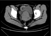
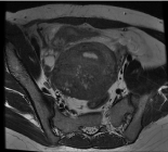
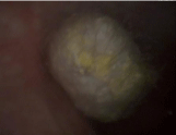
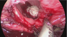
A 36-year-old woman presented to the urology clinic with a 1-year history of dysuria. The patient occasionally felt slight lower abdominal pain, without frequent urination, urgent urination or gross hematuria, no fever. Her medical history was notable for an ovarian endometriosis cyst that had been treated 8 years earlier with surgical excision and 2 times oocyte retrievals 1 year ago. Urine test showed: leukocyte (WBC) 2265.70/uL (normal range: 0 to 25 / uL), urinary protein (PRO)+ -, urine occult blood (BLD) 2+. Blood test showed hemoglobin 84g/L (normal range: 115 to 150 g/L). Computed Tomography (CT) showed a lesion in the urinary bladder whose boundary with the right ovary was not clear with variegated attenuation of fat and local calcification (Figure 1). Therefore, the urologist recommends that the patient go to the gynecology department. After the patient was admitted to the hospital, related examinations were done. Magnetic Resonance Imaging (MRI) showed thickened bladder wall with local incontinence of muscular signals and no enhanced cystoid lesion in the bladder (Figure 2). Serum test showed the carbohydrate antigen 125 228.5U/ml (normal range: 0 to 30.2U/mL), carbohydrate antigen 199 40.96U/mL (normal range: 0 to 37U/mL). After 3 days of antibiotic treatment, the patient underwent cystoscopy which revealed a mass with multiple hairs and floc deposition over it (Figure 3). What exactly is the mass in the patient’s bladder? The patient said that dysuria was related to menstruation. We all know that endometriosis is a disease with high recurrence. Considering that the patient had undergone surgical excision of overian endometriosis and egg retrieval operations and the patient’s symptom appeared after the egg retrieval and CT and MRI demonstrated a mass in the bladder passing through the bladder wall connected to the ovary and the uterus, we suspected that endometriosis of the ovary might have invaded the bladder through tiny egg retrieval needle, leading to bladder endometriosis. The fact that there was a cyst with a diameter of 4cm on the right ovary before egg retrieval and a history of dysmenorrhea also corroborated this hypothesis. However, through CT and cystoscopy, the mass did not seem to be a bladder endometriosis. The patient was treated with laparoscopic surgery. The greater omentum adhered to the bladder, uterus and adnexa uteri. After separation, we observed that the uterus was as big as 2 months pregnant, the posterior wall and the right round ligament thickened obviously, and cyst with a diameter of 2cm was seen in the uterus below the right round ligament, and the same cyst was seen in the right ovary. We removed the bladder tumor, part of the bladder wall and the cysts in the right ovary and the uterus (Figure 4 and Video). Chocolate-like liquid was seen during the resection of the cysts. The histopathologic analysis reported mature cystic teratoma, chronic inflammation of bladder wall mucosa with granulation tissue hyperplasia, endometriosis cyst on the ovary and uterus and adenomyosis. Immunohistochemistry showed CK (+), S-100 (+), GFAP (+), NeuN neurons (+), Desmin smooth muscle (+). The positive rate of Ki-67 was about 2%. The patient was treated with Gonadotrophin-releasing hormone agonists (GnRH-a) after the operation to reduce disease recurrence and the woman recovers well after operation and is being followed up in outpatient clinic. The patient did not go to the reproductive clinic again due to fear of oocyte retrievals.

    Figure 1: CT image demonstrating a lesion in the urinary bladder whose
boundary with the right ovary was not clear with variegated attenuation of fat
and local calcification. CT: Computed tomography.

    Figure 2: MRI image showing thickened bladder wall with local incontinence
of muscular signals and no enhanced cystoid lesion in the bladder. MRI:
Magnetic resonance imaging.

    Figure 3: Cystoscopy showing a mass in the bladder with multiple hairs and
floc deposition over it.

Figure 4: Image showing the removed bladder tumor.
Discussion
This case is worth thinking about. First of all, did the teratoma of this patient arise from the bladder or the ovary? The pathogenesis of teratomas is not clear, and the extragonadal teratomas are poorly understood. The most accepted theory to explain the existence of ovarian teratomas is the parthenogenetic activation of oocytes [1]. Extragonadal teratomas may develop in any central place as a result of reproductive cell migration during the germ cell cycle, from supernumerary ovaries, and when the mature cystic teratoma of the ovary is spontaneously amputated, re-attaching in a different place [2]. For this patient, we did not find a teratoma on the ovary during the operation, so we were not sure whether it was the primary teratoma in the bladder, the teratoma formed by the residual ovarian tissue in the bladder after oocyte retrievals, or bladder erosion of overian teratoma. The second point is about the complications of oocyte retrievals. Levi-Setti PE et al. reviewed 23,827 oocyte retrievals and described hemoperitoneum (0.26%) as the most frequent complication, followed by pelvic pain (0.06%), anesthetic complications(0.06%), pelvic infection (0.04%), vaginal wall bleeding(0.01%) and bladder lesions(0.01%). They reported that two cases of bladder trauma (0.008%) were diagnosed as hematuria and vesical hematoma, but neither of the cases required intervention and both patients were discharged from the hospital after 1 day of observation [3]. Cases of bladder hematoma after oocyte retrieval procedures have been reported previously [4-6]. In this case, we think that the severe pelvic adhesions and endometriotic cysts on the uterine wall might result from the puncture of the wall of the chocolate cyst of the ovary. The lesson from this case is that we should try not to pass through the bladder when taking ova, especially for those patients with chocolate cysts, not only because of some recent complications like bladder bleeding, but also for possible long-term complications, for example, the cyst fluid may aggravate pelvic adhesion and active endometrium may plant in pelvic organs such as the uterus or even the bladder forming new endometriosis leisions. The current guideline also recommends ovarian cystectomy, instead of drainage or coagulation [7]. However sometimes the pelvic adhesion in patients with endometriosis is so serious that it is difficult to avoid passing through the bladder and puncturing the cyst fluid when retrieving the eggs. In these cases, is oocyte recovery under laparoscopic monitoring a better choice?. The last point is the choice of the surgical way. In this case, maybe we could adopt less invasive surgical method such as cystoscopic surgery to remove the teratoma avoiding laparoscopic surgery and bladder wall resection. Injury of surrounding pelvic structures during an oocyte retrieval procedure may not necessarily present immediately after the procedure. Patients may return with serious complications a week or more later, sometimes even a year after the procedure. They may even visit other departments, so iatrogenic injuries should be kept in mind.
References
- Oliveira FG, Dozortsev D, Diamond MP, Fracasso A, Abdelmassih S, Abdelmassih V, et al. Evidence of parthenogenetic origin of ovarian teratoma: case report. Hum Reprod. 2004; 19: 1867-1870.
- Kakuda M, Matsuzaki S, Kobayashi E, Yoshino K, Morii E, Kimura T. A Case of Extragonadal Teratoma in the Pouch of Douglas and Literature Review. J Minim Invasive Gynecol. 2015; 22: 1311-1317.
- Levi-Setti PE, Cirillo F, Scolaro V, Morenghi E, Heilbron F, Girardello D, et al. Appraisal of clinical complications after 23,827 oocyte retrievals in a large assisted reproductive technology program. Fertil Steril. 2018; 109: 1038- 1043.
- Modder J, Kettel LM, Sakamoto K. Hematuria and clot retention after transvaginal oocyte aspiration: a case report. Fertil Steril. 2006; 86: 720.e1- 720.e2.
- Jayakrishnan K, Raman VK, Vijayalakshmi VK, Baheti S, Nambiar D. Massive hematuria with hemodynamic instability--complication of oocyte retrieval. Fertil Steril. 2011; 96: e22-e24.
- Souza MdCBd, Souza MMd, Antunes RdA, Tamm MA, Silva JBd, Mancebo ACA. Bladder hematoma: a complication from an oocyte retrieval procedure. JBRA Assist Reprod. 2019; 23: 75-78.
- Hwang H, Chung Y-J, Lee SR, Park H-T, Song J-Y, Kim H, et al. Clinical evaluation and management of endometriosis: guideline for Korean patients from Korean Society of Endometriosis. Obstet Gynecol Sci. 2018; 61: 553- 564.