Case Series
Austin J Obstet Gynecol. 2022; 9(1): 1198.
Clinical Presentation of Intestinal Obstruction in Pregnancy and Management Challenges: Case Series
Jay Lodhia1,2*, Bariki Mchome2,4, Joylene Tendai1, Adnan Sadiq2,3, Kondo Chilonga1,2, Samwel Chugulu1,2 and David Msuya1,2
1Department of General Surgery, Kilimanjaro Christian Medical Centre, P O Box 3010, Moshi Tanzania
2Kilimanjaro Christian Medical University College, Faculty of Medicine, P O Box 2240, Moshi Tanzania
3Department of Radiology, Kilimanjaro Christian Medical Centre, P O Box 3010, Moshi Tanzania
4Department of Obstetrics and Gynaecology, Kilimanjaro Christian Medical Centre, P O Box 3010, Moshi Tanzania
*Corresponding author: Jay Lodhia, Department of General Surgery, Kilimanjaro Christian Medical Centre, P O Box 3010, Moshi Tanzania
Received: May 19, 2022; Accepted: June 21, 2022; Published: June 28, 2022
Abstract
Introduction: Abdominal emergencies during pregnancy (excluding obstetric emergencies) occur on one out of 500-700 pregnancies. Since they are relatively rare patients should be referred and managed in tertiary level centres where surgical, obstetrical and neonatal cares are available. To our understanding there are no detailed descriptive series of occurrences of intestinal obstruction in pregnancy emanating from (Low and middle income countries) LMIC including Tanzania.
Case Presentation: Herein, we present four cases of intestinal obstruction in pregnancy that was managed at a tertiary level centre in a Northern Tanzania. We share experiences and challenges in the surgical management and short term surgical outcomes.
Conclusion: Clinical presentations may be atypical and misleading due to pregnancy-associated anatomical and physiologic alterations which often lead in diagnostic uncertainty and therapeutic delays with increased risk of maternal and fetal morbidity.
Keywords: Diagnosis; East Africa; Intestinal Obstruction; Pregnancy
Background
Intestinal obstruction (IO) in pregnancy is a challenging and an uncommon non-obstetric surgical pathology with high fetomaternal morbidity and mortality1. The incidence ranges from 1 in 5000 to 1 in 660002. This complexity poses a great challenge to the surgeon and obstetrician on the decision making on the diagnostic and therapeutic options1. Herein we describe four cases of intestinal obstruction in pregnancy and our experience at a resource-limited setting.
Case Presentation
Case 1
A 29-year-old G2P1L1 with an gestation age of 24 weeks and 6 days by date, presented with a one-week history of abdominal pain associated with distension, vomiting and constipation. There was no history of abdominal trauma or vaginal discharge. She was on iron and folate supplements. Past obstetric history includes a cesarean section due to eclampsia.
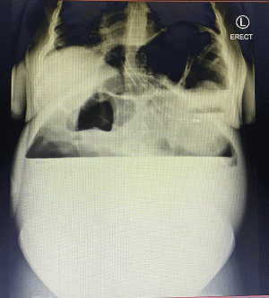
On examination, she was lethargic, febrile (T 38oC), mildly pale and dehydrated,. Her blood pressure of 112/81 mmHg, pulse rate of 136 bpm, saturating at 96% in room air. Her abdomen was symmetrically distended with a pfannenstiel scar, tense and tender on palpation, and hypertympanic percussion note on the upper quadrants with no bowel sounds on auscultation. She had a naso-gastric tube in situ draining fecal content. Other systems were unremarkable. The blood work-up reported a hemoglobin of 9.4 g/dl, leukocyte count of 9.17 X10⁹/L, sodium of 132.23 mmol/l, potassium of 2.52 mmol/l, creatinine of 42 umol/l and urea of 2.50 mmol/l. Abdominal USS reported an impression of a gaseous abdomen, minimal ascites with a viable intrauterine pregnancy of 24 weeks. An erect plain abdominal X-ray was done that was suggestive of intestinal obstruction with a differential of perforated hollow viscus (Figure 1). She was kept nil orally and on intravenous fluids for resuscitation.

    Figure 1: Erect abdominal X-ray showing large air-fluid level suggestive of

intestinal obstruction.
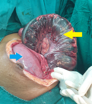
The obstetrics team was consulted and an obstetric ultrasound reported a live singleton intrauterine pregnancy at gestational age of 24 weeks and 3 days, adequate liquor and fetal heart rate of 139 bpm. A decision for expeditious laparotomy was reached after joint discussion (Obstetricians and Surgeons). Intra-operatively, a gravid uterus with 500mls of amber colored ascites was encountered. A 360 degrees anticlockwise sigmoid volvulus was found twisted around its gangrenous mesentery and grossly distended (Figure 2). The stomach, small bowels, caecum, ascending, transverse and descending colon were distended. The volvulus was untwisted, sigmoid colon resected and hartmann’s colostomy raised. The patient received 450mls of whole blood intra-operatively. Her control hemoglobin was 11.5 g/dl day two post operatively.

Figure 2: Gangrenous sigmoid volvulus (Yellow) and gravid uterus (Blue).
Post operatively, the patient was nursed in the surgical ICU, nil orally, antibiotics and analgesics as per hospital protocol. She was also reviewed by the obstetricians that initiated Nifedipine 10 mg twice daily. One day post laparotomy patient had spontaneous abortion of a male fetus of 800 g. The psychology team was involved during the course of treatment, giving her mental health support and was to be seen regularly in their clinic for continued sessions.
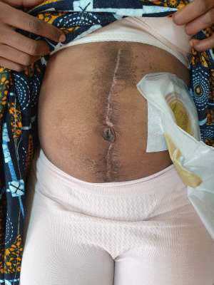
The patient was discharged on the 8th day post operative with stable vitals, and a functional colostomy. She was reviewed 6 weeks later at the surgical and obstetric outpatient unit whereby she had no abdominal symptoms, histology of the sigmoid colon was unremarkable and she was initiated oral combined contraceptives to regulate her menses. Her colostomy (Figure 3) was closed and 6 weeks later was reviewed again at the surgical outpatient unit whereby she was clinically stable hence discharged.

Figure 3: Healed midline incision with colostomy in situ.
Case 2
A 39-year-old G10P9L9 with a gestation age of 23 weeks presented with a 3-day history of sudden onset of abdominal pain associated with distension, absolute constipation and vomiting after every oral intake. There were no significant past medical or surgical history. Her previous obstetric history was essentially uneventful with all previous vaginal deliveries. On general examination, she was ill-looking, mildly pale, mildly dehydrated and febrile to touch. Her initial blood pressure was 140/82 mmHg, axillary temperature of 37.7oC, pulse rate of 122 beats/min, respiratory rate of 20 breaths per cycle saturating at 96% on room air. She has a naso-gastric tube that was draining dark gastric contents. Her abdomen was grossly distended with generalized abdominal pain, absent bowel sounds and a positive blumberg sign. Routine blood tests revealed leukocyte count of 15.40 x 10⁹/L, haemoglobin of 8.0 g/dl and a platelet count of 224 x 109/L, serum sodium of 134.92 mmol/l, potassium of 2.44 mmol/l, serum creatinine of 39 umol/l, BUN of 4.56 mmol/l and liver enzymes within range.
An abdominal ultrasound revealed dilated bowel loops with intraluminal free fluid & internal echoes, prominence of valvulae conniventes, extra luminal free fluid and insufficient peristalsis movements. The uterus was gravid with a single-tone pregnancy of 23 weeks. A diagnosis of small bowel obstruction with peritonitis in pregnancy was made. An emergency laparotomy was performed revealing approximately one litre of foul smelling hemorrhagic ascites and twisted terminal ileum around its mesentery. Approximately 40cm of the terminal ileum was gangrenous including the ileocecal junction, and a fecaloma was found in the ascending and transverse colon. . The gangrenous segment of the bowel was resected, ileocaecal junction closed, ileostomy raised, lavage done and the fascia was closed but the skin was left open. The resected terminal ileum was sent for histology analysis which was insignificant. A drain was left in situ and the patient received 450mls of whole blood intraoperatively.
Post operatively, the patient was nursed in the surgical ICU and the obstetric team initiated Indomethacin 25mg twice daily for 24 hours for tocolytic effect. The Fetal Heart rate (FHR) was 124 bpm. The patient started oral sips on the day-3 post operative and was tolerating feeds well. On day she developed a burst abdomen and was prepared for re-laparotomy. The abdomen cavity was thoroughly cleaned, the ileostomy was refashioned and tension-sutures were applied. She received 450 mls of whole blood. She continued to fair well in the general surgery unit and was discharged after 18 days with gestational age of 27 weeks and 1 day. Prior discharge she was reviewed by the obstetrician and obstetric ultrasound was uneventful with fetal heart rate of 142.
After a week she attended her routine ANC clinic, whereby she was moderately pale, mildly sclera jaundiced and had bilateral lower limb non-tender pitting edema. Her blood pressure measured 97/46 mmHg, pulse rate of 123 beats per minute and saturating at 97% on room air. She was admitted to the obstetric ward with a diagnosis of high risk pre-term pregnancy not in labor. Her blood work revealed hemoglobin of 8 g/dl, platelets of 108 x 109/l, sodium 127 mmol/l and potassium of 3.7mmol/l, AST of 23.31 U/l and ALT 10.67 U/l. Dexamethasone was initiated at 6gm twice daily for 2 days for maturation of fetal lungs and she received 2 units of whole blood during her stay. The ileostomy was patent and the midline incision clean and dry. The patient was then discharged after 2 days on oral iron and ferrous supplements and her control hemoglobin was 9.6 g/dl.
Four weeks later, she attended ANC whereby her ultrasound revealed gestation age of 31 weeks with adequate amniotic fluid. She was not jaundiced and not pale with resolved lower limb edema. Her ileostomy was functioning well and was discharged. She delivered her baby at a peripheral hospital hence details of the gestation age, newborn’s vitals and weight were not obtained.
She was then admitted two months post delivery for ileostomy closure. Her haemoglobin was 10.1 g/dl, platelet count of 258 x 109/L and normal serum sodium and potassium levels. Intraoperatively multiple adhesions were encountered, small bowel was atrophied, transverse colon with fecaloma in the ileostomy was mobilized, adhesiolysis performed and standard right hemicolectomy done. Ileal-transverse end-to-end anastomosis and lavage done, abdominal drain was left in situ. During her stay in the ward, patient has an uneventful recovery and was discharged 4-days post laparotomy and was scheduled to visit the surgical out-patient clinic after 2 weeks but the patient was lost to follow-up.
Case 3
A 34-year-old female, G7P6L5 was admitted at the obstetrics department in her 29th week of pregnancy by booking, with complaints of sudden onset of left upper quadrant abdominal pain for 2 weeks. She past medical and surgical history was uneventful and delivered previously vaginally. On general examination, the patient was moderately pale, alert, Abdominal examination revealed tenderness on the left upper quadrant, a gravid uterus at a fundal height of 29 weeks, and fetal heart rate of 142 beats per minute.
Obstetrics ultrasound recorded a biophysical profile of 8/8 with the placenta lying fundal anterior. A full blood count on admission reported hemoglobin of 6.4g/dl, normal leucocyte and platelet counts. She tested negative for malaria and HIV. Second day in the ward she started having recurrent feculent vomiting associated with absolute constipation. Her plain abdominal x-ray revealed multiple air-fluid levels with empty rectum in pregnancy. She was kept nil orally, catheterized, and a nasogastric tube was inserted. Her blood work reported normal electrolyte count, serum sodium of 135.11mmol/l and potassium of 3.38mmol/l. An emergency laparotomy was performed. Intraoperative findings included clear peritoneal fluid with gravid uterus, ileal-ileal intussusception of 60 cm telescoped, about 160 cm from the ileocecal junction, a constricting tumor like mass and enlarged mesenteric lymph nodes. The tumor was resected with 5 cm on each side, an ileo-ileal end-to-end anastomosis was done, patency was established and vent was closed. Lavage was done and the abdomen was closed in layers. The sample was sent for histopathology that concluded well differentiated invasive adenocarcinoma of the ileum (pT2N0Mx) with free margins. She received 450 mls of whole blood intra-operatively and another 450 mls post operatively. A post operative diagnosis of Intestinal obstruction secondary to an intussusception due to an ileal tumor was established.
The patient had an uneventful recovery and was discharged on the 13th day post operative whereby she reported positive fetal kicks and was tolerating meals. She was then reviewed after four weeks at the surgical out-patient unit whereby she was clinically stable, not pale and tolerating oral feeds well with no constipation. She was on oral hematinics. Unfortunately she was later lost to follow up.
Case 4
A 24-year-old female, prime gravid with a gestational age of 20 weeks by dates, was referred to our centre due to six day history of generalized abdominal pain that was of sudden onset and progressive and aggravating by taking oral meals. This was associated with nonprojectile vomiting and not passing stools. She denied abdominal trauma and did not experience any vaginal discharge during the course of her illness. Her past medical history was uneventful. She has been attending antenatal clinic, received iron and folate supplements and been tested negative for HIV and malaria. She has been reported to have normal blood sugars and blood pressures.
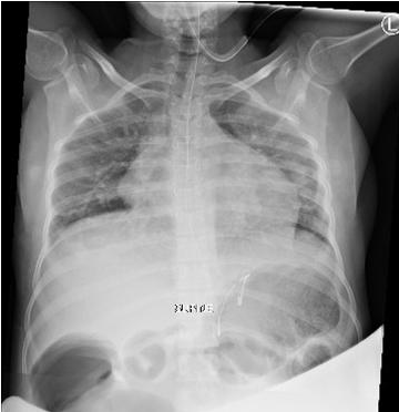
Upon examination, she was fully conscious, mildly pale with no lower limb edema. She has a nasogastric tube in situ that was draining gastric contents. Her BP measured 100/72 mmHg, pulse was 122, axillary temperature of 36.4oC and saturation at 94% on room air. Her abdomen was grossly distended with no scars, generalized tenderness with a palpable gravid uterus and exaggerated bowel sounds on auscultation. Rectal examination revealed empty rectum and other systems were essentially normal. Her leucocyte count was 19 x 109/L, haemoglobin of 8 g/dl and a normal platelet count. Her serum creatinine was 30 μmol/L and BUN of 1.70 mmol/L. She had an abdominal ultrasound that showed dilated bowel loops, abdominal x-ray showed dilated bowels with no air-fluid levels and chest x-ray was normal (Figure 4).

Figure 4: X-ray showing no chest abnormalities with some dilated loops.
She was taken for an emergency laparotomy whereby a gravid uterus was found with clear ascites, and a compound volvulus (ileosigmoid knotting) was found with gangrenous small bowel and sigmoid loop within the volvulus. Hence resection of 20 cm of the terminal ileum and sigmoid done, end-to-end anastomosis of the small bowel done to restore continuity, Hartmann’s colostomy was raised of the sigmoid colon. Blood loss of 700 mls was predicted hence 800 mls of whole blood was transfused. The patient was nursed in the intensive care whereby she recovered and faired well. Control serum sodium and potassium were normal, serum creatinine was 27 umol/L BUN of 3.30 mmol/L and hemoglobin of 9 g/dl. On day 4 she expelled a fetus with no signs of life. She continued to fair well with functioning colostomy, nutritional and psychological support.
Two months later, she was admitted to the surgical unit for an elective closure of her colostomy. Her haemoglobin was 11.2 g/dl with normal parameters and serum creatinine of 78 umol/L. The distal loopogram was normal and the procedure went well with good recovery. Three weeks later she was reviewed at the outpatient unit whereby she was doing well with no abdominal symptoms hence discharged.
Discussion
Pregnancy complicated by intestinal obstruction is rare with high fetomaternal morbidity and mortality; maternal mortality of 6% and fetal mortality of 26%2. Adhesions accounts for 54.6%, intestinal volvulus accounts for 24.5%, intussusceptions in 5.1%, caricoma in 3.7% and hernias in 1.4% of acute bowel obstructions in pregnancy2. Decision making on the diagnostic and therapeutic options is a challenge to the multidisciplinary team keeping in mind bowel ischemia or strangulation as a complication of delayed intervention which can lead to the maternal morbidity and fetal loss. Reduction of complications depends on early detection unfortunately accurate diagnosis is usually difficult in pregnancy [1].
Zhao et al. reported a rare cause of IO in pregnancy being reverse rotation of the midgut nevertheless the management is similar to that of all kinds of obstruction. The authors reported that symptomatic management can be opted for those without sepsis and intestinal ischemia with gastrointestinal decompression, parenteral nutrition, antispasmodic agents, but in those with complications, emergency surgery should not delay as in with our cases whereby the patients delayed to present to the tertiary centre and had features of sepsis [2].
Small bowel obstruction (SBO) is a common cause of IO but rarely occurs in pregnancy. The most common cause of SBO is adhesions, accounting in about 70% of cases3. Although this entity carries significant risk to the fetus and mother, with an overall fetal loss of 17% and maternal mortality rate of 2%3. Diagnosis again is difficult as the symptoms are attributed to pregnancy and there can be a reluctance to request X-rays owing to the risks of ionising radiation hence leading to delayed initial management [3].
Generally there are various etiologies of abdominal-pelvic pain in pregnancy that leads to surgery, however the anatomic and physiological alteration of pregnancy itself often lead to atypical clinical presentations hence making it difficult for surgeons and obstetricians to choose investigations and initiate prompt management since two lives are at risk therefore such cases should be managed in specialized centres [4]. The signs and symptoms of IO are generally similar to that of non-pregnant hence initial medical treatment (NPO, bowel decompression by NGT, fluid and electrolyte repletion) is same, followed by a midline laparotomy is recommended depending on the height of uterus [4].
Most cases of IO in pregnancy are reported in the third trimester whereas on the contrary three index cases were in their second trimester and only one in third [5]. Ghahremani et al also state in their report that in pregnancy diagnosis is delayed hence surgery leading to increased morbidity, similar to the index cases whereby accurate diagnosis were made intraoperatively. Sigmoid volvulus can be managed endoscopically if in first trimester but difficult later as higher risk of bowel perforation due to the large uterus displacing the bowel loops [5]. It was evident in the first case that diagnosis by plain abdominal x-ray was not conclusive and due to the delayed presentation the bowel loop was gangrenous warranting resection unlike in the case reported by Ghahremani et al.
Ileosigmoid knotting (ISK) in pregnancy is rare with unknown incidence [6]. The prognosis is said to be poor and common in multiparous women in their third trimester6. In the index case (four) the surgery and outcome was a success unfortunately had an abortion and on the contrary she was prime gravida and in her second trimester. Probable cause of ISK in the index case could be due to anatomic factors like long mesentery of the small bowels and a long sigmoid with a narrow paddicle [6]. Management is surgery whereby untwisting the bowel loops is adequate if viable, but in the index case whereby the bowel loops were gangrenous due to the late presentation bowel resection is inevitable [6].
Conclusion
The appropriate diagnosis and management of intestinal obstruction in pregnancy are of importance as it is associated with maternal and fetal mortality. The diagnosis and treatment is similar to a non-pregnant patient. Once there is clinical suspicion, a multidisciplinary consultation should be made to ensure prompt surgical intervention.
Consent
Written informed consent was obtained from the patients for publication for this case series; additionally, accompanying images have been censored to ensure that they are de-identified. A copy of the consent is available on record.
Competing Interest
The authors declare they have no competing interests.
Funding
There was no funding towards this research.
Authors’ Contributions
JL, BM and JT conceptualized and drafted the manuscript. AS reviewed the radiological films. JL, BM and DM were the lead surgeons. JT, KC and SC reviewed the medical records. All authors have read and approved the final manuscript.
Acknowledgement
The authors would like to thank the patients for permission to share their medical history for educational purposes and publication.
References
- Udigwe GO, Eleje GU, Ihekwoaba EC, Udegbunam OI, Egeonu RO, Okwuosa AO. Acute Intestinal Obstruction Complicating Abdominal Pregnancy: Conservative Management and Successful Outcome. Case Reports in Obstetrics and Gynecology. 2016; 2016: 1-4. doi:10.1155/2016/2576280.
- Zhao X, Wang X, Li C, Zhang Q, He A, Liu G. Intestinal obstruction in pregnancy with reverse rotation of the midgut: A case report. World Journal of Clinical Cases. 2020; 8(16): 3553-3559. doi:10.12998/wjcc.v8.i16.3553.
- Webster PJ, Bailey MA, Wilson J, Burke DA. Small bowel obstruction in pregnancy is a complex surgical problem with a high risk of fetal loss. Annals of the Royal College of Surgeons of England. 2015; 97(5): 339-344. doi:10.1 308/003588415X14181254789844.
- Bouyou J, Gaujoux S, Marcellin L, Leconte M, Goffinet F, Chapron C, et al. Abdominal emergencies during pregnancy. Journal of visceral surgery. 2015; 152(6): S105-S115. doi:10.1016/j.jviscsurg.2015.09.017.
- Ghahremani S, Razmjouei P, Layegh P, Tavakolian A, Ghazanfarpour M, Shoaee F, Moeindarbary S. A case of sigmoid volvulus in pregnancy: a rare emergency in pregnancy. International Journal of Pediatrics. 2020; 8(1): 10743-7.
- Maunganidze AJ, Mungazi SG, Siamuchembu M, Mlotshwa M. Ileosigmoid knotting in early pregnancy: A case report. International Journal of Surgery Case Reports. 2016;23:20-22. doi:10.1016/j.ijscr.2016.03.022.