Case Report
Austin J Obstet Gynecol. 2022; 9(2): 1205.
Labiaplasty for Symptomatic Grade III-BCD1 Labia Minora
Ng KWR*, Mashod IA and Huang Z
Department of Obstetrics and Gynaecology, National University Hospital, Singapore; National University Singapore
*Corresponding author: Ng KWR, Department of Obstetrics and Gynaecoloy, National University Hospital Singapore; National University Singapore, 1E Kent Ridge Road, NUHS Tower Block, Level 12, Singapore
Received: September 12, 2022; Accepted: October 06, 2022; Published: October 13, 2022
Abstract
A 16-year-old girl presented with symptomatic symmetrically enlarged butterfly-shaped Grade III (4.1–6 cm, measured from the hymen to the distal edge (cranial/caudal)-BCD [1] (‘B’ – Cosmetic bother, ‘C’ –Clitoral hood involvement, ‘D’ –Dyspareunia due to labia minora or Discomfort with physical activity) labia minora of 4 years duration. She was a virgointacta, hence ‘D’ was Discomfort from physical activity. She had an uncomplicated surgical reduction direct excision labiaplasty1 10 years ago. At her six week review, her labiaminora had completely healed with very satisfactory results.
Keywords: Non-laser; Functional¹; Cosmetic¹; Labiaplasty¹; Stage III labiaminora¹
Case Presentation
This 16-year-old school girl presented to the obstetrics and gynaecology clinic of a tertiary university hospital with her mother 10 years ago for bilateral symmetrically enlarged labiaminora of 4 years duration. She was a virgointacta. Her enlarged labiaminora had affected her daily life, including wearing of underwear, when walking and especially during physical activity. She avoided wearing shorts because of discomfort and swimming for embarrassment of revealing her enlarged labia.
She attained menarche at 12 years of age. Her menses were regular: 7 days flow every 30 days, not heavy with no dysmenorrhoea nor inter-menstrual bleeding. She had no past medical or surgical history of note, apart from childhood bronchial asthma, eczema and Amoxicillin allergy.
She was first referred to us by our Accident and Emergency (A&E) Department when she was 12 years of age for unexplained pain, swelling and oedema of her labia minora, 2 days after her A&E presentation. This resolved with oral Serratiopeptidase and antihistamine. She was subsequently reviewed regularly by our paediatric gynaecologists for her consistently similar sized enlarged labia minorafor the next four years.
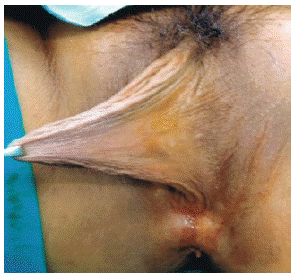
When she saw us again with her mother at 16 years of age, physical examination revealed: Height 1.59m., Weight 69.8kg and BMI 27.6 kg/m2. Examination of her heart, lungs and abdomen were normal. Breast and external genitalia examination revealed Tanner stage 5 and 4 (sparse pubic hair – Figure 1-4) respectively [2,3]. There was no clitoramegaly with enlarged clitoral hood (Figure 2). Both labiaminora were symmetrically enlarged, width measuring 6 cm, 5.5 cm caudally and length 3 cm [supero-inferiorly] on the lateral [outer] side (Figure 1), protruding far beyond her normal labia major a (Figure 1 and 2). Her hymen was intact and her posterior commissure was raised by 10 mm on parting the vagina (Figure 2 and 3 left). Hence a speculum and vaginal examination were not performed.

    Figure 1: Labia minoraheld together with Ellis forceps (lateral view);

Symmetrical Stage III (width of superior border 6 cm, inferior border 5.5 cm,

length 3 cm).

    Figure 2: Labia minora (frontal view); Symmetrical Stage III with intact hymen

and enlarged litoral hood.
The patient was very sure of her request for surgical correction of her bilateral symmetrically enlarged labia minora affecting her daily life and quality of life. She was counseled extensively about the complications of labiaplasty (Discussion). Preoperative full blood count, renal function and electrocardiography were normal.
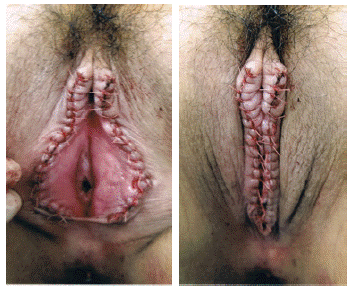
She had anunevenful general anaesthesia and uncomplicated bilateral labiaplasty. Each symmetrically enlarged labia minora was reduced surgically by direct excision [1,4,5] using electrical cautery to excise the lateral margins, after holding both labia minora together using Ellis forceps and marking the latter with a marker pen. The edges of the excised labia minorawere gently approximated using 4-0 Polyglactin (Figure 3). The duration of surgery was 59 minutes. She was served oral Paracetamol and Mefenamic acid for pain, She was able to pass urine well with normal post-void residual urine and discharged home well the next day.

    Figure 3: Stage III symmetrical Labia minora after surgical reduction by

direct excision using electrical cautery and edges sutured with interrupted

4-0 Polyglactin.
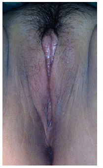
She was reviewed after one, six, ten and twenty weeks postoperatively and remained well; both labia minora had healed completely at six weeks follow-up (Figure 4) and she was very satisfied with the result.

Figure 4: Well healed Labia minora at 6-week review.
The histology was reported as gross description: two pieces of corrugated skin measuring 5.0 x 3.0 x 0.5 cm and 5.0 x 2.5 x 0.5 cm; microscopic description: fibrovascular tissue lined by stratified squamous epithelium showing mild hyperplasia, with occasional hair follicles and sebaceous glands. There was no HPV changes, significant inflammation, vulvar intraepithelial neoplasia/dysplasia or malignancy.
Discussion
There are numerous publications that use the terminology hypertrophy to describe enlargement of the labia minora. Histologically, hypertrophy describes enlargement of an organ resulting from an increase in the size of its constituent cells [1]. However, no study has shown an increase in size of the labia minora cells in women with hypertrophy. The Joint Report on Terminology for Cosmetic Gynecology developed by the International Urogynecological Association (IUGA) and the American Urogynecologic Society (AUGS) recommends substituting hypertrophy with labia minora elongation– Defined as any dimension or morphology at which a woman experiences physical discomfort that causes distress and impairs quality of life. This should be considered a medical condition separate from cosmetic gynecology [1].
The American College of Obstetricians and Gynecologists (ACOG) Committee Opinion defined elective female genital cosmetic surgery as the surgical alteration of the vulvovaginal anatomy intended for the cosmesis in women who have no apparent structural or functional anatomy [6]. It added that the safety and effectiveness of these procedures have not been well documented [6].
The Joint Report on Terminology for Cosmetic Gynecology developed by IUGA and AUGS defined cosmetic gynecology as the elective intervention to alter the aesthetic appearance of the external genitalia or modify the genital organs and the elective functional vaginal procedures (in the absence of pathology) with the goal of improving a person’s quality of life [1].
The indications of labiaplasty include: excessively larger (wider and longer) labia minora protruding much more outside the labia majora causing discomfort and pain in daily life (wearing of underwear, bikini, shorts, jeans, tight clothing), walking, running, exercising, vaginal coitus; asymmetrically enlarged labia minora; causing embarrassment, depression and distortion of body image.
The complications of labiaplasty must be discussed extensively with the patient: Bleeding, haematoma, infection, abscess; asymmetry in the size and shape of the labia minora; dissatisfaction with either the labia minora being not narrow enough or too narrow; scarring, pain, dyspareunia and recurrent urinary tract and vaginal infection.
It is of utmost importance to inform the patient that there is a wide variation of normality of the labia minora. A review of normative datasets for female genital anatomy revealed a wide variation in the length (5-100 mm) and width (1-60 mm) of the labia minora [7]. In another study of 657 asymptomatic Caucasian women aged from 15 to 84 years, the labia minora length was 5.5-100 mm(mean 42.5 mm, SD 16.3); width was 1.5-56.6 mm (mean 13.7 mm, SD 7.7) [8]. A British artist, Jamie McCartney cast over 400 women volunteers’ genitalia in plaster of Paris to educate women of the wide variation of the shape and size of their vulva [9]; mainly because of his concern in the rising trend of cosmetic labiaplasty.
It is also important to emphasize to the patient that the function of the labia minora is to cover and protect the external urethral meatus and urethra as well as the vagina from trauma, pain and infection.
Gynecological functional and/or cosmetic surgeons must be cautious of not being overzealous and excise too much labia minora resulting in a smaller (narrower and shorter) labia minora. It is safer to err on the side of caution to excise less labia minora as it heals by fibrosis and further unintended post-operative reduction in size.
Apart from direct excision, the other surgical reduction methods for labiaplasty include: de-epithelization, central wedge, W-plasty, Z-plasty and composite reduction[1]. The alternative therapeutic approach to surgical reduction is energy-based, which includes radio frequency and laser [1].
References
- Writing Group Chair: Garcia Bobby. Joint Report on Terminology for Cosmetic Gynecology. Developed by the Joint Writing Group of the International Urogynecological Association and the American Urogynecologic Society. Int Urogynecol J (2022) 33:1367– 1386.
- Tinneri JM. Growth of Adolescence. Oxford: Blackwell Scientific Publications. 1962.
- Marshall WA, Tanner JM. Variations in pattern of pubertal changes in girls. Arch Dis Child. 1969; 44: 291-303.
- Pardo J, Sola V, Ricci P, Guilloff E. Laser labioplasty of labia minora. Int J Gynaecol Obstet. 2006; 93: 38–43.
- Placid OJ, Arkins JP. A prospective evaluation of female external genitalia sensitivity to pressure following labia Minora reduction. Plast Reconstr Surg. 2015; 136: 442e-52e.
- American College of Obstetricians and Gynecologists (ACOG) Committee Opinion #795: elective female genital cosmetic surgery. Obstet Gynecol. 2020; 135: e36-42.
- Hayes JA, Temple-Smith MJ. What is the anatomic basis of labiaplasty? A review of normative datasets for female genital anatomy. Aust New Zeal J ObstetGynaecol. 2022: 1-8. httpsL//doi.org/10.1111/ajo.13298.
- KreklauA, Vaz I, Oehme F, Strub F, Brechbuhi R, et al. Measurements of a ‘normal vulva’ in women aged 15-84: a cross-sectional prospective single- Centre study. BJOG AnInt J ObstetGynaecol. 2018; 125: 1656-61.
- Jamie McCartney. The great wall of vagina. 2008.