Case Report
Austin J Obstet Gynecol. 2022; 9(3): 1210.
A Case Report of En Caul Vaginal Delivery of 31 Gestational Weeks Fetus with Podalic Breech Presentation
Moughdeb AA, Taifour W*, Ibrahem A and Abbassi H
Obstetrics and Gynecology Hospital, Damascus University, Damascus Syria
*Corresponding author: Wessam Taifour Obstetrics and Gynecology Hospital, Damascus University, Damascus Syria
Received: November 08, 2022; Accepted: December 23, 2022; Published: December 30, 2022
Abstract
Delivering fetus completely enclosed in an amniotic sac in caesarean section or in vaginal delivery is called en caul delivery, it mostly occurs before 36 gestational weeks. We report en caul podalic breech vaginal delivery of 31 weeks fetus, but unfortunately the newborn dead 4 days after delivery because of prematurity complications. We discus pro and con for en caul delivery and caesarean section in premature fetuses, concluding that a breech intact membrane vaginal delivery may be a good and safe way to avoid birth trauma and unwanted poorly formed lower uterine segment caesarean section consequences.
Keywords: Fetus; Vaginal delivery; En caul; Podalic breech; Prematurity
Introduction
Vaginal en caul birth is the rarest subtype of En caul deliveries which occur when a mostly premature fetus is delivered contained within an amniotic sac. Vaginal and abdominal en caul birth occurs in less than 1 in 80,000 live births [1]. The ideal mode to deliver a premature fetus is still controversial, although caesarean section may reduce the risk of fetus death or birth trauma especially with preterm breech fetus, the caesarean incision is performed in a poorly formed lower uterine segment so obstetricians may face difficult cesarean births and may lead to serious maternal morbidity [2]. On the other hand, vaginal preterm en caul birth has some benefits and the fetus could be protected within the surrounding amniotic fluid from birth trauma.
Case Report
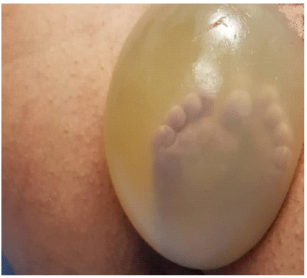
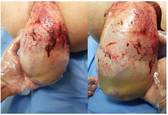
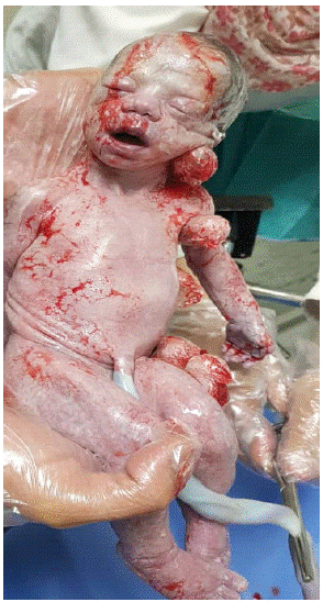
We report a case of an 18-year-old Syrian primigravida, with no significant previous medical history, presented with a preterm singleton gestation at 31, 5 weeks. All fetal parameters were normal during routine check-ups in early neonatal period. The patient presented with painful contractions (4 contractions/ 10 minutes, each lasted for 45 seconds). The BMI was 27, 4 Kg/m2, and the vital signs were within normal limits (blood pressure 112/76 mmhg, pulse 84 beats per minute). Vaginal examination showed a fully dilated and effaced cervix with intact bulging membranes, and the presenting part of the fetus was footling breech, with feet prolapsed in the vagina inside of the still intact amniotic sac. An emergent lower segment caesarean section was planned. Antenatal steroid therapy was not administered due the rapid progression of delivery, neither oxytocin nor tocolytics were introduced. The patient was transported to the operation room and before the induction of the general anesthesia, the bulging membrane with two feet inside were apparent outside of the introitus of the vagina (Figure 1), so then the decision was made to try a vaginal delivery due to the rapid progression of labor and in order to prevent umbilical cord prolapse. We didn’t perform an episiotomy, the membranes were cautiously preserved intact during the active phase until delivery was completed and during that time the fetus was monitored by the obstetric team using a Doppler device. We applied Marchall maneuver, the neonatal legs and torso were allowed to hang by its weight (Figure 2 & 3), the fetal head was then swept in an arc over the maternal abdomen, and the head was slowly born in the process. Nevertheless, the membranes were ruptured after the gentle extraction of the head (Figure 4). The amniotic fluid was clear and the newborn’s Apgar scores were 7, 9, and 10 at 1, 5, and 10 minutes respectively. A small laceration was noted near the fourchette and was sutured using a 2-0 chromic suture. Slow IV infusion of 20 IU of oxytocin in 500 ml saline was given, and the fundus of the uterus was firm. The mother was healthy and got discharged on day two postpartum. As for the newborn, she continued to suffer from mild dyspnea and episodes of apnea that responded to pain stimulation. However, on the fourth day, she developed an episode of apnea that didn’t respond to stimulation, and nasotracheal intubation was performed, and the newborn was put on mechanical ventilation with no apparent improvement. Ventilatory support was stopped after discussion with the parents, and the baby developed an episode of apnea that didn’t respond to CPR, and death was announced after 15 minutes.

    Figure 1: Footling breech presentation with intact amniotic sac outside

the introitus of the vagina.

Figure 2&3: The neonatal legs and torso hanging by its weight.

Figure 4: The neonate after vaginal birth.
Discussion
Breech presentation is a lot of common among preterm fetuses than term infants, being 21% at 25-26 weeks' gestation, compared with 3-4% at term [3]. In most cases, cesarean delivery was shown to have higher neonatal survival rates than vaginal delivery in births among extremely preterm infants [4], because evidence from observational studies suggests that the very preterm breech fetus which delivered vaginally is likely associated with a small but significant increase in adverse outcome and by doing a caesarean section these outcomes may be avoided. The preterm fetal head circumference to abdominal circumference ratio is larger than a term fetus, thus the preterm breech head is more likely to be entrapped in a partially dilated cervix, resulting in birth trauma, beside the higher possibility of cord prolapse that could lead to acute asphyxia from compression of the umbilical cord [5,6]. However, caesarean delivery in infants born so late in the second trimester is probably achieved by a classical vertical incision due to poorly formed lower uterine segment at this age [7], leading to a series of unwanted caesarean sections in subsequent pregnancies. Procedures that involving a classical or T-shaped uterine incision have a greater risk (4–9%) of uterine rupture during a subsequent pregnancy than those undergoes transverse lower uterine incision (0.2– 1.5%) [8]. However, by using the En-caul method vaginal birth can be achieved without compromising the fetus, and in the same time avoiding caesarean delivery would diminish maternal morbidity. En caul premature delivery has manifold benefits, intact amniotic sac protects the fetus against mechanical forces, trauma and bruising injuries with strong uterine contractions and during labor. Other benefits include the opportunity to finish a course of steroids, high cord pH, increased extremely preterm infants' 5-minute Apgar scores, protection from cord prolapse, and lower the risk of entrapment of the head in the setting of an insufficiently dilated cervix [9,10]. en caul deliveries would be 1- 2% or roughly 1 in 80,000 of all vaginal deliveries if no membranes were artificially ruptured (ARM, Amniotomy) [11,12]. The majority of en caul deliveries occur in premature neonates [9,11,12]. The caul itself is generally harmless to the fetus and can be easily removed. However, using sharp forceps or scissors where the situation requires rapid rupturing of membranes can damage the delicate neonatal skin with the Sharp instrumentation and may lead to a permanent scarring, so caution must be taken. Vaginal en caul delivery with simple spontaneous preterm labor had many benefits: completing a steroid course, higher cord pH, and higher 5- minute Apgar score. The retrospective cohort study compared vaginal delivery with or without intact membranes of extreme preterm delivery (24+0 to 26+6 gestational weeks) showed that the en caul delivery method was associated with considerably higher arterial cord pH values [13]. Nevertheless, severe respiratory distress is still a major neonatal complication in preterm infants [13].
Conclusion
It is still recommended to do a caesarean section in active labor or delivery associate with severe fetal growth restriction, placenta abruption, severe preeclampsia, or massive antepartum hemorrhage and the preterm fetus is healthy and viable.
Although there are no randomized trials to confirm this, conducting preterm delivery with intact membranes appears to cause less labor and delivery trauma. Therefore, performing artificial rupture of membranes especially in cases of early preterm deliveries should be only for a clear indication.
The method of vaginal delivery “en caul” may represent a safer alternative to caesarean delivery in extremely preterm infants in breech presentation with intact membranes while monitoring fetal heart rate and in absence of a clear indication to caesarean section.
References
- Malik R, Sarfraz A, Faroqui R, Onyebeke W, Wanerman J. Extremely preterm (23 weeks) vaginal cephalic delivery en caul and subsequent postpartum intraventricular hemorrhage and respiratory distress: a teaching case. Case Reports in Obstetrics and Gynecology. 2018; 2018; 5690125.
- Richmond JR, Morin L, Benjamin A. Extremely preterm vaginal breech delivery en caul. Obstet Gynecol. 2002; 99: 1025–30.
- Hickok DE, Gordon DC, Milberg JA, Williams MA, Daling JR. The frequency of breech presentation by gestational age at birth: A large population-based study. Am J Obstet Gynecol. 1992; 166: 851–2.
- Narayan H, Taylor DJ. The role of caesarean section in the delivery of the very preterm infant. Br J Obstet Gynaecol. 1994; 101: 936–8.
- Robilio PA, Boe NM, Danielsen B, Gilbert WM. Vaginal vs. cesarean delivery for preterm breech presentation of singleton infants in California: a population-based study. J Reprod Med. 2007; 52: 473-9.
- Kayem G, Baumann R, Goffinet F, Abiad SE, Ville Y, et al. Early preterm breech delivery: is a policy of planned vaginal delivery associated with increased risk of neonatal death? Am J Obstet Gynecol. 2008; 198: 289.e1-289.e6.
- Halperin ME, Moore DC, Hannah WJ. Classical versus low-segment transverse incision for preterm caesarean section: Maternal complications and outcome of subsequent pregnancies. Br J Obstet Gynaecol 1988; 95: 990–6.
- ACOG practice bulletin. Vaginal birth after previous cesarean delivery. Number 5, July 1999 (replaces practice bulletin number 2, October 1998). Clinical management guidelines for obstetriciangynecologists. American College of Obste-tricians and Gynecologists. Int J Gynaecol Obstet. 1999; 66: 197–204.
- Z Jin, X Wang, Q Xu, P Wang, W Ai. Cesarean section en caul and asphyxia in preterm infants. Acta Obstetricia et Gynecologica Scandinavica. 2013; 92: 338-341.
- B Stoelinga, J Bienstock, C Goodwin, N Hueppchen. En caul delivery of extremely preterm infants: Does it make a difference?. American Journal of Obstetrics & Gynecology. 2006; 195: S75.
- Greenstein J, Panzo W, O’Connor J, et al. En Caul delivery. The Journal of Emergency Medicine. 2016; 50: 333-334.
- Abouzeid H, Thornton JG. Pre-term delivery by Caesarean section ’en caul’: a case series. European Journal of Obstetrics & Gynecology and Reproductive Biology. 1999; 84: 51-53.
- Low JA, Panagiotopoulos C, Derrick EJ. Newborn complications after intrapartum asphyxia with metabolic acidosis in the preterm fetus. Am J Obstet Gynecol. 1995; 172: 805–10.