Case Report
Austin Oncol Case Rep. 2022; 5(1): 1016.
An Interesting Case of Synchronous Low-Grade Peritoneal Mesothelioma and Ovarian Endometrioid Adenocarcinoma
Chao ES¹ and Abu-Shahin FI²*
¹College of Medicine, Texas A&M Health Science Center, USA
²Department of Hematology/Oncology, Houston Methodist Hospital, USA
*Corresponding author: Eugene S Chao, College of Medicine, Texas A&M Health Science Center, 12330 N. Gessner Rd Apt 124, Houston TX 77064, USA
Received: February 01, 2022; Accepted: February 25, 2022; Published: March 04, 2022
Abstract
Synchronous mesothelioma with other primary malignancies has previously been reported. However, the finding of a low-grade peritoneal mesothelioma with a synchronous ovarian endometrioid adenocarcinoma remains unreported. We report a case of a 57-year-old woman presenting with distended abdomen. CT abdomen and pelvis and subsequent MRI revealed a large pelvic mass. On pathology of the exploratory laparotomy specimen, stage IIIB FIGO grade 3 right ovarian endometrioid adenocarcinoma, as well as stage 1A left ovarian endometrioid borderline tumor, were discovered. Incidentally, a low-grade mesothelioma, epithelioid type, was discovered spanning omentum, right upper paracolic gutter left upper paracolic gutter, and umbilicus. Post-surgery, chemotherapy was initiated primarily to treat the ovarian adenocarcinoma.
Keywords: Peritoneal mesothelioma; Ovarian adenocarcinoma
Introduction
Mesothelioma is a rare primary tumor of serosal membranes, with around 3,300 cases diagnosed in the United States each year. Most commonly arising from the pleura, mesothelioma can also arise from the mesothelium of peritoneum, pericardium, and tunica vaginalis [1,2]. Between 10 and 15 percent (around 600 cases diagnosed in the US) of mesothelioma are peritoneal [3,4]. Most peritoneal mesothelioma is high grade and aggressive, presenting with diffuse, extensive spread throughout the abdomen3. Low-grade peritoneal mesothelioma involving few peritoneal surfaces are yet rarer, usually arising from women with no history of asbestos exposure [4,5]. Furthermore, the synchronous appearance of indolent peritoneal mesothelioma along with other primary malignancies remains unknown.
Case Presentation
A 57-year-old woman presented to her primary care physician (6/28) with concerns about tachycardia and abdominal swelling. She had episodes of elevated heart rate, shortness of breath, and fatigue. Additionally, she reported abdominal swelling, lower extremities edema, and significantly distended abdomen. Patient was in her baseline state of health prior, with only history of endometriosis and uterine cysts. Family history significant for pancreatic cancer from maternal grandfather at age 50.
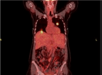
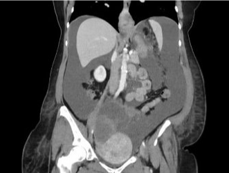
A CT of abdomen and pelvis with contrast (6/28) was performed, multiple pulmonary nodules were present in the lung bases, the largest measuring 1.7cm anteriorly in the lingula, 1.8cm in the right lower lobe (Figure 1a). Diffuse centrilobular emphysematous changes were present. Marked, severe, extensive ascites were found throughout the abdomen and pelvic. The liver, gallbladder, biliary duct, spleen, pancreas, adrenal glands, kidneys, abdominal aorta, retroperitoneal or mesenteric lymph nodes, and bowels were negative for any significant pathology. On pelvis, a large, ill-defined solid and cystic mass with extensive septations appearing to originate in the right lower pelvic and extending superiorly into the midline of the pelvis and upper abdomen was discovered (Figure 1b). Subsequent MRI of pelvis (7/7) confirmed a large septated solid and cystic midline pelvic mass measuring 14x15x21cm.

    Figure 1a: Multiple bilateral pulmonary nodules of varying size demonstrating

abnormal metabolic activity are consistent with metastatic disease.

    Figure 1b: There is a large ill-defined solid and cystic mass with extensive

septations which appears to originate in the right lower pelvis and extends

superiorly into the midline of the pelvis and upper abdomen. The solid

component measures at least 6 x 4 cm the overall mass is difficult to

adequately measure as it blends with the ascites but measures at least 18cm.
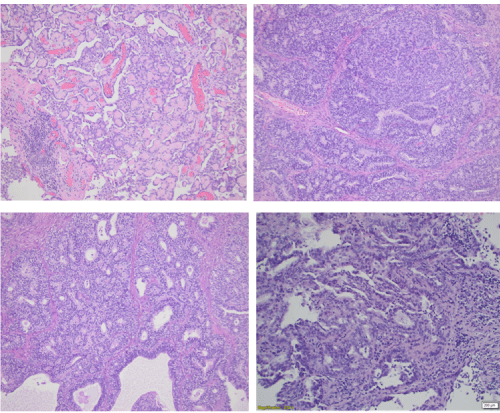
An exploratory laparotomy (8/2) was performed to resect the pelvic mass as well as possible metastases. A right pelvic mass densely adherent to rectum, rectosigmoid colon, posterior uterus, and bilateral retroperitoneal spaces with distal pelvic retroperitoneal fibrosis with pelvic bilateral hydronephrosis was resected. A left pelvic mass with borderline endometrioid and mucinous neoplasm in background of endometriosis was also resected. Pathology (8/2) revealed Stage IIIB FIGO grade 3 right ovarian endometrioid adenocarcinoma with infiltrative pattern, as well as stage 1A left ovarian endometrioid borderline tumor with mucinous metaplasia arising in a background of endometriosis. In addition, incidental pathology findings involved malignant mesothelioma, epithelioid type, low grade (negative for carcinoma or tumor) on omentum, right upper paracolic gutter peritoneum tumor nodule, left upper paracolic gutter peritoneum tumor nodule, and umbilical nodule (Figure 2).

Figure 2: The mesothelioma.
Patient was started on chemotherapy (9/17) using Carboplatin/ Taxol for two cycles. On 10/29, chemotherapy regimen was switched to Carbo/Docetaxel due to infusion reaction. Chemotherapy was switched to Carbo/Doxil/Avastin for the last three cycles. Last cycle was completed on 12/17/2021. Scheduled cycle #5 for 1/17/2022. Natera genetic testing returned negative. Navigational bronchoscopy on 12/3/2021 was performed to biopsy the lung lesions. Pathology confirmed metastatic adenocarcinoma of ovarian origin.
Discussion
While peritoneal mesothelioma occurs in a fraction of mesothelioma cases, there have been previous reports of synchronous peritoneal mesothelioma and other primary malignancies [6-11]. Cases include synchronous peritoneal mesothelioma with pleural mesothelioma6, colonic adenocarcinoma [7], rectal carcinoma [8], urothelial carcinoma [9], hepatocellular carcinoma [10], and breast carcinoma [11]. Findings of a synchronous ovarian endometrioid adenocarcinoma and a peritoneal mesothelioma have not been reported so far.
Moreover, among these studies, a majority report on the more frequently occurring malignant peritoneal mesothelioma, and only one reports on a synchronous appearance of a low-grade peritoneal mesothelioma variant [8]. Low-grade peritoneal mesothelioma is rare and cytologically indistinguishable from diffuse peritoneal mesothelioma [12]. It is characterized by solitary, well-circumscribed mass with no evidence of diffuse spread [12,13]. It is associated with better prognosis, and surgical excision may be curative [12,13]. Recent studies have suggested that for epithelioid peritoneal mesotheliomas, certain histopathologic features, such as tubulopapillary architecture, low nuclear pleomorphism, low mitotic index, and low composite nuclear grade, are associated with better prognosis [14]. While the molecular pathogenesis of malignant peritoneal mesothelioma remains an open question, studies have proposed recurrent alterations in epigenetic regulatory genes BAP1, NF2, SETD2, and DDX3X as potential genetic drivers [15], and thus candidates for targeted therapies.
To the best of our knowledge, this is the first case report of synchronous ovarian endometrioid adenocarcinoma and coexistent primary low-grade peritoneal mesothelioma. Due to the rarity of low-grade peritoneal mesothelioma in general, and its synchronous presentation with ovarian endometrioid adenocarcinoma in particular, the clinical course, prognosis, pathogenesis, and genetics remain an open question.
Conclusion
In conclusion, we report an unusual case of a 57-year-old woman with 2 synchronous malignancies, an ovarian endometrioid adenocarcinoma with evidence of extensive disease in the abdomen and possible metastases to the lung, as well as a low-grade peritoneal mesothelioma involving few peritoneal surfaces. The low-grade nature and small sites of involvement of the peritoneal mesothelioma may suggest an indolent behavior. Given the rarity of such a combination, its clinical course, prognosis, pathogenesis, and genetics remain unclear.
References
- Tandon RT, Jimenez-Cortez Y, Taub R, Borczuk AC. Immunohistochemistry in Peritoneal Mesothelioma: A Single-Center Experience of 244 Cases. Arch Pathol Lab Med. 2018; 142: 236-242.
- Smith-Hannah A, Naous R. Primary peritoneal epithelioid mesothelioma of clear cell type with a novel VHL gene mutation: a case report. Hum Pathol. 2019; 83: 199-203.
- Kim J, Bhagwandin S, Labow DM. Malignant peritoneal mesothelioma: a review. Ann Transl Med. 2017; 5: 236.
- Hassan R, Alexander R. Nonpleural mesotheliomas: mesothelioma of the peritoneum, tunica vaginalis, and pericardium. Hematol Oncol Clin North Am. 2005; 19: 1067-1087.
- Nishikawa Y, Nomura A, Yuba Y, Yazumi S. Education and imaging. Gastrointestinal: indolent case of malignant peritoneal mesothelioma with massive ascites. J Gastroenterol Hepatol. 2014; 29: 662.
- Del Gobbo A, Fiori S, Gaudioso G, Bonaparte E, Tabano S, Palleschi A, et al. Synchronous pleural and peritoneal malignant mesothelioma: a case report and review of literature. Int J Clin Exp Pathol. 2014; 7: 2484-2489.
- Xie W, Green LK, Patel RA, Lai S. A case of unsuspected peritoneal mesothelioma occurring with colonic adenocarcinoma masquerading as peritoneal metastases. Case Rep Pathol. 2014; 2014: 838506.
- Jatzko GR, Jester J. Simultaneous occurrence of a rectal carcinoma and a diffuse well differentiated papillary mesothelioma of the peritoneum. Int J Colorectal Dis. 1997; 12: 326-328.
- Basatac C, Aktepe F, Saglam S, Akpinar H. Synchronous presentation of muscle-invasive urothelial carcinoma of bladder and peritoneal malign mesothelioma. Int Braz J Urol. 2019; 45: 843-846.
- Uemoto J, Hoshi N, Hirabayashi K, Hoshi S, Onodera K, Nishi T, et al. Collision tumors of hepatocellular carcinoma and malignant peritoneal mesothelioma. Med Mol Morphol. 2013; 46: 177-183.
- Prathibha S, Beckwith H, Kratzke RA, Klein M, Kne A, Tuttle TM. Synchronous breast carcinoma and peritoneal mesothelioma. Breast J. 2021; 27: 550-552.
- Carr NJ. New insights in the pathology of peritoneal surface malignancy. J Gastrointest Oncol. 2021; 12: S216-S229.
- Allen TC, Cagle PT, Churg AM, Colby TV, Gibbs AR, Hammar SP, et al. Localized malignant mesothelioma. Am J Surg Pathol. 2005; 29: 866-873.
- Chapel DB, Schulte JJ, Absenger G, Attanoos R, Brcic L, Butnor KJ, et al. Malignant peritoneal mesothelioma: prognostic significance of clinical and pathologic parameters and validation of a nuclear-grading system in a multiinstitutional series of 225 cases. Mod Pathol. 2021; 34: 380-395.
- Joseph NM, Chen YY, Nasr A, Yeh I, Talevich E, Onodera C, et al. Genomic profiling of malignant peritoneal mesothelioma reveals recurrent alterations in epigenetic regulatory genes BAP1, SETD2, and DDX3X. Mod Pathol. 2017; 30: 246-254.