
Case Report
Austin Oncol Case Rep. 2023; 6(1): 1017.
Axillary Metastasis from Occult Breast Cancer and Synchronous Lung Cancer: A Case Report
Khrisat T, Marques ICDS and McCalla SG*
Department of General Surgery, Lincoln Medical Center, USA
*Corresponding author: McCalla SGDepartment of Surgery, Suite 620, Lincoln Medical Center, 234 East 149th St., Bronx, NY 10451, USA
Received: February 06, 2023; Accepted: March 14, 2023; Published: March 21, 2023
Abstract
This is an 84-year-old woman found to have on exam two Right axillary masses by her Primary Care Physician. The patient was then sent to our institution for further evaluation, which surprisingly revealed a lung mass, but no primary breast mass.
After biopsies, both areas revealed different immunohistochemistry. She underwent further treatment, with resection of both the lung and axillary tumors.
This is an interesting case that report highlights the importance of having high index of suspicion of breast cancer in the setting of axillary mass and, the importance of differentiating associated synchronous versus metastatic cancer.
Keywords: Occult breast cancer; Axillary lymph nodes; Synchronous lung
Introduction
Multiple primary malignant tumors are a term used to refer to the presence of two or more primary cancers of different origins in the same patient. The reported incidence of this condition ranges from 0.73 to 11.7% [1] and it’s further classified as synchronous or metachronous depending on the time of diagnosis. It is synchronous when the time between diagnosis of the first and second primary tumors is less than 6 months and metachronous when that period is more than 6 months.Occult breast cancer was first described in 1907 by Halstedas “cancerous axillary glands with non-demonstrable cancer of the mamma” [2]. Due to advancement in diagnostic techniques, occult breast cancer has shown to be an uncommon diagnosis, with a reported incidence of 0.3-1% of all breast cancer patients [3]. Women with breast cancer are at increased risk of subsequent primary malignancies, specifically lungcancer [4]. This paper describes a case of occult breast cancer with a synchronous diagnosis of lung cancer.
Case Report
An 84-year-old female with past medial history of hypertension, chronic kidney disease, and osteoporosis. On physical examination, a right axillary lymphadenopathy was noted, without any palpable breast mass.
The patient reports that she first noticed the axillary mass was about a year ago. Her previous mammogram in 2015, at the age of 77, was negative for any malignancy. The investigation then proceeds with a new diagnostic mammogram and bilateral ultrasonography (US) examination.
US revealed “markedly abnormal lymph nodes with thickened cortex” in the right axilla without any mass in the breast (Figure 1).
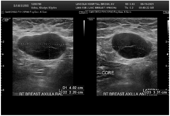
Figure 1: Initial breast Ultrasound.
Figure 2 showed non-specific findings on the right breast with a request for a second look ipsilateral mammogram. Second look imaging remained negative for masses. On her metastatic work up, a 1.1 cm right lung upper nodule was noted on the chest CT (Figure 3).
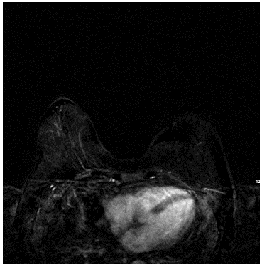
Figure 2: Breast MRI – no lesion found.
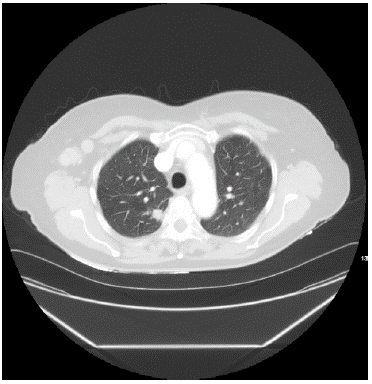
Figure 3: Right upper lung nodule.
Interventional radiology was consulted, and a CT guided biopsy of the node was done. Surprisingly, the node was consistent with lung adenocarcinoma (Figure 4). Immunohistochemistry showing Thyroid Transcription Factor-1 (TTF-1) positive and a Programmed Death-Ligand 1 (PD L1) score of 40%. A Positron Emission Tomography (PET-CT) (Figure 5) was negative for me tastasis and the case was discussed on our tumor board given the presence of two primary synchronous cancers.
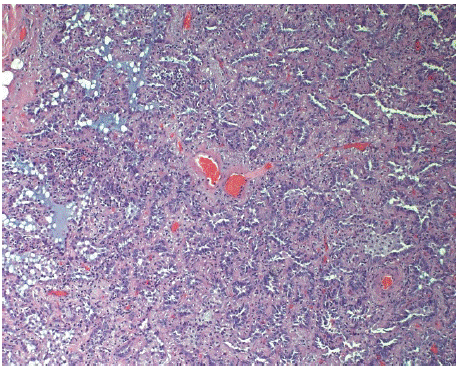
Figure 4:
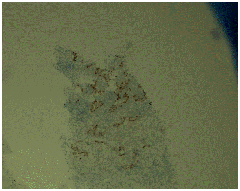
Figure 5: Lung biopsy.
For her lung cancer (T1N0M0 stage IA) her options included upfront surgical resection versus stereotactic radiation. The patient expressed preference for surgical intervention. For her breast cancer (cT0N2M0), a decision was made to offer neoadjuvant therapy followed by axillary dissection and radiation.
Patient refused neoadjuvant chemotherapy, however, agreed to hormonal therapy only. Anastrozole was initiated in December 2021 and in the subsequent month, the patient underwent right video assisted thoracoscopy, with right upper lobe posterior segmentectomy and lymph node sampling. On examination of the specimen, all margins were clear, and all lymph nodes were negative. The patient was then scheduled to proceed with axillary dissection 3 months from her thoracic procedure however, her post operative course was complicated by pleural effusions requiring thoracentesis on two separate occasions. Finally, seven months after her thoracic surgery, the patient had returned to her baseline status and was cleared to proceed with axillary dissection. She was taken Anastrozole in the interim time, with no clinical response appreciated on physical examination. She underwent lymph node dissection in July 2022 and 2 out of 14 lymph nodes showed poorly differentiated breast carcinoma (Figure 6). At the time of this writing, she continues to follow in our institution and remains only on Anastrozole. She was again offered chemotherapy and radiation treatment after the axillary resection but continued to decline it.
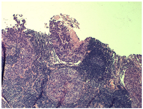
Figure 6: Axillary lymph node with breast carcinoma.
Discussion
Previous studies looking at cancer registries have showed that there is an increased risk for lung cancer after breast cancer [5,6], however, most of the cases are reported following radiotherapy and possibly related to an interaction between radiotherapy and previous smoking status. Our case describes a synchronous lung adenocarcinoma with an occult breast cancer in a non-smoking patient.
Multiple primary malignant tumors rarely occur, and, with the consistent increased availability of diagnostic techniques, occult breast cancer is also a rare instance. In the presence of a suspicious lesion, it remains critical to distinguish between another primary lesion from metastatic lesions. Aside from breast cancer, many other adenocarcinom as have been shown to metastasize to the axillary lymph nodes [7], highlighting the need for further diagnostic work-up.
Our patient first presented with an isolated axillary mass. The first step is to clarify whether they are benign or malignant and then to confirm the origin. The presence of both ER and PR has a crucial role in determining whether the primary disease behind an axillary metastatic lymphnode is breast cancer. In our case, immunohistochemistry was essential to identify the origin of the metastasis.
In a previous study involving five synchronous cases of both lung and breast cancers, all patients underwent concurrent surgery for both cancers. Breast cancer surgery consisted of partial or total mastectomy with/without axillary lymph node dissection while lung cancer surgery consisted of pulmonary lobectomy with lymph node dissection. Authors concluded that concomitant surgery may be feasible and safe [8]. In our case, the decision of separate procedures was individualized to the patient and the potentially benefits of neoadjuvant therapy for her breast cancer.
The treatment of occult breast cancer was traditional radical or modified radical mastectomy. However, a clinical trial performed at Memorial Sloan Kettering Cancer Center (MSKC), where thirty-eight patients with occult breast cancer were treated as per current National Comprehensive Cancer Network (NCCN) guidelines with either Axillary Lymph Node Dissection (ALND) and whole breast radio therapy or ALND and mastectomy. At their median follow up of 7 years, there was no local or regional recurrence in either arm. This study suggests that in the present of occult breast cancer, and then ALND followed by ipsilateral breast radiotherapy is a reasonable option rather than a mastectomy [9]. Therefore, this was the proposed treatment for our patient.
Although there is little additional evidence to guide the use of neoadjuvant chemotherapy, Offri et all have reviewed current data, suggesting that neoadjuvant chemotherapy is areasonable option for occult breast cancer patients, particularly for those with a high gradeHER2 [9]. Our patient did not agree to receive neoadjuvant therapy and it remains unclear whether that would have provided clinical remission in her axillary node.
References
- Aydiner A, Karadeniz A, Uygun K, Tas S, Tas F, et al. Multiple primary neoplasms at a single institution: differences between synchronous and metachronous neoplasms. Am J Clin Oncol. 2000; 23: 364-370.
- Halsted WS. I. The Results of Radical Operations for the Cure of Carcinoma of the Breast. Annals of surgery. 1907; 46: 1-19.
- Walker GV, Smith GL, Perkins GH, Oh JL, Woodward W, et al. Population-based analysis of occult primary breast cancer with axillary lymph node metastasis. Cancer. 2010; 116: 4000-4006.
- Kim JY, Song HS. Metachronous double primary cancer after treatment of breast cancer. Cancer Res Treat. 2015; 47: 64-71.
- Evans HS, Lewis CM, Robinson D, Bell CM, Møller H, et al. Incidence of multiple primary cancers in a cohort of women diagnosed with breast cancer in southeast England. Br J Cancer. 2001; 84: 435-440.
- Prochazka M, Granath F, Ekbom A, Shields PG, Hall P. Lung cancer risks in women with previous breast cancer. Eur J Cancer. 2002; 38: 1520-1525.
- Copeland EM, McBride CM. Axillary metastases from unknown primary sites. Annals of surgery. 1973; 178: 25-27.
- Shoji F, Yamashita N, Inoue Y, Kozuma Y, Toyokawa G, et al. Surgical Resection and Outcome of Synchronous and Metachronous Primary Lung Cancer in Breast Cancer Patients. Anticancer Res. 2017; 37: 5871-5876.
- McCartan DP, Zabor EC, Morrow M, Van Zee KJ, El-Tamer MB. Oncologic Outcomes After Treatment for MRI Occult Breast Cancer (pT0N+). Annals of surgical oncology. 2017; 24: 3141-3147.