Rapid Communication
J Ophthalmol & Vis Sci. 2016; 1(1): 1001.
Intravitreal Bevacizumab Injections Can Release Vitreomacular Adhesions in Eyes with Persistent: Diabetic Macular Edema and Focal Vitreomacular Adhesions
Al Judaibi RM¹, Al Shamsi H¹ and Ghazi NG1,2*
¹Vitreoretinal Division, King Khaled Eye Specialist Hospital, Saudi Arabia
²The Department of Ophthalmology, University of Virginia, Charlottesville, USA
*Corresponding author: Nicola G. Ghazi, Cleveland Clinic Abu Dhabi, Abu Dhabi, UAE
Received: January 22, 2016; Accepted: February 12, 2016; Published: February 22, 2016
Abstract
Purpose: To report cases of persistent Diabetic Macular Edema (DME) with focal Vitreomacular Adhesions (VMA) that responded to Intravitrealbevacizumab (IVB) injection with separation of the VMA and complete or significant resolution of DME.
Methods: Five eyes of five patients were diagnosed with persistent DME and VMA based on typical clinical, angiographic, and spectral-domain Optical Coherence Tomography (OCT) findings. A trial of IVB was attempted prior to Proceeding with Pars Planavitrectomy (PPV) to address the VMA.
Results: Prior to initiation of IVB therapy, all 5 eyes had cystoid macular edema with perifoveal vitreous separation and unifocal point VMA at the fovealcenter by SD-OCT. Following one to four injections all the eyes showed complete separation of the VMA and complete or significant resolution of DME.
Conclusions: A trial of intravitreal therapy, such as IVB, should be attempted in eyes with persistent DME and focal VMA before proceeding to PPV.
Keywords: Diabetic macular edema; Intravitrealbevacizumab; Vitreomacular traction
Abbreviations
DME: Diabetic Macular Edema; VMA: Vitreomacular Adhesions; CMT: Central Macular Thickness; V/A: Visual Acuity; IVB: Intravitrealbevacizumab; PPV: Pars Planavitrectomy; SD_OCT: Spectral-Domain Optical Coherence Tomography; anti-VEGF: Anti- Vascular Endothelial Growth Factor; No.: Number; IVAI: Intravitrealavastin Injections; μm: Microns
Introduction
DME is one of the most common causes of impaired vision in diabetic patients [1]. It is thought that the vitreous plays an important role in DME in some patients through Vitreomacular Adhesion/ Traction (VMA/VMT) and likely other mechanisms [2]. Pars Planavitrectomy (PPV), an invasive procedure, has been used in cases of persistent DME secondary to VMT [3,4]. However, its role remains controversial. Intravitreal Injection Of Bevacizumab (IVB), an anti- Vascular Endothelial Growth Factor (anti-VEGF) agent, shows an increasing evidence for a beneficial effect in treating DME in general [1,5]. However, in eyes with VMA the role of anti-VEGF agents is less well defined [3].
We have previously shown that eyes with persistent DME and VMA may have focal or plaque VMA [2]. It is our impression that focal VMA may resolve with intravitreal injections without the need for PPV. We report 5 such eyes herein.
Materials and Methods
We report five eyes of five patients that were diagnosed with persistent DME and VMA following focal laser therapy based on typical clinical, angiographic, and Spectral-Domain Optical Coherence Tomography (SD-OCT, Spectralis, Heidelberg, Germany) findings. A trial of IVB was attempted prior to proceeding with (PPV) to address the VMA. Each eye received IVB (1.25mg/0.05ml) using sterile ophthalmic techniques. SD-OCT was performed for every eye at baseline prior to injections and then at each follow up visit.
Results
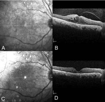
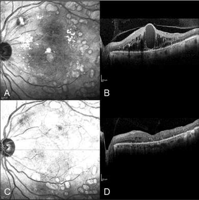
The age of the patients ranged from 54 to 73 years (mean= 61.8 years). All patients were females except for one. The follow up ranged from two months to 14 months (mean=8 months). The baseline Visual Acuity (VA) ranged from 20/300 to 20/30 (median= 20/100) and the Central Macular Thickness (CMT) ranged from 444 to 688 μm (mean=569.4). All eyes disclosed cystoid macular edema with perifoveal vitreous separation and a focal adhesion of the posterior hyaloid at the fovea on SD-OCT. Following one to four injections of IVB (mean=2.8), SD-OCT showed complete separation of the VMA in all eyes with complete or significant resolution of the intraretinal fluid. The VA and CMT at last follow up ranged from 20/200 to 20/40 (median =20/60) and 191 to 427 μm (mean=312.6) respectively (Table 1) (Figure 1&2).
Patient No.
Age
years
Gender
Eye
No. of IVAI
CMT
Before
(Ám)
CMT After
(Ám)
V/A Before
V/A After
I
60
M
OD
1
688
386
20/200
20/200
II
64
F
OS
4
657
191
20/300
20/200
III
73
F
OS
4
533
427
20/100
20/60
IV
54
F
OD
1
444
246
20/30
20/40
V
58
F
OD
2
525
313
20/50
20/40
Table 1:  Demographic and statistical measurement of the cases.
No.: Number; IVAI: Intravitrealavastin Injections; μm: Microns; CMT: Central Macular Thickness; V/A: Visual Acuity

    Figure 1:  Patient (II); (A+C) fundus images, (B+D) spectral domain optical
coherence tomographyimages. (A+B) before treatment; (C+D) after 4
intravitrealavastin injections.
Note: Resolution of the focal vitreomacular adhesion and macularedema
following therapy.

    Figure 2:  Patient (III); (A+C) fundus images, (B+D) spectral domain optical
coherence tomographyimages. (A+B) before treatment; (C+D) after 4
intravitrealavastin injections.
Note: Separation of the posterior hayloid face with marked resolution of the
macular edema.
Disscussion
Resolution of the focal VMA following IVB may be due to the mechanical effect of the injection on the vitreous, the pharmacological effect of bevacizumab on retinal swelling in DME, or both. We believe that both mechanisms are involved here such that two synergisticprocesses may result following the injection. The intravitreal injection of a placebo was shown to result in separation of focal VMA within 28 days in approximately 10% of eyes in the lacebo group in the MIVI study [6]. This may be attributed to the vitreous changes and liquefaction that may result from the mechanical effect of the needle and liquid injected into the vitreous body. In addition, it is well know that intravitreal anti-VEGF agents lead to resolution or reduction of macular edema in a significant number of treated eyes [1,7]. The vitreous changes pull the hyaloid away from the retina (mechanical effect) while the resolution of retinal swelling (pharmacological effect) retracts the retinal surface away from the hyaloid. These two synergisticprocesses may lead to resolution of the VMA following IVB.
Until the role of ocriplasmin is better defined in eyes with diabetic retinopathy; our findings suggest that a trial of intravitreal therapy, such as IVB, should be attempted in eyes with persistent DME and focal VMA before proceeding to PPV. The mechanical effect of the intravitreal injections may help in the separation of the VMA. This, together with the pharmacological effect of the injected agent, may lead to improvement of the DME as seen in the cases reported herein.
References
- Arevalo JF, Sanchez JG, Wu L, Maia M, Alezzandrini, Brito M, et al. Primary intravitreal bevacizumab for diffuse diabetic macular edema: the Pan-American Collaborative Retina Study Group at 24 months. Ophthalmology. 2009; 116: 1488-1497.
- Ghazi NG, Ciralsky JB, Shah SM, Campochiaro PA, Haller JA. Optical coherence tomography findings in persistent diabetic macular edema: the vitreomacular interface. Am J Ophthalmol. 2007; 144: 747-754.
- Harbour JW, Smiddy WE, Flynn HW Jr, Rubsamen PE. Vitrectomy for diabetic macular edema associated with a thickened and taut posterior hyaloid membrane. Am J Ophthalmol. 1996; 121: 405-413.
- Lewis H, Abrams GW, Blumenkranz MS, Campo RV. Vitrectomy for diabetic macular traction and edema associated with posterior hyaloidal traction. Ophthalmology. 1992; 99: 753-759.
- Al Shamsi H, Ghazi NG. Diabetic macular edema: new trends in management. Expert Rev ClinPharmacol. 2012; 5: 55-68.
- Stalmans P, Benz MS, Gandorfer A, Kampik A, Girach A, Pakola S, et al. MIVI-TRUST Study Group. Enzymatic vitreolysis with ocriplasmin for vitreomacular traction and macular holes. N Engl J Med. 2012; 367: 606-615.
- Michaelides M, Kaines A, Hamilton RD, Fraser-Bell S, Rajendram R, Quhill F, et al. A prospective randomized trial of intravitrealbevacizumab or laser therapy in the management of diabetic macular edema (BOLT study) 12-month data: report2. Ophthalmology. 2010; 117: 1078-1086.