
Case Report
J Ophthalmol & Vis Sci. 2016; 1(1): 1010.
Volume Displacement Technique: Lower Lid Laxity in Anophthalmic Sockets
Raizada K* and Raizada D
International Prosthetic Eye Center, Telangana, India
*Corresponding author: Raizada Kuldeep, International Prosthetic Eye Center, Telangana, India
Received: October 10, 2016; Accepted: November 09, 2016; Published: November 14, 2016
Abstract
Lower lid Laxity is a very common problem, among the prosthetic eye users, in the paper we explain various types of lid laxity and management by change the design of a custom ocular prosthesis can improve the lower lid laxity.
Keywords: Volume displacement technique; Lower lid sagging; Anophthalmic socket; Custom artificial eyes; Conformers; Entropion; Orbital implant
Abbreviations
PESS: Post Enucleation Socket Syndrome
Introduction
Loss of an eye, a disastrous condition that not only leads to loss of vision but also cause cosmetic blemish. These conditions include phthisis bulbi, atrophic bulbi, sightless deformed globe and an ophthalmic socket [1]. Enucleation and evisceration can be performed either placing or without placing an orbital implant. Final outlook depends on the symmetrical appearance with a prosthetic eye or a prosthetic shell (Figure 1).

    Figure 1:  Shows anophtalmic socket and post fitted with custom ocular
Prosthesis (1A,1B).
Socket deformities such as lower lid sagging and soft tissue displacement were commonly seen in the past, when the eye removal surgery techniques were traditional and performed without placing an orbital implant. Another reason is using stock eyes prosthesis that give rise to socket deformities in the long run due to ill-fitting. Aging and scarring of the lower fornix is other contributing factors?
Eyelid laxity can be encountered in some prosthetic eye users that may be associated with ectropion, entropion and lower lid sagging. This can happen due to laxity of medial and lateral canthal tendon, lower lid tarsal plate displacement, weak orbicular is muscle and mal positioning of inferior tarsus muscle. The most common cause for the displacement or laxity of lateral canthus in prosthetic eye users are include weight of the prosthesis on lower eyelid and thick lateral edge of the prosthesis.
Lower lid laxity in anophthalmic sockets or prosthetic eye users can be classified as:
- Involutional -anatomical tissue and muscle displacement in prosthetic eye users leads to Horizontal Eye Lid Laxity & Lateral Eye Lid Laxity [2]
- Cicatricial
- Paralytic
- Inflammatory
- Mechanical
-Migrated orbital implant
-Migrated soft tissue
-Cyst in lower eye lid
-Large size of eye prosthesis or excessive volume replaced with prosthetic eye
-Excessive volume loss in socket
-Post Enucleation Socket Syndrome (PESS)
Distraction test (Pinch test) is simply test which can help you in understanding the various kinds on Lid laxity for Involutions as well as cicatriacal Lid Laxity types.
Distraction test (Pinch test)
In Distraction Test (Pinch test), “Lower lid is pinched between thumb and index finger and pulled away to see how far it stretches [3,4]. A normal lid can be pulled away not more than 3 mm, anything more than 3 mm confirms lower lid laxity (Figure 2).

Figure 2: Shows to perform distraction test (2A,2B).
Horizontal lid laxity is common among anophthalmic patients, giving rise to lower lid sagging and lateral canthus displacement. Lower lid can be pulled away towards temple by 10 mm or more, which can be confirmed by Distraction test, however lateral Canthal laxity have more rounder appearance of lateral canthus and pull lid medially should move medially more than 2 mm, medial canthal laxity is very rare in prosthetic eye users [2-4].
Cicatrical lower lid laxity: The most common etiologic factor in cicatricial lower lid Laxity also caused ectropion and due to shortening of the anterior lamella, causes include mechanical, chemical, or thermal injury [2,5] (Figure 3).
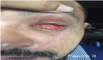
Figure 3: Shows the left cicatrical lower lid laxity.
To know the actual cicatrical lower lid laxity simply one can perform upward Distraction test, Place thumb beneath lateral canthus, push lid laterally and superiorly, excessive tension on lid margin indicates contraction, where the lower lid is tethering to inferior orbital rim and when patient open the eye lids it pull the lower lid downwards [2,6].
Paralytic lower lid laxity: There are very rarely, it happen either by trauma where facial never also got involved and patient also lost an eye, leads to hemi facial Paresis or Palasy, where , the half of face have no function, whatever you do, it remains unfunctional and due to facial or 7th CN Palsy [7] (Figure 4).

Figure 4: Shows lower lids laxity with entropion.
Lid laxity-inflammatory
Lower lids sagging also can be due to various dermatological conditions such as acne rosacea, atopic dermitis, eczema, herpes zoster infection, actinic damage, ichtyosis though these are temporary and disappear once the cause is cured [8] (Figure 5).

Figure 5: Shows lower eye lad laxity due to inflammatory eye lids.
Mechanical lower lid laxity: There are many a times where it is mechanical due to many reasons, such as migrated orbital implant, displaced or migrated soft tissue inferiorly, due to cyst formation in lower lid (temporary), while the main concerns one are due to large size of prosthesis, heavy weight of eye pros-thesis, excessive volume replacement in eye prosthesis, however PESS, Post Enucleation Socket Syndrome Remains the mostly spoken cause for lower lid sagging [9,10].
Migrated orbital implant: following an eye removal surgery in case of Enucleation, Migration is possible when a spherical Non Porous Implant is used, as many of the country still the cost is concern when it comes to have surgery in blind eye, spherical implants can be migrated in any sites, but the most common one is inferior temporal as it gives a bumpy looking condition when there is prosthesis in place and also leads lower lid sagging [9,10] (Figure 6A).

    Figure 6a:  Left Eye shows Inferior Temporal displaced orbital implant, which
gives the mechanical lower lid sagging.
Migrated soft tissue: Many Condition when primary placing an implant is not possible due to Infection in the socket before surgery as surgeon choice are not to put implant, there is usually migration of soft tissue in lower fornix, which makes prosthesis to be slip away and make it prosthesis failure [9,10] (Figure 6B).
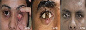
    Figure 6b-d:  B) Left Eye shoes the migrated soft tissue in lower fornix,
which is also responsible for Lower Eye Lid sagging, C) Right Eye Lower
Eye Lids shows external holdelum in anopthalmic socket, which make the
weight of lower eye lid and also when prosthesis in in-place, it gives a illusion
of lower lid sagging. D) Right Eye Post Enucleation with implant, shows the
lower lid sagging and wide opening gf prepare following a custom ocular
Prosthesis, which are results of excessive volume of eye prosthesis.
Cyst in lower eye lid: Chalazion specially in Lower lid can cause temporary lids heavy and cause the lid to look bulky & saggy [9,10] (Figure 6C).

    Figure 6e-f:  E) Following enucleation without any implant, all the tissue
and lids leave no support and comes to gross asymmetric appearance. F)
Case of Post Enucleation socket syndrome, where there is no implant, and
prosthesis is not correcting the defect of upper lid ptosis, sulcus defect, as
well as lower lid sagging.
Large size of eye prosthesis: patients as well as eye care provider always keen to fill the maximum volume, to make symmetrical appearance, however adding the volume beyond the critical point, turns to be very disappointing and leads temporary or permanent lids sagging and damage to orbicularis muscle [9,10] (Figure 6D).
Excessive volume loss in socket: having lost an eye and not able to have an orbital implant been unfortunate choice for many patients where their desire to have a symmetrical balanced prosthesis is a big problem and ultimate it make the prosthesis very heavy and leads lower lid sagging [9,10] (Figure 6E).
Post Enucleation Socket Syndrome (PESS): Enucleation without using an orbital implant often causes enophthalmos, deep upper eyelid sulcus, ptosis and laxity of the lower lid [9,10] (Figure 6F).
Treatment modalities for lower lid laxity and sagging include
Surgical: There the surgical procedure to correct the associated conditions of lid laxity depends on the underlying anatomic factors responsible for the mal-position. Surgical procedures should be directed at correcting the horizontal and vertical instability of the lid by medial and lateral canthal tendon stabilization tarsal strip procedure or other horizontal lid shortening procedures, everything or inverting sutures, plication or reinsertion of the lower lid retractors, hard palate or skin grafting, tumor excision, or combined techniques. Lateral cathoplasty or Lateral Tarsal Strip procedure can be performed to tighten the lax tissue of the lower lid, however it should be done after all efforts are made to restore the appearance non-surgically [11-14].
Non surgical: Lower lid sagging can be improved to a satisfactory level using volume displacement technique. This can be implemented during fabrication of a custom prosthetic eye (details of the fabrication technique is described under Case 1). Advantages of this technique include: it is a noninvasive procedure, no discomfort to the patient and any further changes required to improve the appearance in the follow-up visits could be undertaken. Most of the condition of lower lid laxity and sagging can be handled successfully and satisfactory results could be achieved with the new prosthesis.
The aim of this study was to report a case series of 10 patients with lower lid laxity who are using the custom eye prosthesis.
Case 1
A 65 years old male presented to us with complain of poor cosmetic and function problem with his eye prosthesis, on history he has revealed that he underwent enucleation for tumor in eye in back 1960 somewhere else and later 12 years later second surgery for lower lid sag as lower lid was unable to hold eye prosthesis, on examination in right eye there was no orbital implant and socket was deep but have severe sulcus deformity as well as lower lid sagging and eye prosthesis was hypotropic and down displace about 10 mm.
We discuss with the patient about surgical option vs non-surgical, as patient was on medical treatment for diabetes type II hypertension and cardiac treatment, patient refuse to undergo any new surgical intervention.
In the fabrication process, firstly the socket impression was obtained using hydrophilic colloid and a wax prototype is duplicated [1]. “The wax prototype was modified to match the size, shape and antero-posterior protrusion by either carving the excess wax or building-up the melted wax”.
Anterior apex position of wax Shape was removed and placed 3-4 mm, superior from its original position, so all weight and volume moves upwards and leaves min pressure and weight on lower lid. in case of there is sulcus defect and ptosis, depression avpve the iris upper part can be made and superior ridge can be made as we did in this case. If needed small amount of lower lid feather can be done to reduce further pressure on lower edge of shape of model of prosthesis. There will be minimum or no alteration in posterior surface as else new shaped does not sit on the socket and can be failure as excessive modified shape may not be stable (Figure 7A-7E).
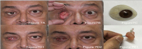
    Figure 7a-e:  A) Patient pre condition with old eye prosthesis, showing the
lower eye lid as well as other defects. B) Patient socket picture. C) Patient
eye prosthesis picture. D) Patient image with socket expander immediate as
well after 1 week (Figure 7D1, 7D2). E) Patient new prosthesis wax shape
new prosthesis front and back profile (Figure 7E1, 7E2, 7E3).
It was possible to successfully improve the level of the socket and correct the lower lid sagging using this volume displacement technique. The final wax pattern was invested in dental stone and a custom con-former was fabricated [1]. The patient was asked to do squeezing exercises 25 times a day and reviewed after a week. The fabrication of a prosthetic eye was then undertaken by obtaining the iris-corneal centeration over the custom conformer and processing with white Poly Methyl Metha Acrylate under High Pressure and Temperature, once the shapes achieved, pigmentation and veining was done further to give life like appearance to match with fellow eye [1]. The patient was very much satisfied with his new look (Figures 7F1,7F2) and followed up after 1 year intervals and his condition

    Figure 7f:  Final results with new and old prosthesis and after 1 years of
follow up.
was stable. He was pleased with his new prosthesis and express his gratitude with the new look.
Plan was as standard except to manipulate the volume as condition was severe.
- Impression of eye
- Realignment of soft tissue with custom conformer as it exactly fits over the defect.
- Eye crunching exercise was given for 25 times a day for 1 weeks
- Review the condition
- Make an eye prosthesis
- Follow-up 3 month and 1 years
Case 2
A 24 years Female history of loss of her left eye due to open globe injury and treated to restore sight in 2004, within 2 years time she developed phthisis bulbi, she start using eye prosthesis in 2007, she came for review in 2016 at our center, on examination she was having deeps socket, upper lid ptosis with sulcus deformity and low lid sagging.
She was keen on non-surgical options, we had common consultation with our in-house surgeon for the opinion, she was given a choice, for enucleation with implant and later ptosis correction, as patient was working women and from other state, she did not had plan to undergo surgery immediately, however she opt for trial of new volume displaced custom artificial eyes, we have follow the same protocol as explained in earlier case.
Patient was pleased with her new prosthesis (Figures 8A- 8C).
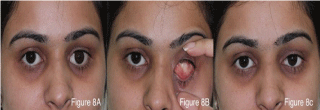
    Figure 8a-c:  A) Patient pre condition with old eye prosthesis, showing the
lower eye lid as well as other defects. B) Patient socket picture. C) Final
results with new prosthesis.
Case 3
A 35 years old male from Middle East lost his right eye following eye infection and had not able to have any orbital Implant and underwent several eye prosthesis, on discussion with patient, patient reveals that he doesn’t seek any more surgical intervention and looking to have a better eye prosthesis. His right eye lower lid was 4.5 mm lax with new volume displaced eye prosthesis, his condition and appearance improved to drastic way (Figures 9A-9C).
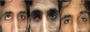
    Figure 9a-c:  A) Patient pre condition with old eye prosthesis, showing the
lower eye lid as well as other defects. B) Patient socket picture. C) Final
results with new prosthesis.
Patient appreciates the visible difference and comfort in his look and comfort.
Materials and Methods
Case were reviewed from 2013-2015 Sep, age, gender, eye, cause for loss of eye, usages of prosthetic eye, socket condition was noted.
Results
Case series of 10 cases who underwent the new prosthesis for lower lid sagging, using the volume displacement technique, mean age of the patient was 39.5 Years (27-63 years), while all the patients had an ophthalmic socket, while there was only 60% were male patient, both eye did not had any difference, was 50% each, while the average use of the eye prosthesis patient were using was 15,7 years, while the range was 1 year to 61 years , All the patient had lower lid laxity, while only 20% of case had orbital implant, there was only 1 case who had lower lid laxity with entropion one case with ciactrical following fire cracker injury, i case was with migrated inferior temporal implant, 40% of cases who had lower lid laxity on presentation was due to poor design of custom ocular prosthesis. The entire patient underwent new volume displaced new custom prosthesis and noticed significant difference in their appearance as well as in lower lid laxity.
Discussion
Horizontal & Lateral Eye lid Laxity is very common disorder among the patients who use the prosthetic eye, often seen usually due to frequent removal of eye prosthesis. Mainly condition had been seen following eye removal surgeries such as Enucleation and Evisceration over years as patients develops Post Enucleation Socket Syndrome [15] a well known syndrome. More over commonest one are the people who use the ill fitted eye prosthesis or stock eyes due to frequent secretion and mucous discharges, which is one of the main reasons for the frequent removal of eye prosthesis.
Volume displacement method is an effective way to correct lower lid sagging also correct other underlying defects such as sulcus defect, ectropions in lower eye lids. It not only manages even distribution of the soft tissue volume, but also reduces the weight and pressure points on the lower lid, but also enhances the cosmoses drastically. Most importantly, the patient doesn’t have to undergo any invasive procedure, which can come into play when these modalities fail (Figures 10A,10B).
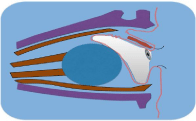
    Figure 10a:  Figure Shows Ptosis, Deep Sulcus Defect, tilting of prosthesis
as well as lower lid sagging, a classic sign of Post Enucleation Socket
Syndrome.
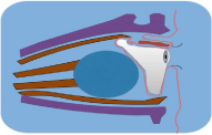
    Figure 10b:  Figure Shows that change the volume displacement, not only
improve the sulucs defect but also, improve the tilting of eye prosthesis as
well as major effect come on lower lid sagging.
In the case series, it was noted 80% cases had no orbital implant and had complex and big size of eye prosthesis results in lower lid laxity, which can be taken care at early stage at the time of surgery or with secondary implant so new prosthesis can be light weight and leave min pressure on lower lids.
It was also seen one case with excessive volume of prosthesis which is pushing the lower eye lids too much, ocularist should be careful in fabricating the eye prosthesis in order not to have not much volume, as it looks good at the times of delivering and later on patient develop lower lid laxity however up to certain extent with the use of hollow eye prosthesis it can be taken, care, as in this case there was implant was already there in socket, carefully fabrication and use of volume displacement technique it can be taken care and it improved the lid sagging in such case.
In one case patient has migrated implant, where the prosthesis was pushing the lower lids and also giving the bulge on lateral side of socket, these condition can be handle with vaulting the area of implant position and thinning the edge of eye prosthesis does improve the appearance of such case.
Conclusion
Volume displaced technique can be very useful in correcting the lower lid and socket level position. Skills of the Ocularist and experience, plays a major role in mastering this technique.
References
- Raizada K, Rani D. Ocular prosthesis Cont Lens Anterior Eye. Epub. 2007; 30: 152-162.
- Miller DG, Hesse RJ. Involutional entropion of the upper lid. Ophthal Plast Reconstr Surg. 1990; 6: 16-20.
- Raflo GT. Horizontal laxity of the lower lid. Plast Reconstr Surg. 1991; 87: 203.
- Morax S, Herdan ML. The aging eyelid. Schweiz Rundsch Med Prax. 1990; 79: 1506-1511.
- Piskiniene R, Eyelid malposition: lower lid entropion and ectropion. Medicina (Kaunas). 2006; 42: 881-884.
- Hurwitz JJ. Senile entropion: the importance of eyelid laxity. Can J Ophthalmol. 1983; 18: 235-237.
- Bergeron CM, Moe KS. The evaluation and treatment of lower eyelid paralysis. Facial Plast Surg. 2008; 24: 231-241.
- Bergeron CM, Moe KS. The evaluation and treatment of lower eyelid paralysis. Facial Plast Surg. 2008; 24: 231-241.
- Raizada K. Lower Lid Laxity in Geriatric Patients for Joint session at of American Academy of Ophthalmology (AAO) and American Society of Ocularists at Sand Expo, Las Vegas. USA. 2015.
- Ansari Z, Singh R, Alabiad C, Galor A. Prevalence, risk factors and morbidity of eye lid laxity in a veteran population. Cornea. 2015; 34: 32-36.
- Weiss RA, McCord CD, Ellsworth RM. Reconstruction of the anophthalmic socket: lower eyelid malposition and canthal tendon laxity. Adv Ophthalmic Plast Reconstr Surg. 1990; 8: 192-208.
- Mahé E, Harfaoui-Chanaoui T, Chappey C, Banal A, Chi TQ. Blepharoplasty: various technics in surgery of eyelid aging. Ann Chir Plast Esthet. 1990; 35: 134-140.
- Georgescu D. Surgical preferences for lateral canthoplasty and canthopexy. Curr Opin Ophthalmol. 2014; 25: 449-454.
- McCord CD, Boswell CB, Hester TR. Lateral canthal anchoring. Plast Reconstr Surg. 2003; 112: 222-237.
- Keserü M, Große Darrelmann B, Green S, Galambos P. Post enucleation socket syndrome-new and established surgical solutions Klin Monbl Augenheilkd. 2015; 232: 40-43.