Research Article
J Ophthalmol & Vis Sci. 2016; 1(1): 1011.
Relationship between Pseudo Exfoliation Syndrome (PEX) and Hyperlipidemia in Patients Over 40 Years Visiting Ardabil City Hospital
Masoumi R, Ojaghi H* and Zamanian T
Faculty of Medicine, Ardabil University of Medical Science, Iran
*Corresponding author: Habib Ojaghi, Faculty of Medicine, Ardabil University of Medical Science, Iran
Received: December 12, 2016; Accepted: December 22, 2016; Published: December 28, 2016
Abstract
Introduction: Pseudo Exfoliation Syndrome (PEX), as a common disease of old age, is of noticeable prevalence in Iran. There are conflicting reports about the association of hyperlipidemia with this syndrome. This study aimed at determining the relation between PEX and hyperlipidemia.
Materials and Methods: This prospective cross-sectional study was done with 100 patients being diagnosed with PEX. Initially, a checklist was completed according to the patients’ responses to questions. Then the patients were referred to the lab for blood samples (for checking the lipid profile). The collected data were analyzed by statistical methods in SPSS16.
Results: The gender of 58% of the patients was male, and of 42% was female with an average age of 62.7 years. Of all patients, 59% had hypertension, 14% had diabetes mellitus, 25% had a history of heart disease, and 61% had a history of hyperlipidemia. The majority of cases (64%) had bilateral involvement, and 33% had symptoms of increased intraocular pressure in their eyes. The mean triglyceride level in patients was 181.64±21.23 mg/dl, the mean cholesterol level was 221.31±32.12 mg/dl and the mean LDL 138.86±13.27 mg/dl.
Conclusion: The results indicated that the prevalence of hyperlipidemia disorder, especially triglyceride, in patients with Pseudo exfoliation syndrome was high.
Keywords: PEX; hyperlipidemia; Glaucoma; Cataract
Introduction
Pseudo Exfoliation Syndrome (PEX) was first described in 1917 by Lindberg [1]. The most obvious clinical symptom of the syndrome is deposition of white flakyexfoliative material on the anterior surface of the lens and most parts of anterior segment and its angle. PEX syndrome is closely related to the development of open and closeangle glaucoma at older ages, and is the most common cause of openangle glaucoma in many communities. PEX prevalence has been reported in various ways regarding race, age, gender and geographical area [1-3]. The prevalence of the disease varies from zero in Siberia, 4% in the UK, 4.7% in Germany and 6.3% in Norway to 21% in Iceland [4-6]. The incidence of this syndrome is such closely associated with age that its incidence is very rare before age 40 while, after age 50, it shows a twofold increase per decade of age [7,8].
The incidence rate of cataract and the amount of cataract surgery complications in patients with PEX syndrome is more than ordinary people. The detailed examination of pupils in most cases gives clue to diagnosis of the disease and in suspicious cases, opening the pupil and observing the lens surface changes can be of greater help. In addition, angle hyper pigmentation and pigment dispersion in different parts of the anterior segment allows accurate diagnosis in most cases [9- 11].
The risk factors for the disease haven’t been properly identified yet. Since this exfoliative material, in addition to eye, may be detected in different organs and parts of the body like skin, viscera and other parts of the body. Some studies have found that PEX is associated with cardiovascular diseases, hearing loss and coronary heart disease [12- 15]. The aim of this study was to investigate the relationship between PEX with hyperlipidemia in patients over 40 years old visiting Alavi Clinic in Ardabil City.
Methods and Materials
This cross-sectional study was undertaken with a sample including 100 patients with PEX which were over 40 years of age and had visited ophthalmology clinic in Alavi hospital at Ardabil city. Initially, a checklist was completed based on the patients’ responses to some questions. Subsequently, the patients were referred to the laboratory for collecting blood samples (for checking the lipid profile). The collected data were analyzed in SPSS (version 16) using statistical methods. The significance level for all tests was set at 0.05.
Results
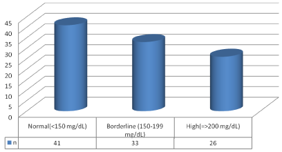
Of the total patients, 58% were male and 42% were female. The mean age of patients was 62.7±18.3 years. The majority of patients belonged to the (60-70) age range. Moreover, 39% were housewives, and 51% were of low level of literacy. The number of those patients who had a history of hypertension was 59, that only 31 of whom were taking hypertension drugs regularly. Of all patients, 14 patients had a history of diabetes, of which 2 patients had Diabetes Mellitus in Childhood (DMT1). 25 patients had a history of heart disease of which7 patients had a history of recent MI. 61 patients had a history of hyperlipidemia of whom 38 patient were taking lipid-lowering drugs on a regular basis. The rest of them either hadn’t consumed drugs or had taken them irregularly. In most of patients (64%), both eyes were involved. 73 patients had already visited ophthalmologist and received medicine. 33 percent reported symptoms of increased intraocular pressure. The average triglyceride level was 181.6±21.3 mg/dl, and most of the patients (41%) had normal triglyceride level (Figure1).
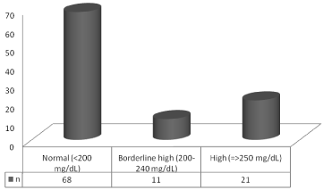
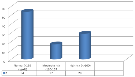
The average cholesterol level was 221.31±32.12 mg/dl, and the majority of patients (68%) had a normal cholesterol level (Figure 2). The average level of LDL in patients was 134.86±13.27 mg/dl, and the majority of patients under study 54% had normal LDL level (Figure 3).

Figure 1: Triglyceride levels in patients.

Figure 2: Cholesterol levels in patients.

Figure 3: LDL levels in patients.
Discussion
In this study, most of participated patients were male with an average age of 62.65±18.34 and most of them have a history of hyperlipidemia which was in line with other study results. [12-15] in the study conducted by Nouri & Kouchaki [12] the results showed that 72 patients (69.2%) were male with the mean age of 68.9 years. Of the studied 150 eyes, 66 eyes (44%) were with glaucoma. In Mirzaee’s study, [13] of the 1850 eyes undergone surgery, 422 eyes (8.2%) were diagnosed with PEX and the disorder was bilateral in 224 cases (53%). Of all samples, 247 cases (58.5%) were male and 175 (47.4%) were female. The rigidity of the pupil was seen in 21 cases (5%), the lens vibration in 52 people (12.5 %), high intraocular pressure in 40 cases (9.5%), sector iridectomy in 42 cases (10% percent), Sphincterotomy in 4 cases (1%) and vitreous loss in 23 cases (3.5%). In Maanaviat and et al. study [14] of the 400 patients with diabetes mellitus who were over 50 years, 24 patients (6%) had PEX syndrome. As the results indicated, the incidence of PEX syndrome increased as one grew older. And this increase was significant (P=0.007). But no significant relationship was detected between the prevalence of PEX and gender, duration of suffering from diabetes or retinopathy. And the prevalence of glaucoma in patients with PEX syndrome was 14.8%. In the study done by Citirik et al. [15]. it was shown that the mean age of patients was 60.6 years, 57% of the patients were male, and 62% had a history of heart disease, 30% diabetes, 54% hypertension and 48% had hyperlipidemia which was similar to our study results. In their study, no significant association was found between hypertension, diabetes, hyperlipidemia and PEX, but cardiovascular disease was significantly prevalent among the patients with this syndrome. In the study performed by French et al. [16]. it was observed that among patients with PEX syndrome, 96.8% were male, 45.6% were above the age 80, with the mean age of 77.1 years, 77.7% of the patients had hypertension, 72.6% hyperlipidemia, 33.4% diabetes mellitus, and 38% cardiovascular disease which was in line with our study results. Our study data analysis showed that similar to other studies, hypertension and hyperlipidemia in patients with PEX syndrome was higher than those in the control group [5,16,17]. Asadi-amoli et al. [17] in their study reported that diabetes and hypertension, hyperlipidemia, and cardiovascular disease were more common in patients with the PEX syndrome than the control group, but this difference was not statistically significant. In the study undertaken by Miyazaki et al. [5] it was found that the incidence of the disease increased significantly as the patients become older and only hypertension was significantly seen in these patients, whereas diabetes, hyperlipidemia, smoking, alcohol consumption and BMI had no significant relationship with this syndrome. In Altintas et al. study [9], the mean age of patients with the PEX syndrome was 69.2 years, the gender of 72.2% of them were male and similar to our study result most of them had hyperlipidemia. Among gender, hypertension, hyperlipidemia, diabetes, cardiovascular disease which were assessed in patients with PEX syndrome, none had significant rise in the patients with PEX syndrome. The study of Roedl et al. [6] discovered no significant association between the risk factors like age, sex, hypertension, diabetes mellitus, atherosclerosis, hyperlipidemia and PEX syndrome.
Conclusion
The results showed that disorders in the lipid profile are of high prevalence among patients with PEX syndrome and checking lipid profile in these patients at the time of diagnosis is necessary.
References
- Lindberg JG. Clinical investigations on depigmentation of the pupillary border and translucency of the iris. Academic Dissertation, Helsinki. Acta Ophthalmol Suppl. 2000; 190: 66.
- Ashton N, Shakib M, Collyer R, Blach R. Electron microscopic study of pseudo exfoliation of the lens capsule: I. Lens capsule and zonularfibers. Invest Ophthalmol. 2001; 4: 141-153.
- Aasved H. Intraocular pressure in eyes with and without fibrillopathia epithelio capsularis. Acta Ophthalmol. 2009; 49: 601-610.
- Krause U. Frequency of capsular glaucoma in central Finland. Acta Ophthalmol. 1973; 51: 235-240.
- Miyazaki M, Kubota T, Kubo M, Kiyohara Y, Iida M, Nose Y, et al. The prevalence of pseudoexfoliation syndrome in a Japanese population: the Hisayama study. J Glaucoma. 2005; 14: 482-484.
- Roedl JB, Bleich S, Reulbach U. vitamin deficiency and hyperhemosisteinemia in pseudoexfoliation glaucoma. J Neural Transm. 2007; 114: 571-575.
- Layden WE, Shaffer RN. Exfoliation syndrome. AM J Ophthalmol, 1947; 78: 835-841.
- Ritch R. Exfoliation syndrome and occludable angles. Trans Am ophthalmol Soc. 2004; 92: 845-944.
- Altintas O, Maral H, Yüksel N, Karabas VL, Dillioglugil MO, Caglar Y. Homocysteine and nitric oxide levels in plasma of patients with pseudoexfoliation syndrome, pseudoexfoliation glaucoma and primary open-angle glaucoma. Graefes Arch Clin Exp Ophthalmol. 2005; 243: 677-683.
- Sunde OA. Senile exfoliation of the anterior lens capsule. Acta Ophthalmol Suppl. 2006; 45: 7-85.
- Tarkkanen A. Pseudoexfoliation of the lens capsule. Acta Ophthalmol Suppl, 2002; 71: 1-98.
- Nouri M, Koucheki B. Related factors with Gloucoma and IOP in patients suffering to PEX in Feyz and Farabi hospitals in Esfahan 1998-1999. Bina. 2001; 6: 214-222.
- Mirzaei M. Study prevalence of PEX and its side-effects in patients with Cataract. Medical J of Tabriz University. 2003; 37: 65-68.
- Maanaviat M, Rashidi M, Miratashi A, Shapouri H. Prevealene of PEX in diabetic patients in YAZD research center. J of Yazd Medical University. 2007; 15: 3-8.
- Citirik M, Acaroglu G, Batman C, Yildiran L, Zilelioglu O. A possible link between the pseudoexfoliation syndrome and coronary artery disease. Eye (Lond). 2007; 21: 11-15.
- French DD, Margo CE, Harman LE. Ocular pseudoexfoliation and cardiovascular disease: N Am J Med Sci. 2012; 4: 468-473.
- AsadiAmoli F, Ghobadi A, Fakhraie G. Serum Homocysteine Levels in Pseudoexfoliative Glaucoma and Primary Open Angle Glaucoma. Iranian Journal of Ophthalmology. 2012; 24: 19-24.