Research Article
J Ophthalmol & Vis Sci. 2017; 2(1): 1014.
Profile of Pathology in Patients with Orbital Diseases
Shrestha GB¹*, Karmacharya PC², Shrestha JK¹ and Shrestha GS¹
¹BP Koirala Lions Center for Ophthalmic Studies, Maharjgunj Medical Campus, Nepal
²BP Eye Foundation, Maharjgunj, Nepal
*Corresponding author: Gulshan Bahadur Shrestha, Department of Ophthalmology, Institute of Medicine, Kathmandu, Nepal
Received: January 06, 2017; Accepted: February 15, 2017; Published: February 20, 2017
Abstract
Background: A spectrum of diseases can involve the orbit. However, the reported incidence of orbital diseases shows great variation, depending on the nature of the study.
Aim: The present study aims to prepare a profile of orbital pathology in patients with orbital diseases presenting to the Out-Patient Department (OPD) of BP Koirala Lions Centre for Ophthalmic Studies (BPKLCOS) and Eye-Ward of Tribhuvan University Teaching Hospital (TUTH).
Methods: In a descriptive cross-sectional study, 47 patients who presented to the OPD of BPKLCOS and the Eye Ward of TUTH from March 2003 to August 2004 and who underwent pathological examination of the lesions were included into the study. Assessment included detailed clinical examination of anterior and posterior segment of eyes, evaluation of proptosis, measurement of globe displacement and detailed histopathological investigations.
Clinical diagnosis and pathological diagnosis of each case were also compared.
Results: Among 47 patients, male female ratio was 2.1:1 and comprised of 42.6% children. The common symptoms at presentation were lump over eyelids and periorbital region (85.1%), ocular and periorbital pain (46.8%), diminution of vision (44.7%) and protrusion of eyeball (38.3%). Superior orbit (51%) was most commonly involved. Orbital lesions were malignant in 31.9%. The commonest clinical diagnoses were dermoid cysts (29.8%) and lacrimal gland tumours (14.9%). On pathological examination, cystic lesion (48.9%) was the most common. Clinico-pathological correlation was present in 72.3%.
Conclusion: Benign cystic lesions are the commonest orbital pathology. Malignant orbital diseases are more common in older population.
Keywords: Orbital lesion; Dermoid cysts; Benign; Malignant; Lacrimal gland tumour
Introduction
A spectrum of diseases can involve the orbit [1]. The understanding of orbital diseases demands a clear concept of normal orbital anatomy and physiologic function. Besides that, a comprehensive knowledge of the structural relationships among the numerous anatomic systems, that are crowded into such small space, is required. Only with these foundations, can the clinician identify and characterize pathologic states [2]. Any pathology occurring in the orbit can arise from any of the tissues of the orbital contents. Alternatively, pathology may spread into the orbit from adjacent structures or distant sources via vascular pathways, or it can be manifestation of systemic disease [3].
Orbital diseases, even though not very common as refractive error or conjunctivitis, have major importance in ophthalmology. They usually have cosmetic blemishes, usually lead to deterioration in visual status and sometimes even cause mortality.
Though several publications have addressed the incidence of orbital diseases, there is a greater variation depending on the nature of the study such that they could be the cases referred to the ocular oncology unit, [4] the cases of certain age group, [5-7] the cases of certain race or geographic areas particularly tropical countries, [5] and the cases from the other specialties like neurosurgery or otorhinolaryngology.8,9 However, definitive diagnosis of orbital diseases comes only after pathological examination. Array and frequency of orbital diseases seen in these studies is expected to be different from those seen in general ophthalmic practice. So, the present study has been undertaken to prepare a profile of pathology in patients with orbital diseases, presenting to the Out-Patient Department (OPD) of BP Koirala Lions Center for Ophthalmic Studies (BPKLCOS) and/or admitted in the Eye-Ward of Tribhuvan University Teaching Hospital (TUTH) and all cases undergoing pathological examination of the lesion.
Methods
Study design and sample size
In the hospital based descriptive study, all cases of orbital diseases presented to the out-patient department of BP Koirala Lions Center for Ophthalmic Studies and the Eye Ward of Tribhuvan University Teaching Hospital from March 2003 to August 2004 were enrolled into the study and they were sent for pathological examination of the lesion.
All patients received a detailed explanation of the procedure involved in the study and provided informed consent. The approval of the implementation of the study was received from the ethics review committee of Institute of Medicine, Kathmandu. The study protocol adhered to the provision of the Declaration of Helsinki for research involving human subjects.
Assessment
- A detailed history was taken from the patient and/or patient’s relatives including patients’ age, gender, ethnic group, occupation, patient’s location according to ecological zone and development region of Nepal, chief complaints, presenting and associated symptoms, treatment history, family history and personal history
- Visual Acuity was assessed with internally illuminated Snellen vision chart with multiple optotypes, E-chart for illiterates and the Catford Drum for children.
- Extraocular Movements (EOM) were assessed in all cardinal gazes with the help of torch light.
- Examination of periorbital region was performed with the torchlight.
- Proptosis, if present, was measured with the Hertel’s exophthalmometer. The findings of palpation, pulsation and periorbital changes were also recorded in proptosis cases.
- Vertical and horizontal displacement of the globe was measured with millimeter scale. Externally visible or palpable lesion was measured with millimeter scale. Cutaneous sensation was assessed in the relevant cases.
- Anterior segment examination was performed with the help of the Haag-Streit 900 slit lamp in appropriate magnification & illumination.
- Dilated fundus examination was performed with binocular indirect ophthalmoscope and +20.0 D lens and with the Haag-Streit 900 slit lamp and +90.0 D Lens.
- The orthoptic test, the Hess screen charting, the diplopia charting and the visual field examination were performed whenever indicated.
Investigations
- Complete Blood Count (CBC), Thyroid function tests (T3, T4, TSH), X-ray of chest and orbit, CT scan of head and orbit, MRI Scan of brain and orbit were performed as per requirements.
- The ENT, Neurosurgery, Neuromedicine, Paediatrics, Radiology and Pathology consultation were taken as per requirements.
- Pathological examinations of the lesion, using appropriate techniques (like Incisional biopsy, Excisional biopsy, Aspiration smear) were performed in all cases.
Data analysis
- Data were analysed using Statistical Package for Social Science (SPSS) 11.0.1 version. Data were presented in number, frequency and mean.
- Shields JA. Diagnosis and Management of Orbital Tumours. Philadelphia Saunders. 1989; 89-388.
- Yanoff M, Duker JS. Ophthalmology. Mosby. 1998.
- American Academy of Ophthalmology. Orbit, Eyelids, and Lacrimal System. Section 7. Basic and Clinical Science Course. 1999-2000.
- Shields JA, Shields CL, Scartozzi R. Survey of 1264 patients with orbital tumours and simulating lesions. Ophthalmology. 2004; 111: 997-1008.
- Johnson TE, Senft SH, Nasr AM, Bergqvist G, Cavender JC. Pediatric orbital tumours in Saudi Arabia. Orbit. 1990; 9: 205-215.
- Kodsi Sr, Shetlar DJ, Campbell RJ, Garrity JA, Bartley GB. A review of 340 orbital tumours in children during a 60-year period. Am J Ophthalmol. 1994; 117: 177-182.
- Demirci H, Shields CL, Shields JA, Honavar SG, Mercado GJ, Tovilla JC. Orbital tumours in the older adult population. Ophthalmology. 2002; 109: 243-248.
- Seregard S, Sahlin S. Panorama of orbital space-occupying lesions. The 24-year experience of a referral centre. Acta Ophthalmol Scand. 1999; 77: 91-98.
- He Y, Song G, Ding Y. Histopathologic classification of 3476 orbital diseases. Chung Hua Yen Ko Tsa Chih. 2002; 38: 396-398.
- Dallow RL, Pratt SG. Approach to Orbital Disorders and Frequency of Disease Occurrence. In: Albert DM, Jakobiec FA. Principles and Practice of Ophthalmology. Philadelphia WB Saunders. 1994; 3: 1881-1890.
- Henderson JW, Campbell RJ, Farrow GM, Garrity JA. The tumour survey. Orbital Tumour. 3rd edn. 1994; 43-52.
- Shields JA, Bakewell B, Augsburger JJ, Flanagan JC. Classification and incidence of space-occupying lesions of the orbit. A survey of 645 biopsies. Arch Ophthalmol. 1984; 102: 1606-1611.
- Reese AB. Bowman lecture: Expanding lesions of the orbit. Trans Ophthalmol. 1971; 91: 85-104.
- De Rosa G, Zeppa P, Tranfa F, Bonavolonta G. Acinic cell carcinoma arising in a lacrimal gland. First case report. Cancer 1986; 57: 1988-1991.
- Jang J, Kie JH, Lee SY, Lew H, Hong SW, Spoor TC, et al. Acinic cell carcinoma of the lacrimal gland with intracranial extension: a case report. Ophthal Plast Reconstr Surg. 2001; 17: 454-457.
- Rosenbaum PS, Mahadevia PS, Goodman LA, Kress Y. Acinic cell carcinoma of the lacrimal gland. Arch Ophthalmol. 1995; 113: 781-785.
Results
Only a total of 47 patients including 32 males (68.1%) and 15 females (31.9%), which completed the study protocol, were considered for further analysis. Mean age of males and females was 24.32 years and 18.80 years respectively with a range of 14 months to 76 years (Table 1).
    Demographic Profile
    No
    Frequency


    Age (years)
    =15
    20
    42.5


    16-45
    21
    44.7


    >45
    6
    12.8


    Sex
    Male
    32
    68.1


    Female
    15
    31.9


    Ecologic distribution
    Mountain
    1
    2.1


    Hill
    28
    59.6


    Terai
    18
    38.3


    Administrative Development region
    Eastern
    4
    8.5


    Central
    24
    51.1


    Western
    12
    25.5


    Mid western
    5
    10.6


    Far western
    2
    4.3


    Occupation
    Farmer
    10
    21.3


    Student
    21
    44.7


    Housewife
    4
    8.5


    Others
    12
    25.5
Table 1: Demographic distribution of subjects.
Mode of presentation is shown in (Table 2). Majority of subjects presented with swelling periorbital area and mass of eyelids (85.1%) followed by pain in orbital, periorbital and ocular area (46.8%) and diminution of vision (44.7%). Time of presentation to the hospital ranged from 10 days to 26 years, with the mean of 4.65 years. Lesion was located in superior part (51.1%) in majority of subjects followed by central part (21.3%) and inferior part (19.1%). Right eye was the mostly affected eye (55.3%).
    Presentation
    Characteristics
    Number
    Frequency


    Symptoms at presentation
    Swelling/Mass of eye lids/periorbital region
    40
    85.1


    Ocular/orbital/periorbital pain
    22
    46.8


    Diminution of vision
    21
    44.7


    Protrusion of eyeball
    18
    38.3


    Restricted ocular movement
    16
    34.0


    Drooping of upper eyelid
    11
    23.4


    Double vision
    2
    4.2


    Time of presentation
    = a month
    8
    17


    = a year
    16
    34


    >a year
    23
    49


    Anatomical location
    Diffuse/central orbit
    10
    21.3


    Supero-lateral
    9
    19.1


    Superomedial
    9
    19.1


    Superior
    6
    12.8


    Inferolateral
    1
    2.1


    Inferomedial
    5
    10.6


    Inferior
    3
    6.4


    Medial lateral
    3
    6.4


    Lateral
    1
    2.1


    Laterality
    Right
    26
    55.3


    Left
    19
    40.4


    Both
    2
    4.3
Table 2: Mode of presentation.
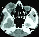
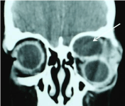
Clinical diagnosis is presented in (Table 3). Individual clinical diagnoses of the patients were grouped into broad clinical diagnoses according to presumed pathological involvement. The most common clinical diagnosis was the dermoid cyst (29.8%) followed by the lacrimal gland tumour (14.9%) (Figure 1A) and non-specific orbital cyst (12.8%) (Figure 2A).

    Figure 1a: Axial CT Scan image of orbit showing right lacrimal gland
pleomorphic adenoma (arrow) of 30-year-old man with 5-year history of
gradually progressive inferomedial proptosis of right globe.
    Clinical Diagnosis
    Number


    Dermoid cyst
    14 (29.8)


    Lacrimal gland tumour
    7 (14.9)


    Orbital cyst
    6 (12.8)


    Retinoblastoma with extra-ocular extension
    3 (6.4)


    Vascular tumour
    2 (4.3)


    Lymphoproliferative diseases
    2 (4.3)


    Optic nerve glioma
    2 (4.3)


    Osteoma
    2 (4.3)


    Basal cell carcinoma with orbital extension
    2 (4.3)


    Mucocele
    2 (4.3)


    Malignant growth
    1 (2.1)


    Squamous cell carcinoma with orbital extension
    1 (2.1)


    Foreign body granuloma
    1 (2.1)


    Sarcoma
    1 (2.1)


    Abscess
    1 (2.1)
Table 3: Clinical diagnosis.

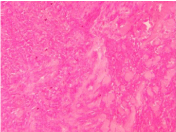
    Figure 1b: Histopathologic slide photograph of the pleomorphic adenoma
(10×10 magnification).
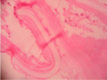
Pathological diagnosis is presented in (Table 4). The most common pathological diagnoses were the cystic lesions (48.9%) (Figure 2B) followed by the secondary tumours (21.2%), the lacrimal gland lesions (6.4%) (Figure 1B), the inflammatory lesions (6.4%) and the vasculogenic lesions (4.2%). The specific pathological diagnoses were the dermoid cyst (23.4%) followed by the non-specific orbital cyst (10.6%), the parasitic orbital cyst (6.4%), the squamous cell carcinoma (6.4%) and the retinoblastoma with extraocular extension (6.4%). Among these pathological conditions, malignant lesion was present in 15 patients (31.9%).
    Specific Diagnosis
    No (%)


    Cystic lesions
    Dermoid cyst
    11 (23.4)


    Orbital cyst
    5 (10.6)


    Parasitic cyst
    3 (6.4)


    Retention cyst
    2 (4.3)


    Epidermoid cyst
    1 (2.1)


    Sudoriferous cyst
    1 (2.1)


    Secondary tumour
    Squamous cell carcinoma
    3 (6.4)


    Retinoblastoma with extracular extension
    3 (6.4)


    Malignant round cell tumour
    1 (2.1)


    Sebaceous gland carcinoma
    1 (2.1)


    Basal cell carcinoma
    1 (2.1)


    Acinic cell carcinoma
    1 (2.1)


    Lacrimal gland lesion
    Adenoid cystic carcinoma
    2 (4.3)


    Pleomorphhic adenoma
    1 (2.1)


    Inflammatory lesion
    Orbital abscess
    1 (2.1)


    Foreign body granuloma
    1 (2.1)


    Chronic inflammatory lesion
    1 (2.1)


    Vasculogenic lesion
    Hemangiopericytoma
    1 (2.1)


    Lymphangioma
    1 (2.1)


    Optic nerve lesion
    Optic nerve glioma
    2 (4.3)


    Lymphoid lesion
    Non-Hodgkin’s lymphoma
    2 (4.3)


    Fibro-osseous lesion
    Fibrous dysplasia
    1 (2.1)


    Myogenic lesion
    Rhabdomyosarcoma
    1 (2.1)
Table 4: Pathological diagnosis.

    Figure 2a: Coronal CT Scan image of orbit showing right orbital hydatid cyst
(arrow) of a 13-year-old boy with a history of gradually progressive non-axial
proptosis of right eye secondary to orbital hydatid cyst.

    Figure 2b: Histopathologic slide photograph of the hydatid cyst showing its
wall (40×10 magnification).
Agreement between clinical and pathological diagnosis was present in 34 cases (72.3%) and absent 13 cases (27.7%) (Table 5).
    Clinical Diagnosis
    Pathological Diagnosis


    1.
    Bilateral Orbital Lymphoma
    Chronic Inflammatory Lesion


    2
    B/L Lacrimal Gland Tumour
    Sudoriferous Cyst


    3.
    Lacrimal Gland Tumour
    Sebaceous Gland Carcinoma


    4.
    Lacrimal Gland Tumour
    Acinic Cell Carcinoma


    5.
    Lacrimal Gland Tumour
    Non-Hodgkin’s Lymphoma


    6.
    Dermoid Cyst
    Parasitic Cyst


    7.
    Dermoid Cyst
    Epidermoid Cyst


    8.
    Dermoid Cyst
    Simple Orbital Cyst


    9.
    Haemangioma
    Haemangiopericytoma


    10.
    Haemangioma
    Lymphangioma


    11.
    Mucocele
    Rhabdomyosarcoma


    12.
    Sarcoma
    Non-Hodgkin’s Lymphoma


    13.
    Osteoma
    Orbital Cyst
Table 5: Cases without clinico-pathological correlation.
Discussion
The study presents a profile of pathology in patients with orbital diseases. In our study males (68.1%) preponderance was quite higher in orbital pathology than females. This finding didn’t align with the present studies by Shield, et al. [4] and Dallow, et al. [10] where they reported female preponderance by 57% and 66% respectively. These studies included all the cases of orbital lesions where as we had to exclude the cases where pathological examination was apparently not possible. In the Henderson, et al. [11] orbital tumour series reported nearly equal sex predilection. The difference in the gender ratio may be attributed to the difference in the nature of the current study. There is no age immune to orbital diseases as it was reported from as early as 14 months to 78 years encountering dermoid cysts in children to malignant conditions in elderly. The Dallow, et al also reported the wide range of age distribution from birth to 92 years [10].
In this study, significant proportion of subjects was children aged below 15 years (42.5%). In the Henderson, et al. study, [11] of orbital tumour series, only 15% of the study population was children. In the Dallow et al study, [10] only 25% of the cases were aged below 30 years and the most of the patients (52%) were in 41-70 years of age range. This difference could possibly be due to variation in geographic and demographic distribution.
The mode of presentation of patients depends upon the type and extent of the lesion. In this study (Table 2), by far the commonest symptom presented was swelling, mass of eyelid in periorbital region (85.1%). In Henderson’s series, [11] proptosis was the commonest presentation (79%). In the study carried out by Demirci, et al. [7], mass (26%) was the commonest clinical feature at presentation. In that study, investigators had included only one clinical feature as the main clinical feature at presentation in each patient. In term of the anatomical location, majority of the patients (51%) had orbital lesion in the superior half of the orbit in our study. This finding was comparable to the study by Demirci, et al. [7]. It reported the tumour in the superior half of the orbit (53%) among 217 cases. Right-sided involvement (55.3%) was higher than left-sided involvement (40.4%). One case was clinically diagnosed as the bilateral orbital lymphoma, which showed chronic inflammatory lesion only with no evidence of malignancy on excisional biopsy. This patient later developed chronic renal failure. Another case was diagnosed clinically as bilateral ectopic lacrimal gland, which on excision biopsy, turned out to be sudoriferous cyst. Almost equal orbital lesion in right and left side was reported in the Demirci, et al. study [7].
In this study (Table 3), the commonest clinical diagnosis, after clinical examination and radiological investigations was the dermoid cyst (29.8%), the lacrimal gland tumour (14.9%) and non-specific orbital cyst (12.8%). In our study, the most common pathological diagnosis (Table 4) was the cystic lesions (48.9%) and the secondary tumours (21.2%). Our finding was consistent with the Shields et al study (1984) of 645 biopsies of orbital lesions [12]. In the study, orbital cystic conditions were the commonest type (30.0%). Investigators who have included cystic lesions in their orbital series have had a high incidence, where as those who have excluded such dermoid cysts have reported a low incidence.
Finding of our study did not match with other large-scale studies. In the Shields et al study4 among 1264 patients with orbital tumours and simulating lesions referred to an oncology service, the largest major group was vasculogenic lesions (17%) followed by the secondary orbital tumours (11%) and the inflammatory lesions. The most common diagnosis was the lymphoid tumours (11%) and the idiopathic orbital inflammation (11%). Cystic lesions constituted only 6% of total cases. Among them, the dermoid cyst constituted only 2% of the total orbital lesions. Possibly, a typical small subcutaneous dermoid cyst sometimes is managed by a general ophthalmologist, paediatric ophthalmologist, or oculoplastic surgeon and the patient is less likely to be managed in an oncology service for their less incidence of the dermoid cyst [4]. In our study, we referred patients for pathological investigations after detail clinical examination. Some studies reported thyroid related orbital diseases the most common orbital diseases [10]. However, granuloma was also reported to be the commonest type in a literature [13].
In term of malignant potential of the orbital pathology, 31.9% orbital pathology in this study was malignant. Though, the malignant orbital pathology was noted above 31 years, all the cases above 60 years were malignant. This finding was also comparable to the other studies. Shields, et al. [4] reported 36% orbital pathology was malignant and the incidence of malignancy increased as the age of the subjects advanced. So the malignant lesions were 20% in children (0-18 years), 27% in young adults and middle-aged patients (19-59 years) and 58% in older patients (60-92 years). Similarly, Dallow, et al. [10] reported 70% of the orbital diseases were benign. There was a significant agreement between clinical diagnosis and pathological diagnosis by 71.7%. However, in 13 cases (28.3%) clinical diagnosis did not match with pathological diagnosis. In the Kim, et al. study [14] the agreement was 60.8% between preoperative CT diagnosis and postoperative histopathological diagnosis of orbital tumours. In our study, one case of dermoid cyst turned out to be epidermoid cyst after pathological examination. Out of two cases of haemangioma in clinical diagnosis was turned out to be haemangiopericytoma and lymphangioma each. One case of lacrimal gland tumour turned out to be non-hodgkin’s lymphoma after pathological examination. Another case of lacrimal gland tumour showed acinic cell carcinoma with unknown origin after pathological examination. Acinic cell carcinoma is common in salivary glands, particularly parotid glands but there are few case reports [14-16] indicating acinic cell carcinoma of the lacrimal gland. So, this case might also be the rare occurrence of acinic cell carcinoma of the lacrimal gland itself. In the other eight cases, clinical diagnosis did not match with pathological diagnosis.
In summary, orbital diseases are relatively uncommon entities that affect people of wide age range. Benign orbital lesion was common in younger patients where as malignant orbital lesion was common in older patients. Dermoid cyst was the most common orbital lesion. Though there was a firm agreement between clinical diagnosis and pathological diagnosis, histopathologic investigation proved to be effective.
References