
Research Article
J Ophthalmol & Vis Sci. 2021; 6(1): 1047.
Associations of Optic Nerve Head and Anterior Segment Parameters in Normotensive Saudi Females
Zeraid FM1, Hakami AW1 and Osuagwu UL2*
¹Department of Optometry & Vision Sciences, King Saud University, Saudi Arabia
²Diabetes Obesity & Metabolism Translational Research Unit (DOMTRU), Australia
*Corresponding author: Uchechukwu L. Osuagwu, School of Medicine, Diabetes Obesity & Metabolism Translational Research Unit (DOMTRU), Western Sydney University. Parkside crescent, Campbelltown 2560 NSW, Australia
Received: February 01, 2021; Accepted: March 03, 2021; Published: March 10, 2021
Abstract
Objectives: To determine the relationship between anterior segment parameters and the optic nerve head (ONH) parameters of females.
Methods: Anterior segment, optic nerve head parameters, Intraocular Pressure (IOP) and Axial Length (AL) of 99 participants (aged 20.1±1.3 years) were obtained. Differences between refraction groups and relationship between anterior segment and ONH parameters were assessed as well as linear regression for factors associated with ONH and anterior segment parameters after adjusting for cofounders.
Results: SER (-0.12 Acknowledgements: The authors acknowledge the support of the Research Centre, of the Female Scientific and Medical Colleges, Deanship of Scientific Research at King Saud University.0.58D), AL (23.5±0.8 mm), IOP (17.8±2.0 mmHg), anterior segment parameters and optic disc parameters were similar between refraction groups (p>0.20 for all).
Age was correlated with anterior chamber volume (r=-0.19), while SER was correlated with AL (r=-0.26), anterior chamber volume (r=-0.25) and angle (r=- 0.19) and with the disc area (r=0.25; p<0.02 for all). Myopes had wider anterior chamber depths than emmetropes (mean difference, 2.06, 95% confidence interval: 0.32/3.80 degs, p=0.02) with smaller disc area (-0.21mm², 95% CI: -0.38/-0.03mm²; p=0.021). After correcting for age and refraction, axial length was associated with corneal refractive power (r=-0.60, p<0.001), and anterior segment parameters (p<0.001 for all).
Conclusions: The ONH structure was related to anterior segment morphology including corneal thickness, anterior chamber volume and depth, which is important when interpreting OCT imaging.
Keywords: Cornea; Refraction; Optic nerve head; Cornea thickness; Axial length; Optic disc parameters; Myopia
Introduction
The anterior segment parameters particularly those of the corneal parameters have been implicated as potential determinants of glaucoma risk factors [1,2]. Corneal thickness and/or biomechanical properties including central hysteresis were reported to significantly influence glaucoma development. Corneal hysteresis was shown to have a strong negative correlation with Intra Ocular Pressure (IOP), and was only weakly correlated with Central Corneal Thickness (CCT) in people with glaucoma [3,4]. In the Ocular Hypertensive Treatment Study (OHTS) and the Early Manifest Glaucoma Treatment Study (EMGT), significant relationship was found between CCT and the risk of glaucoma [5,6] with greater risk in older females, those with thick or anteriorly positioned lenses, short axial lengths and those with shallow anterior chambers [7].
The human eyeball gains its structure through the continuity of the cornea, sclera, and lamina cribrosa and during development, the corneal stroma, corneal endothelium, and optic nerve lamina cribrosa differentiate from the neural crest, suggesting that these parameters are related embryologically [8]. Eyes with an optic disc pit had smaller back radius of corneal curvature compared with fellow eyes as shown in previous studies, suggesting the influence of the embryological factors involved in the development of optic disc pit on the development of cornea [9]. Based on this anatomical facts, studies have suggested the possibility of a relationship between the cornea and the optic nerve [1,2,10-12]. Although the main known risk factor for glaucoma onset and progression remains high IOP [13], underestimating the Goldmann applanation tonometry reading appear to only partly explain the relationship between cornea thickness and increased risk of glaucoma. However, the other mechanisms underlying this relationship are unknown.
Corneal thickness has been linked to scleral thickness [14] and the optic disc parameters, particularly optic disc size and Cup/Disc ratio (C/D) are related to glaucoma risk [15], such that large optics discs may be susceptible to glaucomatous damage at low IOPs [16]. Also, the Optic Nerve Head (ONH) appears larger in normal tension glaucoma, than in primary open angle glaucoma [17]. Regarding the corneal power, a study found significant correlations between the optic disc size and both keratometry readings and axial lengths. People with larger optic disc had lower keratometry reading and longer axial lengths compared with those with smaller optic discs [18]. However, there is paucity of data on the impact of other cornea parameters such as corneal volume and corneal refractive powers on ONH, particularly, in healthy people. Past studies have considered only the relationship between CCT and ONH, but the relationship between anterior chamber parameters (depth and volume) and ONH parameters remains unclear. In non-glaucomatous eyes, it has been suggested that the dimensions and shape of the ONH may be related to anterior segment morphology and the authors suggested to consider the anterior segment parameters when evaluating the ONH [2].
Therefore, the present study aims to determine the relationship between anterior segment parameters and the ONH parameters in non-glaucomatous participants. Information from this study will be valuable in determining the impact of corneal parameters as potential determinants of the risk of glaucoma.
Materials and Methods
Ethics
This research followed the tenets of the Helsinki Declaration of Helsinki. Ethical approval was obtained from the College of Applied Medical Sciences Research Ethics Committee (# CAMS 024-3940) and all participants provided informed consent prior to participation in this study, after explanation of the study protocol.
Study population
The participants were recruited from female university students who presented to the outpatient optometry clinic of the department for their routine eye examination. Participants had best corrected VA equal to/better than 20/20, Spherical Equivalent Refraction (SER) within ± 1.5D; cylinder power <1D; and IOP <22mmHg; Participants that had open anterior chamber angle on slit lamp examination, and normal ONH appearance determined by intact neuro-retinal rim was intact, without splinter haemorrhage, notches, localized pallor, or asymmetry of the cupping <0.2 between the eyes, taking into account disc size, were also included. The exclusion criteria included those that had a history of vascular, circulatory (diabetes mellitus, hypertension, or hypotension), autoimmune, or neurological diseases (including migraine), or those with neoplasia, history of glaucoma, uveitis, any ocular surgery, or trauma, any corneal abnormality such as infection or trauma. Participants with an optic disc abnormality (such as drusen or tilted disc) were also excluded in this study.
Measurement
All participants received a comprehensive ocular examination, consisting of medical history, manifest refraction, IOP measurement by Topcon Auto-kerato-refracto-tonometer KR-800, gonioscopy, and slit lamp and fundus examination. Optic disc imaging with a Topcon 3D OCT-2000 (TOPCON Corporation, Tokyo, Japan). Using the Topcon hardware, the following parameters were obtained by an examiner (FA): disc area (mm²), cup area (mm²), rim area (mm²), cup volume (mm³), rim volume (mm³), cup to disc area ratio (C/D area), linear cup to disc ratio (linear C/D), and mean and maximum cup depth (mm).
Another examiner (LO) obtained the anterior segment parameters for all participants using the Pentacam Scheimpflug system (OCULUS Optikgerate GmbH, Wetzlar, Germany) with a quality score >95. The instrument’s software constructs the three-dimensional image of the anterior segment and calculates the anterior chamber parameters. This imaging provides measurements of anterior chamber depth and anterior chamber volume. CCT is determined at the corneal apex point, the corneal volume values and the dioptric powers of the anterior and posterior corneal refractive surfaces (which were positive and negative, respectively) within a span of 3.0mm from the centre was also recorded. A description of the method for acquiring images is shown elsewhere [19]. Axial length measurements were recorded with the IOL Master® 500 (Carl Zeiss Meditec AG Jena, Germany).
Statistical analysis
Data from both eyes of participants are strongly correlated and this introduces interdependency problems during analysis [20] which we tried to avoid by choosing only measurements from one eye of each participant at random. The selection of which eye data to be used for all analysis was by an intern optometrist who was not part of this study. The data was analyzed using SPSS program version 22.0 (IBM Corp. Armonk, NY, USA) and presented using descriptive statistics (mean ± Standard Deviation (SD) and range). The normality of the distribution of variables was confirmed using the Kolmogorov Smirnov test. One-way analysis of variance was used to assess the differences between refraction groups for all variables. As in previous studies [2], the impact of Age, disc area and Spherical Equivalent Refraction (SER) on anterior segment and ONH parameters, respectively were assessed and were adjusted for in the partial correlation, which determined the relationship between anterior segment and ONH parameters, and in the linear regression analysis, which examined the factors associated with both parameters.
Results
Characteristics of the study population
A hundred and forty three females were initially enrolled in this cross-sectional study, only 99 of them met the inclusion criteria. Of those excluded, majority had myopia (SER >-1.50D), one participant had a papilledema. Overall, measurements were obtained from 57 right eyes and 42 left eyes of female participants including 49 myopes (defined as SER that was less than -0.25DS), 44 emmetropes (SER, -0.25 to +0.50 DS) and 6 hyperopes (SER greater than +0.50DS) who were aged between 19-26 years (mean age, 20.1, standard deviation 1.3 years). Figure 1 shows the Topcon OCT images of the optic disc and their corresponding Oculus Pentacam refractive maps for three participants, is shown in (Figure 2). The mean SER, axial length and IOP for the participants were -0.12 ±0.58D (range, -1.50 to +1.00 D), 23.5 ± 0.8 mm (range, 21.5-25.3 mm), and 17.8 ± 2.0 mmHg (range, 13-25 mmHg) respectively.
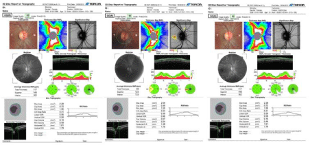
Figure 1: Topcon Ocular Coherence Tomography report of three healthy Arab females.
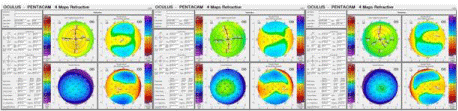
Figure 2: Oculus-Pentacam 4 maps refractive of three healthy Arab females.
Table 1 is the descriptive analysis of the anterior segment and optic disc parameters, which were similar between refraction groups (oneway ANOVA, p>0.20 for all). Post hoc analysis showed significantly wider angles in myopic eyes than emmetropic eyes (mean difference, 2.06 degs, 95 CI: 0.32-3.80 degs; p=0.021) with similar ACD between myopes and hyperopes (mean difference, 1.56 degs, 95% CI -2.06-5.18 degs; p=0.40). The myopic eyes had similar optic disc areas compared with the hyperopic eyes (-0.24, 95% CI:-0.60 to 0.13mm², p=0.20) but significantly smaller disc areas than the emmetropic eyes (-0.21mm², 95% CI: -0.38 to 0.03mm²; p=0.021).
![]()
Variables
Anterior Segment parameters
CCT (�m)
Corneal volume (mm3)
Anterior Chamber Volume (mm3)
Anterior Chamber Depth (mm)
Anterior Chamber angle (deg)
Anterior Corneal Refractive power (D)
Posterior Cornea Refractive power (D)
Optic Nerve Head parameters
Mean (±SD)
42.9 (1.4)
-6.2 (0.2)
560.1 (34.8)
60.9 (6.8)
160.2 (27.6)
2.9 (0.3)
38.4 (4.3)
Axial length (mm)
23.5 (0.8)
-0.60***
0.57***
0.03
0
0.60***
0.49***
0.34***
Disc area (mm2)
2.2 (0.4)
-0.07
0.06
0.11
0.01
-0.15
-0.11
-0.03
Cup Area (mm2)
0.6 (0.4)
-0.07
0.06
-0.11
-0.1
-0.02
0.06
-0.03
Rim Area (mm2)
1.7 (0.4)
0.08
-0.1
0.18*
0.13
-0.22*
-0.19*
0.01
C/D ratio
0.2 (0.2)
-0.08
0.07
-0.14
-0.13
0.02
0.09
-0.03
Linear C/D ratio
0.5 (0.2)
-0.03
0.04
-0.22
-0.14
-0.03
0.04
-0.03
Cup volume (mm3)
0.1 (0.1)
-0.06
0.02
-0.13
-0.11
0.02
0.06
0.1
Rim volume (mm3)
0.7 (0.3)
0.03
-0.06
0.11
0.11
-0.08
-0.03
0.01
Retinal Nerve fibre layer
114.3 (9.7)
0.18*
-0.12
-0.1
-0.1
-0.16*
-0.16
-0.01
*p<0.05; ***p<0.001; CCT: Central Corneal Thickness; D: Diopter.
Table 1: Mean (±standard deviation) of measured parameters (bolded) and the Pearson correlation coefficients (italics) for the relationship between disc parameters and anterior segment parameters.
There was a weak negative correlation between participants’ age and the anterior chamber volume (r=-0.19, p=0.03) but the negative correlation between participants’ age and the anterior chamber volume did not reach significance (r=-0.14, p=0.09). The mean SER was negatively correlated with axial length of the eye (r=-0.26, p=0.005), anterior chamber volume (r=-0.25, p=0.006) anterior chamber angle (r=-0.19, p=0.027) and positive correlations with the optic disc area (r=0.25, p=0.006) in healthy young females. There were no significant correlations between optic disc area and anterior segment parameters in this cohort.
Relationship between ONH and anterior segment parameters
Partial correlation analysis to investigate the relationships between anterior segment and optic nerve head parameters, adjusting for age and SER, found significant correlations between axial length of the eye and all of the anterior segment parameters excluding CCT and corneal volume (p>0.05). There was a negative correlation between axial length and the refractive powers of the anterior (r=- 0.597, p<0.001) and the posterior corneas (r=0.567, p<0.001). CCT was significantly related to the optic disc rim area (r=0.18, p=0.04) and showed a close to significant relationship with linear C/D (r=- 0.163, P=0.06). No other ONH parameter was significantly correlated with CCT.
Both the anterior corneal volume and anterior chamber depth were negatively correlated with the optic disc rim area (-0.22 & -0.19, p<0.03 for both) whereas the relationship between both anterior segment parameters and RNFL was at borderline significance (r=-0.16 for both, p=0.05 and 0.06 respectively). No other ONH Parameter showed significant correlations with the anterior segment parameters in this analysis.
Table 2 presents the odds and adjusted odds ratio of factors associated with ONH and anterior segment relationship. Linear regression analysis revealed no significant relationship between ONH parameters and all measured anterior segment parameters after correction for the cofounding variables of age and refraction.
![]()
    Anterior Corneal Refractive    power
    Posterior Cornea Refractive    power
    CCT
    Corneal volume
    Anterior Chamber Volume
    Anterior Chamber Depth
    Anterior Chamber angle


    OR
    aOR (95CI)
    OR
    aOR (95CI)
    OR
    aOR (95CI)
    OR
    aOR (95CI)
    OR
    aOR (95CI)
    OR
    aOR (95CI)
    OR
    aOR (95CI)


    Disc area
    0.01
    0.03 (-0.12/0.14)
    0.3
    0.16 (-0.53/1.14)
    0.00
    0.15 (-0.00/0.01)
    -0.01
    -0.08 (-0.02/0.01)
    -0.01
    -0.24 (-0.01/0.00)
    0.06
    0.04 (-0.44/0.55)
    0.01
    0.05 (-0.02/0.03)


    Cup area (mm2)
    -0.04
    -0.14 (-0.17/0.08)
    -0.14
    -0.07 (-0.96/0.68)
    0.00
    -0.08 (-0.00/0.00)
    -0.01
    -0.09 (-0.02/0.01)
    0.00
    -0.2 (0.01/0.00)
    0.31
    0.21 (-0.17/0.80)
    8.91E-05
    0.00 (-0.02/0.02)


    Rim area (mm2)
    0.05
    0.17 (-0.07/0.18)
    0.31
    0.16 (-0.51/1.13)
    0.00
    0.18 (-0.00/0.01)
    0.00
    0.01 (-0.02/0.02)
    0.00
    -0.18 (-0.01/0.00)
    -0.15
    -0.10 (-0.63/0.34)
    0.01
    0.10 (-0.01/0.03)


    Cup disc ratio
    -0.02
    -0.18 (-0.07/0.03)
    -0.08
    -0.11 (-0.39/0.24)
    0.00
    -0.12 (-0.00/0.00)
    0.00
    -0.09 (-0.01/0.01)
    0.00
    -0.18 (-0.00/0.00)
    0.13
    0.23 (-0.05/0.32)
    0
    -0.01 (-0.01/0.01)


    Linear Cup disc ratio
    -0.02
    -0.15 (-0.06/0.03)
    -0.08
    -0.12 (-0.39/0.23)
    0.00
    -0.13 (-0.00/0.00)
    0.00
    -0.11 (-0.01/0.00)
    0.00
    -0.23 (-0.00/0.00)
    0.12
    0.21 (-0.06/0.30)
    0.00
    0.02 (-0.01/0.01)


    Cup volume (mm3)
    -0.03
    -0.32 (-0.06/0.01)
    -0.15
    -0.30 (-0.37/0.06)
    0
    -0.12 (-0.00/0.00)
    0.00
    -0.13 (-0.01/0.00)
    0.00
    -0.15 (-0.00/0.00)
    0.06
    0.15 (-0.07/0.19)
    0.00
    0.16 (-0.00/0.01)


    Rim volume (mm3)
    0.01
    0.02 (-0.09/0.11)
    0.03
    0.02 (-0.63/0.69)
    0.00
    0.08 (-0.00/0.00)
    0.00
    0.03 (-0.01/0.02)
    0.00
    -0.09 (-0.01/0.00)
    0.05
    0.04 (-0.34/0.44)
    0.00
    0.04 (-0.02/0.02)


    RNFL
    1.94
    0.27 (-0.82/4. 70)
    5.38
    0.12 (-12.98/23.74)
    -0.01
    -0.02 (-0.08/0.06)
    -0.16
    -0.12 (-0.56/0.23)
    -0.03
    -0.09 (-0.15/0.09)
    -3.28
    -0.10 (-14.06/7.50)
    0.1
    0.05 (-0.41/0.61)


    RNFL: Retinal Nerve Fibre Layer; CCT: Central    Corneal Thickness.
Table 2: Linear Regression analysis between Optic Nerve head and anterior segment parameters. Results were expressed as Odds Ratio (OR) and Adjusted Odds Ratio (aOR, 95% Confidence Intervals, CI). CIs including zero were non-statistically significant.
Discussion
The corneal properties including the thickness and biomechanical properties have been found to be important risk factors for the development of glaucomatous optic nerve damage [5,6,21]. Considering the clinical importance of the ONH parameters in assessing glaucomatous optic neuropathy, this study was undertaken to investigate the relationship between anterior segment and ONH parameters in healthy, non-glaucomatous females. The study found that, with the exception of the corneal parameters (CCT and cornea volume), all anterior segment parameters, showed significant correlation with the axial length of non-glaucomatous eye. Of the optic disc parameters, the optic disc area was significantly correlated with CCT, anterior chamber volume and anterior chamber depth. However, these effects were dependent on the refractive error status and axial length of the eye. No other significant association was found between anterior segment and ONH parameters in this cohort. There have been inconsistencies in published literatures regarding the relationship between anterior segment parameters and ONH parameters in healthy eyes [2,22,23] and in glaucomatous eyes [24,25]. Studies have shown evidence of no correlation between optic disc parameters and corneal measurements or optic disc parameters and anterior chamber depths [23] particularly in non-healthy eyes, and another showed significant negative correlations between corneal thickness and optic disc area after correcting for age and optics disc areas [2]. The study concluded that eyes with thicker corneas have smaller discs and those with deeper anterior chambers had smaller ONHs [2]. Where significant associations were observed in normal eyes, the effects were nullified after adjustment for cofounders. For instance, in non-tension glaucoma patients, Park et al. [26] showed that corneal hysteresis was directly associated with rim area, rim volume, and mean RNFL thickness (P=0.012, P=0.028, and P=0.043) and inversely associated with linear cup-to-disc ratio (P=0.015), after adjusting for age, axial length, CCT, disc area, GAT data, and SE. However, in normal patients, they found no significant associations between corneal biomechanical properties and optic disc parameters. In another study, Cankaya et al. noted significant relationship between CCT and optic disc area, rim area, RV, and RNFL in nonglaucomatous eyes [2] and suggested that the central corneal thickness influences optic nerve structures. Similar significant relationship have been observed in diseased eyes [25,27,28] and in one study, the effects were nullified when the results were evaluated separately for the glaucoma eyes and for the healthy eyes [25]. In a population-based study among Asian Malay adults, lower CCT was related to decreased rim area and increased cup-disc ratio only in patients with primary open glaucoma, and not in those without glaucoma [29]. This later finding was supported by yet another study [30] and was in line with the present result.
This study also assessed the relationship between RNFL thickness and anterior segment parameters and found negative but nonstatistically significant correlation between anterior chamber depth and RNFL. In a population-based Singapore Chinese Eye Study including adults aged 40 to 80 years, average RNFL thickness was independently related to age, disc area, and axial length of the eye [31]. Regarding the relationship between CCT and RNFL thickness, we did not find any significant correlation and this was similar to a previous study [32], where the authors also noted that significant relationships observed in ocular hypertension and glaucomatous eyes may be due to RNFL loss caused by the disease. The significant correlations found between the rim area and the anterior segment parameters were nullified after adjusting for refractive status and axial length of the eye in these study. In the Blue Mountains Eye Study, researchers found larger optic disc in glaucomatous eyes compared with eyes that had ocular hypertension and normal eyes [33], suggesting that the optic disc may be more susceptible to axonal damage than other disc parameters. The differences between studies may be attributed to the methodological differences including the different races and the use of different instruments to evaluate the ONH parameters, cornea and anterior chamber parameters.
The present study has some limitations, including the narrow age range of the participants, inclusion of only female participants, the non-inclusion of glaucomatous eyes, and the small sample size, which should be considered when interpreting the present results of this study, or extrapolating the results to other populations or comparing the results with studies including both genders.
Conclusions
This study showed that the shape and dimensions of the ONH might be related to the anterior segment morphology, such as the corneal thickness, corneal volume and the anterior chamber depth. It is important to know the relationship between anterior segment and ONH parameters as this is a relatively a new consideration with very limited studies. The findings of this study are also important when interpreting SD-OCT measurement, and provide information on the optic nerve head of healthy female participants of Arab origin within the age group. However, additional evidence on these relationships are needed.
Acknowledgements
The authors acknowledge the support of the Research Centre, of the Female Scientific and Medical Colleges, Deanship of Scientific Research at King Saud University.
References
- Hussnain SA, Kovacs KD, Warren JL and Teng CC. Corneal hysteresis and anterior segment optical coherence tomography anatomical parameters in primary angle closure suspects. Clin Exp Ophthalmol. 2018; 46: 468-472.
- Cankaya AB and Ozates S. Relationship between anterior segment and optic nerve head parameters in healthy subjects. Arquivos Brasileiros De Oftalmologia. 2017; 80: 285-289.
- Mangouritsas G, Morphis G, Mourtzoukos S and Feretis E. Association between corneal hysteresis and central corneal thickness in glaucomatous and non-glaucomatous eyes. Acta Ophthalmologica. 2009; 87: 901-905.
- Roberts CJ. Concepts and misconceptions in corneal biomechanics. Journal of cataract and refractive surgery. 2014; 40: 862-869.
- Gordon MO, Beiser JA, Brandt JD, Heuer DK, Higginbotham EJ, Johnson CA, et al. The Ocular Hypertension Treatment Study - Baseline factors that predict the onset of primary open-angle glaucoma. Archives of Ophthalmology. 2002; 120: 714-720.
- Leske MC, Heijl A, Hyman L, Bengtsson B, Dong LM, Yang Z, et al. Predictors of long-term progression in the early manifest glaucoma trial. Ophthalmology. 2007; 114: 1965-1972.
- Wright C, Tawfik MA, Waisbourd M and Katz LJ. Primary angle-closure glaucoma: an update. Acta Ophthalmol. 2016; 94: 217-225.
- Sellheyer K and Spitznas M. Development of the human sclera. A morphological study. Graefe’s archive for clinical and experimental ophthalmology. 1988; 226: 89-100.
- Goktas A and Goktas S. Common embryological links to the optic pit and anterior segment structures. Optometry and vision science: Official publication of the American Academy of Optometry. 2010; 87: 585-587.
- Gaspar R, Pinto LA and Sousa DC. Corneal properties and glaucoma: a review of the literature and meta-analysis. Arq Bras Oftalmol. 2017; 80: 202- 206.
- Kim JM, Park KH, Kim SH, Kang JH and Cho SW. The relationship between the cornea and the optic disc. Eye. 2010; 24: 1653-1657.
- Sarfraz MH, Mehboob MA and Haq RI. Correlation between central corneal thickness and visual field defects, cup to disc ratio and retinal nerve fiber layer thickness in primary open angle glaucoma patients. Pak J Med Sci. 2017; 33: 132-136.
- Saenz-Frances F, Garcia-Feijo J, Janez L, Borrego-Sanz L, de la Casa JMM, Fernandez-Vidal A, et al. Comparing corneal variables in healthy subjects and patients with primary open-angle glaucoma. Invest Ophthalmol Vis Sci. 2011; 52: 3683-3688.
- Oliveira C, Tello C, Ritch R, Liebmann JM and Ritch R. Correlation between central corneal thickness, scleral thickness and refractive error. Investigative Ophthalmology & Visual Science. 2004; 45: U370-U370.
- Hoffmann EM, Zangwill LM and Crowston JG. Optic disk size and glaucoma. Survey of ophthalmology. 2007; 52: 32-49.
- Burk RO, Rohrschneidern K and Noack H. Are large optic nerve heads susceptible to glaucomatous damage at normal intraocular pressure? J Graefe’s Archive for Clinical Experimental Ophthalmology. 1992; 230: 552- 560.
- Kiriyama N, Ando A, Fukui C, Nambu H, Nishikawa M, Terauchi H, et al. A comparison of optic disc topographic parameters in patients with primary open angle glaucoma, normal tension glaucoma, and ocular hypertension. Graefe’s archive for clinical and experimental ophthalmology. 2003; 241: 541- 545.
- Jonas JB, Budde WM and Stroux A. Iris colour, optic disc dimensions, degree and progression of glaucomatous optic nerve damage. Clin Exp Ophthalmol. 2006; 34: 654-660.
- Ambrosio R, Lopes BT and Faria-Correia F. Integration of Scheimpflug- Based Corneal Tomography and Biomechanical Assessments for Enhancing Ectasia Detection. J Refract Surg. 2017; 33: 434-443.
- Armstrong RA. Statistical guidelines for the analysis of data obtained from one or both eyes. Ophthalmic and Physiological Optics. 2013; 33: 7-14.
- Zangwill LM, Weinreb RN and Beiser JA. Baseline topographic optic disc measurements are associated with the development of primary open-angle glaucoma: the Confocal Scanning Laser Ophthalmoscopy Ancillary Study to the Ocular Hypertension Treatment Study. Archives of ophthalmology. 2005; 123: 1188-1197.
- Cankaya A, Elgin U and Batman A. Relationship between central corneal thickness and parameters of optic nerve head topography in healthy subjects. European Journal of Ophthalmology. 2008; 18: 32-38.
- Bourne RRA, Foster PJ and Bunce C. The morphology of the optic nerve head in the Singaporean Chinese population (the Tanjong Pagar study): part 2 - biometric and systemic associations. British Journal of Ophthalmology. 2008; 92: 310-314.
- Pakravan M, Parsa A and Sanagou M. Central corneal thickness and correlation to optic disc size: a potential link for susceptibility to glaucoma. The British journal of ophthalmology. 2007; 91: 26-28.
- Insull E, Nicholas S and Ang GS. Optic disc area and correlation with central corneal thickness, corneal hysteresis and ocular pulse amplitude in glaucoma patients and controls. Clin Exp Ophthalmol. 2010; 38: 839-844.
- Park K, Shin J and Lee J. Relationship between corneal biomechanical properties and structural biomarkers in patients with normal-tension glaucoma: a retrospective study. BMC Ophthalmol. 2018; 18: 7.
- Pakravan M, Parsa A, Sanagou M and Parsa CF. Central corneal thickness and correlation to optic disc size: A potential link for susceptibility to glaucoma. British Journal of Ophthalmology. 2007; 91: 26-28.
- Vergados A, Papaconstantinou D, Diagourtas A, Theodossiadis PG, Vergados I, Georgalas I. Correlation between optic nerve head parameters, RNFL, and CCT in patients with bilateral pseudoexfoliation using HRT-III. In: Seminars in Ophthalmology. 2015: 44-52.
- Wu RY, Zheng YF, Wong TY, Cheung CYL, Loon SC, Chauhan BC, et al. Relationship of central corneal thickness with optic disc parameters: the Singapore Malay Eye Study. Invest Ophthalmol Vis Sci. 2011; 52: 1320-1324.
- Bourne R, Foster P and Bunce C. The morphology of the optic nerve head in the Singaporean Chinese population (the Tanjong Pagar study): part 2-biometric and systemic associations. British Journal of Ophthalmology. 2008; 92: 310-314.
- Cheung CY, Chen D, Wong TY, Tham YC, Wu R, Zheng Y, et al. Determinants of quantitative optic nerve measurements using spectral domain optical coherence tomography in a population-based sample of nonglaucomatous subjects. Invest Ophthalmol Vis Sci. 2011; 52: 9629-9635.
- Mumcuoglu T, Townsend KA, Wollstein G, Ishikawa H, Bilonick RA, Sung KR. Assessing the relationship between central corneal thick retinal nerve fiber layer thicknesses in healthy subjects. Am J Ophthalmol. 2008; 146: 561- 566.
- Healey PR and Mitchell P. Optic disk size in open-angle glaucoma: the Blue Mountains Eye Study. Am J Ophthalmol. 1999; 128: 515-517.