Case Report
J Ophthalmol & Vis Sci. 2022; 7(2): 1066.
Manifestation of Corneal Wound Gap during Cataract Surgery: A Case Report
Wang X1, Li Y1, Zhang C2,3 and Dang G1,2*
1Department of Ophthalmology, Jinan Mingshui Eye Hospital, Zhangqiu District, Jinan City, People’s Republic of China
2Department of Ophthalmology, The First Affiliated Hospital of Shandong First Medical University & Shandong Provincial Qianfoshan Hospital, Lixia District, Jinan City, People’s Republic of China
3State Key Laboratory of Ophthalmology, Zhongshan Ophthalmic Center, Sun Yat-sen University, Tianhe District, Guangzhou City, China
*Corresponding author: Guangfu Dang, Department of Ophthalmology, Jinan Mingshui Eye Hospital, Zhangqiu District, Jinan City, People’s Republic of China; Department of Ophthalmology, The First Affiliated Hospital of Shandong First Medical University & Shandong Provincial Qianfoshan Hospital, Lixia District, Jinan City, People’s Republic of China
Received: February 28, 2022; Accepted: March 19, 2022; Published: March 26, 2022
Abstract
Significance: Intraoperative AS-OCT showed great advantages in the treatment of the patient with traumatic cataract and corneal perforation, and could be used to real-time monitor the changes of corneal wound and the IOL position.
Purpose: To report the change of corneal wound in a patient with traumatic cataract and corneal perforation during the anterior segment-OCT-assisted (ASOCT- assisted) cataract surgery.
Case Presentation: A 48-year-old male patient, whose right eye was injured by a steel wire two months ago, presented with traumatic cataract and corneal perforation. When he was referred to our hospital, the corneal epithelium and part of anterior stroma at the laceration had healed. Therefore, the corneal laceration was not sutured, and AS-OCT was conducted to monitor the corneal laceration and assess the IOL position during cataract surgery. The laceration maintained closed, and no liquid leakage through corneal laceration was found throughout the surgical operation. IOL was placed into the capsular bag, and the visual acuity of the affected eye recovered to 3/10 postoperatively.
Conclusions: Intraoperative AS-OCT could be used to real-time monitor the changes of corneal wound and the IOL position, and showed a significant advantage in the treatment of the eye with traumatic cataract and corneal perforation
Keywords: AS-OCT; Cataract surgery; IOL
Introduction
The incidence of ocular trauma is common despite the anatomical and functional protective mechanisms of the eye from the orbital rim and reflex closure of the lids [1]. Open-globe injuries, a visually and economically devastating cause of vision loss, are estimated to affect 2.8 and 3.7 per 100,000 population annually according to previous studies from New Zealand and Singapore [2,3], and are often accompanied by traumatic cataract. For the patients with open-globe injuries and traumatic cataracts, the change of corneal wound during cataract surgery has not yet been reported.
Anterior segment optical coherence tomography (AS-OCT) is a tool used for assessing different anterior segment eye variables, having the advantage of contactless operation and high scanning speed and precision [4,5]. It could be used to obtain high-resolution anterior segment images in vivo, even in the eyes with corneal scars. In clinical practice, AS-OCT has been widely applied for the diagnosis and treatment of many ocular anterior segment diseases, such as primary angle closure glaucoma, ocular surface neoplasia, corneal dystrophies, and so on, and for improving the accuracy of intraocular lens (IOL) power calculations during cataract treatment [6]. Previous study reported that the operating microscope integrated intraoperative OCT could also be used for guiding cataract surgery and following up the changes of corneal wound [7]. Here we report a patient with traumatic cataract and corneal perforation, whose corneal wound was tracked in real time during cataract operation using a microscope integrated intraoperative OCT.
Case Presentation
A 48-year-old male, whose right eye was injured by a steel wire two months ago, was referred to the outpatient of The First Affiliated Hospital of Shandong First Medical University due to blurred vision in the affected eye. After injury, he went to a local hospital immediately, and the antibiotic eye drops were used. But the corneal laceration was not sutured at that time, and no other ophthalmological treatment was performed before visiting our hospital.
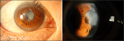
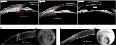
The visual acuity of the affected eye was hand movement. Slitlamp examination showed an about 4mm corneal laceration in the temporal side of the pupil, where the corneal tissue exhibited local nubecula and edema. Iris posterior synechia and white cataract could be observed (Figure 2A and 2B). The presence of intraocular foreign body was excluded by ocular B-scan ultrasound (Figure 2C and 2D) and CT examination (Data not shown). Angle closure at the upper and nasal sides could be found by UBM, and the inferior and temporal anterior chamber angle was narrow (Figure 3A and 3B). The anterior chamber was slightly shallow (Figure 3C). AS-OCT revealed a fullthickness laceration in the temporal side of the central cornea, but the corneal epithelium and part of anterior stroma at the laceration had healed when the patient was referred to our hospital. Furthermore, a membrane adhering to the posterior surface of corneal laceration was found (Figure 3D). However, what the membrane was and where the membrane was from were still not sure. We inferred that it might come from the capsule of the lens or the folded Descemet membrane.

    Figure 1: Clinical Timeline: a 48-year-old male patient diagnosed with traumatic cataract and corneal perforation and treated with AS-OCT-assisted cataract

surgery and IOL implantation.

AS-OCT: Anterior Segment Optical Coherence Tomography.

    Figure 2: Preoperative slit lamp photograph and B-scan images of the affected eye. (A-B) There was a laceration in the temporal side of the pupil, and the corneal

tissue around the laceration exhibited local nubecula and edema. Iris posterior synechia and white cataract were observed; (C-D) No intraocular foreign body was

found by B-scan.

    Figure 3: UBM and AS-OCT images of the affected eye. (A) UBM image showed the angle closure at the upper and nasal sides; (B) The inferior and temporal

anterior chamber was narrow; (C) The anterior chamber was slightly shallow; (D-E) AS-OCT revealed a full-thickness laceration in the temporal side of the central

cornea (D) and a membrane adhering to the posterior surface of corneal laceration (D-E).
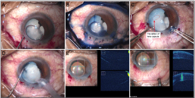
After the eye and general examination, phacoemulsification was performed under topical anesthesia. A typical small incision phacoemulsification and primary IOL implantation was adopted. To avoid the staining of the tissue around the laceration, the filtered air was injected into the anterior chamber before the trypan blue dyeing of the lens capsule (Figure 4A and 4B). After trypan blue staining, we found that a small piece of lens capsule was missing (Figure 4C). Based on this, we deduced that the membrane attached to the corneal laceration should be the missing lens capsule. A bottle height of 90cm was used for irrigation to maintain the relatively low intraocular pressure during phacoemulsification. Corneal laceration, which was not sutured, kept closed and no liquid leakage was found throughout the surgical operation (Figure 4D). We inferred that it might benefit from the reduced irrigation pressure during phacoemulsification and the healing of the corneal epithelium and anterior stroma. Furthermore, due to the affected eye with corneal perforation injury and the opacity of the central zone of cornea, AS-OCT was used to guide surgical operation in monitoring the change of corneal laceration and assessing the IOL position (Figure 4E and 4F). Postoperatively, conventional topical antibiotic and steroid therapy (both Pred Forte and Levofloxacin eyedrops 4 times per day, and Tobradex eye ointment at bedtime) was used to reduce inflammation.

    Figure 4: The process of phacoemulsification and primary IOL implantation. (A-B) The filtered air was injected into the anterior chamber before the trypan blue

dyeing of lens capsule to avoid the staining of the tissue around the laceration; (C) The defect of anterior lens capsule; (D) The process of phacoemulsification;

(E-F) AS-OCT was used to assess the IOL position and monitor the change of corneal laceration.
On the 1st postoperative day, the visual acuity of the affected eye was 0.3. Corneal laceration was closed, and no liquid leakage was found (Figure 5). After the three-month follow-up, the laceration healed well, and the position of IOL was stable. However, the visual acuity had no significant improvement due to the corneal scar caused by the laceration (Data no shown).

    Figure 5: The slit lamp photograph and AS-OCT image of the affected eye postoperatively. Corneal laceration was closed, and no liquid leakage was found after

cataract surgery on the 1st postoperative day.
Discussion
Open globe trauma, defined as a full-thickness laceration of the eye wall, is the major cause for single eye blindness and a subsequent decline in quality of life [7,8]. Traumatic cataract is a common complication of the penetrating ocular injury [9]. Corneal edema and opacity caused by ocular injury significantly increased the difficulty of cataract surgery. AS-OCT, which uses low-coherence interferometry laser to obtain a cross-sectional image and provide objective assessment, can effectively monitor the dynamic changes of the anterior segment of the eye and assess the IOL position in the eyes with corneal edema and opacity [10]. It showed great performance in cataract surgery of the patient with corneal edema and opacity.
AS-OCT has been used to check the abnormality of cornea, anterior chamber angle, and lens, such as open-globe injury, angle closure, lens subluxation, IOL tilt and decentration, and so on [11- 14]. In this case, we found that intraoperative AS-OCT had great advantage in constantly monitoring the changes of corneal laceration and identifying the decentration and tilt of IOL during surgery, especially in the eyes with opaque cornea. All of these could do a great favor for further decision making during the operation and reduce the operative complication.
For open globe injury, the first step of the treatment is to suture the wound, and recover the integrity of eyeball [15]. However, for this patient, his right eye had been wounded for more than two months, when he presented to our hospital. Though the corneal laceration was not sutured, the corneal epithelium and part of the anterior corneal stroma had healed at that time. Plus, corneal laceration suture would include an additional step to remove the surgical stitches. Based on these, we decided not to deal with the corneal laceration. Liquid leakage and corneal wound dehiscence were not observed in the whole operation procedure. We deduced that the corneal laceration with healed epithelium and anterior segment stroma would not split again under the reduced irrigation pressure during phacoemulsification. However, significant scar formation was observed in the area of corneal laceration during follow-up. Previous studies reported that, in the repair of penetrating corneal injury, the posterior gap of the laceration allows aqueous infiltration of the posterior stroma and increases scarring [16]. These demonstrated that surgical repair might be necessary for the corneal wound with divergence of posterior part and within two months after injury.
Furthermore, we noted that the membrane attached to the endothelium surface of the laceration disappeared after cataract surgery. It suggests that this membrane should be the lens capsule. Anterior capsulotomy, which was used to remove the anterior capsule incarcerated into cornea, might need to perform for the healing of corneal wound in the patients with traumatic cataract and capsular/ corneal incarceration [17]. In this case, the capsule incarceration might not be so tight, and the broken capsule spontaneously released from the corneal laceration during phacoemusification. We inferred that it might be benefit from the fluid flow caused by phacoemulsification irrigation/aspiration. According to this, we concluded that some loose incarceration of the anterior capsule into the cornea might be spontaneously released during phacoemulsification.
In summary, intraoperative AS-OCT could be used to realtime monitor the changes of corneal wound and the IOL position in the eyes with traumatic cataracts and corneal injuries, and might do a great favor for further decision making during the operation. Furthermore, corneal wound with healed epithelium and anterior segment stroma would not split again under the reduced irrigation pressure during phacoemulsification.
Declaration
Ethics approval: This study was approved by the Medical Ethics Review Board at The First Affiliated Hospital of Shandong First Medical University & Shandong Provincial Qianfoshan Hospital. All procedures performed in the patient were in accordance with the ethical standards of the institutional and/or national research committee and with the 1964 Helsinki declaration and its later amendments or comparable ethical standards. No identifiable health information was included in this case report.
Funding /Support: This study was supported by the grant from National Natural Science Fund of China (82000853) and Science and Technology Development Project of Medicine and Health in Shandong Province (2009HW071), which afford part of the fee for the patient’s follow-up and the data collection.
Authors’ contributions: Conceptualization: GD; Data Curation: XW and YL; Writing - Original Draft: XW; Writing - Review & Editing: CZ and GD; Funding acquisition: CZ and GD.
Acknowledgements: We would like to thank all the staff in the operating room and the nurses in the ophthalmic ward of Mingshui Eye Hospital for their help in data collection and patient care.
References
- Nichols BD. Ocular trauma: emergency care and management. Can Fam Physician. 1986; 32: 1466-1471.
- Court JH, Lu LM, Wang N, McGhee CNJ. Visual and ocular morbidity in severe open-globe injuries presenting to a regional eye centre in New Zealand. Clin Exp Ophthalmol. 2019; 47: 469-477.
- Wong TY, Tielsch JM. A population-based study on the incidence of severe ocular trauma in Singapore. Am J Ophthalmol. 1999; 128: 345-351.
- Dembski M, Nowinska A, Ulfik-Dembska K, Wylegala E. Swept Source Optical Coherence Tomography Analysis of the Selected Eye’s Anterior Segment Parameters. J Clin Med. 2021; 10.
- Diana MP, Ana B. Tear evaluation by anterior segment OCT in dry eye disease. Rom J Ophthalmol. 2021; 65: 25-30.
- Shan J, DeBoer C, Xu BY. Anterior Segment Optical Coherence Tomography: Applications for Clinical Care and Scientific Research. Asia Pac J Ophthalmol (Phila). 2019.
- Das S, Kummelil MK, Kharbanda V, et al. Microscope Integrated Intraoperative Spectral Domain Optical Coherence Tomography for Cataract Surgery: Uses and Applications. Curr Eye Res. 2016; 41: 643-652.
- Wu S, Bian C, Li X, et al. Controlled release of triamcinolone from an episcleral micro film delivery system for open-globe eye injuries and proliferative vitreoretinopathy. J Control Release. 2021; 333: 76-90.
- Upaphong P, Supreeyathitikul P, Choovuthayakorn J. Open Globe Injuries Related to Traffic Accidents: A Retrospective Study. J Ophthalmol. 2021; 2021: 6629589.
- Tabatabaei SA, Soleimani M, Etesali H, Naderan M. Accuracy of Swept- Source Optical Coherence Tomography and Ultrasound Biomicroscopy for Evaluation of Posterior Lens Capsule in Traumatic Cataract. Am J Ophthalmol. 2020; 216: 55-58.
- Mahmoud A, Messaoud R, Abid F, Ksiaa I, Bouzayene M, Khairallah M. Anterior segment optical coherence tomography and retained vegetal intraocular foreign body masquerading as chronic anterior uveitis. J Ophthalmic Inflamm Infect. 2017; 7: 13.
- Selvan H, Yadav S, Gupta V, Gupta S. Case Report: Cyclodialysis Cleft in a Case of Open-globe Injury and Role of Swept-source Anterior Segment Optical Coherence Tomography in Diagnosis. Optom Vis Sci. 2020; 97: 395- 399.
- Xiao Z, Wang G, Zhen M, Zhao Z. Stability of Intraocular Lens with Different Haptic Design: A Swept-Source Optical Coherence Tomography Study. Front Med (Lausanne). 2021; 8: 705873.
- Fu H, Xu Y, Lin S, et al. Angle-Closure Detection in Anterior Segment OCT Based on Multilevel Deep Network. IEEE Trans Cybern. 2020; 50: 3358- 3366.
- Li X, Zarbin MA, Bhagat N. Pediatric open globe injury: A review of the literature. J Emerg Trauma Shock. 2015; 8: 216-223.
- Dalma-Weiszhausz J, Galvan-Chavez M, Guinto-Arcos EB, et al. Full- versus partial-thickness sutures: experimental models of corneal injury repair. Int Ophthalmol. 2021; 41: 325-334.
- Hamad JB, Sivaraman KR, Snyder ME. Lamellar Dissection Technique for Traumatic Cataract with Corneal Incarceration. Cornea. 2021; 40: 393-397.