
Case Series
J Ophthalmol & Vis Sci. 2023; 8(3): 1084.
Yag Laser Versus Sequential Yag-Argon for Peripheral Iridotomy in Acute Angle Closer Glaucoma
Haddani O1,3*; Khamaily M1,3; Maarouf I1,3; Mouhib L1,3; Razzak A1,2; Bouazza M1,2; Elbelhadji M1,2,3
¹Department of Ophthalmology at International University Hospital Mohamed VI of Casablanca, Morocco
²Department of Ophthalmology at International University Hospital Cheikh Khalifa of Casablanca, Morocco
³University Mohamed VI of Health Sciences, Morocco
*Corresponding author: Haddani O Department of Ophthalmology at International University Mohamed VI, University Mohamed VI of Health Sciences, Clecy street, Oasis, Casablanca, 20104, Morocco. Tel: +212623069606 Email: othman-haddani96@hotmail.fr
Received: April 24, 2023 Accepted: May 25, 2023 Published: May 31, 2023
Abstract
Acute angle closer glaucoma accounts for 10% to 20% of all glaucoma cases. Laser peripheral iridotomy is considered the standard treatment modality for predisposed subjects it can be performed with Argon, YAG or both. The combined method provides the benefits of each laser while minimizing their respective drawbacks.
This is a prospective study over a period of 18 months on 50 eyes: 25 patients with acute angle closer glaucoma: for each patient, a peripheral iridotomy with YAG laser alone on one eye and sequential iridotomy with Argon laser then YAG in the other eye.
The results showed that the YAG only technique is less painful (2.5/10 versus 7.5/10), needs less time (5 minutes) and uses less energy than the combined method. Meanwhile the Argon-YAG technique showed less complication for dark irises.
Ho and Fan [1] did a combined technique for iridotomy in 20 eyes with dark irises. They performed it with only a mean of 4 YAG impacts versus 12 in our study for the combined technique group. In term of complications, they had a 10% hyphema and endothelial opacity; we had none of both complications in our combined technique cases.
The sequential method remains the most widely used, it theoretically offers an advantage over the YAG laser only in the treatment of darker irises with less complications. Meanwhile the YAG only technique is better for the comfort of the patient, it is quicker, brings less pain and delivers less energy to the iris.
Keywords: Peripheral iridotomy; Acute angle closer glaucoma; nd: YAG neodymium; Argon laser; Sequential method; ark irises.
Introduction
Acute angle Closer Glaucoma (ACG) accounts for 10% to 20% of all glaucoma cases.
Unsurprisingly, acute ACG is estimated to cause blindness in two to five times as many people as primary open-angle glaucoma.
Studies have shown that 22% of subjects with suspected angle closure may progress to ACG and 28.5% of susceptible subjects may develop it within 5 years if no treatment is prescribed.
Laser Peripheral Iridotomy (PI) is considered the standard treatment modality for predisposed subjects.
Angle-closure glaucoma is commonly treated by PI.
It consists of perforating the stroma of the iris by focusing on it one or more laser impacts and to create an orifice of sufficient size to allow an unobstructed flow of the aqueous humor from the posterior chamber to the anterior chamber, thus preventing or lifting permanent or definitive apposition of the iris against the trabecular meshwork and thus suppressing the gradient between the chambers to flatten the iris.
This procedure can be carried out using either argon or Nd: YAG (neodymium: yttrium–aluminum–gar- net) lasers [2].
The YAG laser is effective for Caucasian population because of light colored irises.
For dark irises (Asian, African) the YAG laser is less effective because of a highly pigmented, thick stroma and fewer crypts irises [3].
The use of Argon laser (photocoagulation) iridotomy is not as effective in these types of irises, according to studies [1,3,4]. To overcome this limitation, a proposed solution is to use a sequential approach with both argon and Nd:YAG lasers [1,2,5,6]. This method provides the benefits of each laser while minimizing their respective drawbacks. Pretreatment with argon laser causes iris contraction and coagulation of nearby vessels, reducing tissue thickness and minimizing iris hemorrhage [7]. The Nd: YAG laser is then used for the final iris perforation, resulting in minimal pigment dispersion and a low closure rate.
Moroccan population has mostly dark pigmented irises.
Patients and Methods
We used ARGON Laser and YAG Laser.
The objective of the study was to evaluate, in a comparative way, the efficiency and safety of PI by YAG laser alone versus ARGON laser then YAG.
This is a prospective study over a period of 18 months from January 2021 to august 2022 done in the department of ophthalmology at the international university Hospital Mohamed VI on 50 eyes: 25 patients with indications for PI: for each patient, a PI with YAG laser alone on one eye and combined PI with ARGON laser then YAG in the other eye.
We included in our study patients with a narrow angle either an attack of acute glaucoma by angle closure or chronic glaucoma by angle closure.
The data collected were the number of impacts, the energy delivered, the presence or absence of bleeding and hypertonia. We also evaluated per and post-laser pain using the visual analog scale and the time for both procedures.
Results
The average age was 48,2 years old.
The sex ratio was 0,78 with a female predominance.
The average follow-up was 14 months with a 100% functional PI for the 2 methods.
Iridotomy closure was not observed during follow up.
The pain felt during the PI was evaluated by the visual analog pain scale from 1 to 10. It was an average of 2.5/10 [2.02-3.38] for the YAG method alone while in the combined method it was 7.5/10 [6.39-8.17] (Figure 1).
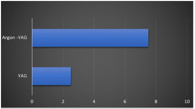
Figure 1: Average pain felt on a scale of 1 to 10 for the 2 methods.
The time of realization of the PI was on average 5 minutes for the YAG method alone while in the combined method it was 13.5 minutes.
Bleeding was more frequent in the YAG technique alone (stage I hyphema) in 15% of patients (Figure 2) and none of the patients in combined technique.
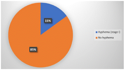
Figure 2: Percentage of hyphema during the YAG method.
Transient hypertonia was observed in both techniques, suppressed by local treatment.
Fewer YAG laser impacts were used in the combined method VS YAG alone: 12 [11.1-12.9] for the combined and 18 impacts [16.15-19.85] for the YAG only method (Figure 3).
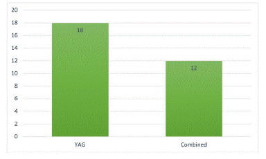
Figure 3: The mean number of YAG impacts in the 2 methods.
The mean energy used by Argon in the Combined eyes was 238 mj [224-252], meanwhile the energy used with the YAG laser was of 134,8 mj [124,8-147,96] (Figure 4).
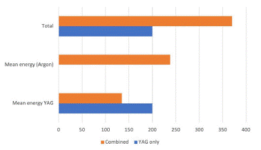
Figure 4: Energy delivered by the 2 types of lasers.
Total energy delivered was 200mJ [169.88, 230.12] in the YAG group alone while it was 370mJ [362.33, 383.67] for the combined laser group. It is an 85% increase in energy delivered by the combined technique (Figure 5).
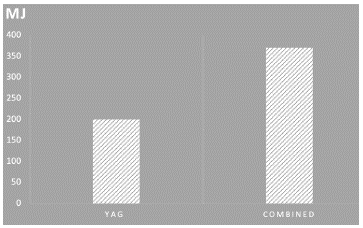
Figure 5: Total energy (mj) delivered in the 2 methods for an efficient iridotomy.
Discussion
The Peripheral iridotomy method needs a preparation by premedication with miotics to facilitate the performance of the iridotomy by stretching the iris. Premedication with alpha-2-agonists reduces the risk of pressure peaks after laser.
During the gesture the use of a focusing lens is highly desirable it improves focusing accuracy (reduces effective spot size by 50%), fixes globe, blocks the upper eyelid and increases the density of energy delivered to the PI site.
The main lens is the Abraham lens (convex plane lens of +66 diopters).
The 12’o clock meridian is to be avoided, because the formation of a gas bubble during argon laser coagulation will prevent the continuation of the iridotomy. The 11’o clock or 1’o clock meridians are generally chosen. Some operators sometimes choose the 3’o clock or 9’o clock meridians.
A flood of aqueous humor (flow sign), pigments, and the deepening of the anterior chamber in the periphery testify to the perforating nature of iridotomy.
If in doubt, OCT imaging of the anterior segment can be performed.
A diameter of 250 to 500μm is sufficient, a smaller iridotomy exposing the risk of closure in mydriasis, a larger iridotomy at a risk of excessive pigment release and pressure peak.
The preferred method of laser iridotomy varies greatly across different geographic locations, and this variation is partially explained by the perceived iris characteristics of the predominant local populations [7,8]. For example, in the UK, the standard technique for laser iridotomy is using a 1064nm Nd: YAG only laser [9], while in Japan, argon only laser iridotomy is the norm [8] and in Singapore, sequential argon-nd: YAG iridotomy is preferred [8,9].
The use of an argon laser causes photocoagulation in tissues by heating them due to absorption by iris pigment. This leads to coagulation of blood and collagen, resulting in tissue shrinkage and charring [2]. However, relying solely on argon laser iridotomy for treatment may not be effective, especially for those with brown eyes, as it has a high failure rate of 20% [10] and subsequent closure rate of up to 30% [10,11]. It can also cause bullous keratopathy many years after laser, by endothelial cells damage [8].
The YAG laser uses photo disruption to impact tissues by emitting nanosecond pulses that remove electrons from atoms in the iris. As a result, a plasma is generated that expand and then rapidly contract, producing a shock wave at the focal point. This process occurs due to the creation of intense pressure waves that disrupt the tissue [12,13].
The YAG laser only technique is effective on light colored eyes because of less energy required to achieve an effective PI.
On the other hand, performing YAG laser iridotomy on individuals with heavily pigmented irises poses certain limitations, as it necessitates the use of elevated levels of laser energy, and results in iris hemorrhages in approximately 40% of cases [14,15,16].
Approximately 1mm exists between the mid-peripheral iris and the endothelium in individuals suffering from angle-closure glaucoma [17,18].
The YAG only technique can cause focal endothelial opacity by cell losses above the iridotomy site in approximatively 35% of the cases [14,19].
These complications depend on the distance cornea-iris and the level of energy used [20].
Pure YAG laser iridotomy often requires high levels of energy to achieve a patent iridotomy in dark irises [2].
Sequential technique using Argon-YAG laser is very effective for dark brown irises with acute angle closure glaucoma. The heavy pigment in the iris absorbs the argon laser energy effectively, resulting in a flattening of the black pigment crust with minimal risk of causing a retinal burn [5]. After the iris has been thinned, subsequent YAG laser treatment in that area is efficient and requires only one-third of the power typically used for pure YAG laser iridotomy [1].
There is little number of studies comparing the sequential technique with the YAG only in dark irises. The cumulative evidence suggests that the combined laser technique has more advantages for dark pigmented irises.
In our study we compared the 2 techniques for a Moroccan population.
In term of parameters, we used for our study the YAG laser with 4-6 mj/ impact until perforation (flow sign).
For the preparation with Argon, we used a time of 0,02-0,05 seconds, with a power of 1000mw, 50μm spots and between 5 to 25 impacts to retract the iris away from the cornea, thin it and limit the risk of bleeding.
The YAG only group showed more complications than the combined group.
15% presented stage 1 hyphema for YAG only and 0% for the Argon then YAG group.
Twice the number of YAG laser impacts was required to enlarge the iridotomy in the YAG alone group (18 impacts) compared to the sequential group (12 impacts). The thicker brown iris absorbs Argon energy well and less YAG energy so fewer impacts were needed to enlarge the iridotomy in patients with sequential PI.
However, we noticed some advantages for the YAG only technique: The pain felt was much less important (mean of 2,5/10 versus 7,5/10 for the sequential technique), less time was needed to perform YAG only (a mean of 5 minutes versus 13,5 minutes for combined technique).
In term of energy, the YAG laser only delivered a mean of 200mj to the irises. Meanwhile, with the sequential technique it was 370mj, so it was approximately 2 times more energy than the YAG only PI.
Ho and Fan [1] did a combined technique for IP in 20 eyes with dark irises. They used Agon laser first with 810mw of power and a mean of 55 impacts for the preparation phase, meanwhile we used a 1000mw power and 25 impacts.
They then performed the IP with only a mean of 4 YAG impacts vs 12 in our study for the combined technique group. This can be explained by the fact that they used much more energy and impacts with the argon laser (mean of 3600mj) than in our study (mean of 238mj for the combined group). They used a mean of 9,4mj for their YAG vs 134,8mj in our study (Table 1).
    Our study

    Ho and Fan [1]


    Number of eyes

    25

    20


    Energy Argon

    1000 mw

    810 mw


    TIme Argon

    0,02-0,05 s

    0,01-0,1 s


    Number of impacts Argon

    5 to 25

    55


    Mean energy Argon

    238 mj

    3600 mj


    Number of impacts YAG

    12

    4


    Mean energy YAG

    132 mj

    9,4 mj
Table 1: Comparison of parameters and energy delivered for the combined method in our study and Ho and Fan [1].
In term of complications, they had a 10% hyphema and endothelial opacity, we had none of both complications in our combined technique cases (Table 2).
    Combined technique

    Ho and Fan (1)


    Hyphema

    0%

    10%


    Endothelial opacities

    0%

    10%


    Pigmentary dispersion

    100%

    100%


    Inflammatory signs

    0%

    0%


    Closed PI

    0%

    5%
Table 2: Complications in the combined technique cases compared to Ho and Fan [1].
This is explained by the fact that we used low energy argon for the preparation phase. De Silva and al [2] performed a 3-stage combined IP: a low energy argon laser using a mean of 114mj, then a high-power Argon (mean of 1087mj) and finally YAG laser to perforate the iris with a mean energy used of 33mj.
In our study we used a mean of 238mj during the argon phase so it is an equivalence of a low Argon laser phase, this is why we needed more YAG impacts (12 impacts) to perform an efficient PI. Robin and Pollack [14] performed only Argon laser iridotomies, the energy used was very high of 12j it is 3 times higher than the energy used for the combined technique in Ho and Fan [1] study and 30 times higher than in our study (Table 3).
    Combined technique

    Ho and Fan [1]

    Robin and Pollack [14]


    Mean energy used Argon (J)

    0,238

    3,6

    12


    Mean energy used YAG (J)

    0,134

    0,0094

    0,0033
Table 3: The energy used by the 2 types of lasers for the combined technique in our study and Ho and Fan [1] study compared to Robin and pollack [14] non-combined laser study.
Conclusion
The combined method remains the most widely used, it theoretically offers an advantage over the YAG laser only in the treatment of darker irises with less complications. Meanwhile the YAG only technique is better for the comfort of the patient, it is quicker, brings less pain and delivers less energy to the iris.
The two techniques are equal in terms of efficiency and effectiveness, and the choice of technique depends on the surgeon, the type of the iris and the patient.
References
- Ho T, Fan R. Sequential argon-YAG laser iridotomies in dark irides. Br J Ophthalmol. 1992; 76: 329e31.
- de Silva DJ, Gazzard G, Foster P. Laser iridotomy in dark irides. Br J Ophthalmol. 2007; 91: 222-5.
- Quigley HA, Silver DM, Friedman DS, He M, Plyler RJ, et al. Iris cross-sectional area decreases with pupil dilation and its dynamic behavior is a risk factor in angle closure. J Glaucoma. 2009; 18: 173e9.
- Lim L, Seah SK, Lim AS. Comparison of argon laser iridotomy and sequential argon laser and Nd:YAG laser iridotomy in dark irides. Ophthalmic Surg Lasers. 1996; 27: 285e8.
- Ho TK, Fan RF. Laser iridotomies in Asian eyes. Ann Acad Med Singapore. 1994; 23: 49-51.
- Naveh-Floman N, Blumenthal M. A modified technique for serial use of argon and neodymium-YAG lasers in laser iridotomy. Am J Ophthalmol. 1985; 100: 485-6.
- de Silva DJ, Day AC, Bunce C, Gazzard G, Foster PJ. Randomised trial of sequential pretreatment for Nd:YAG laser iridotomy in dark irides. Br J Ophthalmol. 2012; 96: 263-6.
- Ang LP, Higashihara H, Sotozono C, Shanmuganathan VA, Dua H, et al. Argon laser iridotomy-induced bullous keratopathy a growing problem in Japan. Br J Ophthalmol. 2007; 91: 1613e15.
- Narayanaswamy A, Kumar RS, Aung T, Foster PJ. Argon laser iridotomy-induced bullous keratopathy. Br J Ophthalmol. 2009; 93: 842.
- Schwartz LW, Rodrigues MM, Spaeth GL, Streeten B, Douglas C. Argon laser iridotomy in the treatment of patients with primary angle-closure or pupillary block glaucoma: a clinicopathologic study. Ophthalmology. 1978; 85: 294–309.
- Quigley HA. Long-term follow-up of laser iridotomy. Ophthalmology. 1981; 88: 218–24.
- Kielkopf JF. Laser-produced plasma bubble. Phys Rev E Stat Nonlin Soft Matter Phys. 2001; 63: 016411.
- Vogel A, Busch S, Jungnickel K, Birngruber R. Mechanisms of intraocular photodisruption with picosecond and nanosecond laser pulses. Lasers Surg Med. 1994; 15: 32–43.
- Robin AL, Pollack IP. A comparison of neodymium: YAG and argon laser iridotomies. Ophthalmology. 1984; 91: 1011–16.
- Del Priore LV, Robin AL, Pollack IP. Neodymium: YAG and argon laser iridotomy. Long-term follow-up in a prospective, randomized clinical trial. Ophthalmology. 1988; 95: 1207–11.
- Fleck BW, Dhillon B, Khanna V, Fairey E, McGlnn C. A randomised, prospective comparison of Nd:YAG laser iridotomy and operative peripheral iridectomy in fellow eyes. Eye. 1991; 5: 315–21.
- Wu SC, Jeng S, Huang SC, Lin S. Corneal endothelial damage after neodymium:YAG laser iridotomy. Ophthalmic Surg Lasers. 2000; 31: 411–16.
- Marraffa M, Marchini G, Pagliarusco A, Perfetti S, Toscano A, et al. Ultrasound biomicroscopy and corneal endothelium in Nd:YAG-laser iridotomy. Ophthalmic Surg Lasers. 1995; 26: 519–23.
- Schwartz LW, Moster MR, Spaeth GL, Wilson RP, Poryzees E. Neodymium-YAG laser iridectomies in glaucoma associated with closed or occludable angles. Am J Ophthalmol. 1986; 102: 41–4.
- Meyer KT, Pettit TH, Straatsma BR. Corneal endothelial damage with neodymium: YAG laser. Ophthalmology. 1984; 91: 1022–8.