Case Report
J Ophthalmol & Vis Sci. 2024; 9(1): 1088.
Diffuse Peripheral Anterior Synechiae Post-Sectoral Selective Laser Trabeculoplasty
Ammar M Khan, MD¹; Mathew M Palakkamanil, MD²; Andrew C Crichton, MD³
1Orbit Eye Centre, Calgary, Alberta, Canada
2Department of Ophthalmology and Visual Sciences, Faculty of Medicine and Dentistry, University of Alberta, Canada
3Division of Ophthalmology, Cumming School of Medicine, University of Calgary, Canada
*Corresponding author: Ammar Khan Orbit Eye Centre, Calgary, Alberta, Canada. Tel: 403-255-5561 Email: amahmoo@ucalgary.ca
Received: January 30, 2024 Accepted: February 04, 2024 Published: March 02, 2024
Keywords: Peripheral anterior synechiae; Selective laser trabeculoplasty; Glaucoma
Case Report
A 57-year-old male from the Philippines was referred to a glaucoma specialist from his comprehensive ophthalmologist for management of Primary Open Angle Glaucoma (POAG). He had been diagnosed with bilateral POAG in the Philippines several years prior, however, was unable to afford the medications that had been prescribed to him. He was known to have progressing POAG in his right eye and advanced POAG in the left eye. His intraocular pressures pre-treatment was in the mid 20’s mmHg. Given his non-compliance to medications, Selective Laser Trabeculoplasty (SLT) was performed in the left eye prior to referral. Laser treatment was performed in the nasal 180 degrees of the left eye with 45 applications at 0.7 mJ (total 31.5 mJ). Ongoing control of intraocular pressures was via Xalatan QHS OU, Cosopt BID OU and Alphagan TID OU.
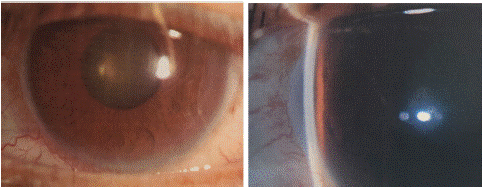
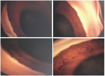
Upon assessment by a glaucoma specialist, his visual acuity was 20/20 and light perception in the right and left eye, respectively. His IOP was 19 mm Hg in the right eye and 37 mmHg in the left eye. Pupillary examination was significant for a left relative afferent pupillary defect. Anterior segment examination was unremarkable in the right eye. In the left eye, he was noted to diffuse peripheral anterior synechiae on gonioscopic examination (Figures 1-2). Posterior segment examination was unremarkable apart from bilateral glaucomatous nerve damage with cup to disc ratios of 0.6 and 1.0 in the right and left eye, respectively. Examination was additionally negative for any uveitis, iris/angle neovascularization, iris atrophy/polycoria or corneal endothelial abnormalities. Of note, there were no evidence of any PAS in his right eye (no prior SLT treatments OD).
Right Eye
Left Eye
Vision (uncorrected)
20/20
Light perception
Intraocular Pressures (mmHg)
19
37
Relative Afferent Pupillary Defect
none
present
Corneal Pachymetry (microns)
532
546
Anterior Segment Exam
Open on gonioscopy
Scattered synechiae
Fundus Examination
WNL
WNL
Cup to Disk Ratio
0.6
1.0
30-2 Humphrey Visual Field
Mean Deviation (MD), Pattern Standard Deviation (PSD)MD: -3.14 dB
PSD: 11.11 dBMD: -32.33 dB
PSD: 2.23 dB
Table 1: Ocular examination and findings at time of referral to glaucoma specialist.

Figure 1: Left eye anterior segment slit lamp photographs.

Figure 2: Left eye gonioscopy photographs indicating 360 degrees of intermittent peripheral anterior synechiae posterior to Schwalbe’s Line.
There was no prior history of ocular trauma or ophthalmic surgery. His past medical history was significant for controlled type II diabetes mellitus and dyslipidemia which was managed with oral metformin and atorvastatin, respectively.
Discussion
Common side effects of SLT include conjunctival hyperemia, transient IOP spike and mild anterior chamber inflammation. PAS is also a known complication of laser trabeculoplasty. Argon Laser Trabeculoplasty (ALT) has a much higher rate of PAS formation ranging between 12 and 47% [1,2]. SLT has lower rate of PAS formation, ranging between 0 and 2.86% as reported in a metaanalysis [3]. SLT uses a Q-switched, 532 mm, frequency doubled Nd: YAG laser to irradiate the melanin content of the trabecular meshwork. As such, there is no collateral thermal energy in nonpigmented nearby structures and no trabecular meshwork scar formation [4]. In comparison to ALT, SLT requires 100 times less energy and consequently, causes minimal damage to the trabecular meshwork with negligible contractile scars [5-7].
Our case represents the first documented report of scattered PAS in all four quadrants after SLT. Namely, our case is unique in that diffuse PAS resulted after only 180 degrees of SLT. Baser et al reported two cases of PAS formation after repeat SLT [9]. In contrast, our case characterizes PAS after a single treatment of SLT, as opposed to repeat SLT.
The pathophysiology for PAS formation after SLT treatment is unknown. Specifically, it is unclear how diffuse, four quadrant PAS formation occurred after sectoral SLT. Only 31.5 mJ (0.7 mJ per shot; 45 shots) of energy was applied to the trabecular meshwork during SLT in this eye. This is in accordance with other authors practices and would not be considered excessive energy or pulses [1,8]. It is possible, however, that SLT therapy created a significant anterior chamber reaction with resulting PAS. Secondly, in comparison to ALT (50 microns), SLT uses a much large spot size (400 microns). This larger spot size could theoretically irradiate other angle structures including the iris and ciliary body, inducing angle inflammation resulting in PAS.
The differential diagnosis for PAS includes iridocorneal endothelial (ICE) syndrome, trauma, epithelial downgrowth, neovascular glaucoma, and inherited conditions such as Axenfeld-Rieger spectrum. Our patient had no history polycoria, iris atrophy or endothelial abnormalities in keeping with ICE syndrome. Furthermore, there was no history of trauma or prior ophthalmic surgeries. There was no evidence of neovascularization of the iris or angle and no preceding retinal ischemic event. The unilateral nature of the PAS in the months after the SLT treatment makes an underlying inherited condition unlikely.
Unfortunately, our patient had persistently high intraocular pressures after SLT treatment compounded with formation of PAS. Due to his advanced glaucoma, cyclophotocoagulation was performed in the same eye to reduce the intraocular pressure. It is likely the PAS formation after SLT further impeded aqueous outflow. Based on our case presentation, it is important to recognize that SLT can rarely cause formation of PAS.
Author Statements
Funding
This research did not receive any specific grant from funding agencies in the public, commercial, or not-for-profit sectors.
Conflict of Interest
No conflicting relationship exists for any authors
Ethical Considerations
This study adhered to the Declaration of Helsinki. Research Ethics Board was contacted, and this study did not require IRB approval.
References
- Rouhiainen HJ, Ter Svirta ME, Tuovinen EJ. Peripheral anterior synechiae formation after trabeculoplasty. Arch Ophthalmol. 1988; 106: 189-91.
- Traverso CE, Greenidge KC, Spaeth GL. Formation of peripheral anterior synechiae following argon laser trabeculoplasty. A prospective study to determine relationship to position of laser burns. Arch Ophthalmol. 1984; 102: 861-3.
- Wong MO, Lee JW, Choy BN, Chan JC, Lai JS. Systematic review and meta-analysis on the efficacy of selective laser trabeculoplasty in open-angle glaucoma. Surv Ophthalmol. 2015; 60: 36-50.
- Latina M, Park C. Selective targeting of trabecular meshwork cells: in vitro studies of pulsed and CW laser interactions. Exp Eye Res. 1995; 60: 359-72.
- Avery N, Ang GS, Nicholas S, Wells A. Repeatability of primary selective laser trabeculoplasty in patients with primary open-angle glaucoma. Int Ophthalmol 2013; 33: 501-6.
- Hong BK, Winer JC, Martone JF, Wand M, Altman B, Shields B. Repeat selective laser trabeculoplasty. J Glaucoma. 2009; 18: 180-3.
- Damji KF, Bovell AM, Hodge WG, Rock W, Shah K, et al. Selective laser trabeculoplasty versus argon laser trabeculoplasty: results from a 1-year randomised clinical trial. Br J Ophthalmol. 2006; 90: 1490-4.
- Wang H, Cheng JW, Wei RL, Cai JP, Li Y, Ma XY. Meta-analysis of selective laser trabeculoplasty with argon laser trabeculoplasty in the treatment of open-angle glaucoma. Can J Ophthalmol. 2013; 48: 186-92.
- Baser E, Akbulut, D. Significant peripheral anterior synechiae after repeat selective laser trabeculoplasty. Can J Ophthalmol. 2015; 50: 36–38.