Research Article
J Ophthalmol & Vis Sci. 2024; 9(1): 1089.
Changes in Intraocular Pressure After Silicone Oil Removal Using Small-Gauge Techniques
Hummel LA; Niespodzany E; Urias E; Puthenparambil L; Banaee T*
Department of Ophthalmology, University of Texas Medical Branch, USA
*Corresponding author: Banaee T Department of Ophthalmology, University of Texas Medical Branch, 301 University Blvd. Galveston, TX 77550, USA. Tel: 440-530-0735; Fax: 979-848-2116 Email: tobanaee@utmb.edu
Received: January 17, 2024 Accepted: February 27, 2024 Published: March 05, 2024
Abstract
Background: Intraocular Pressure (IOP) changes can occur after silicone oil placement and removal. There is limited evidence regarding how using smaller instruments in retinal surgeries may influence these IOP fluctuations.
Objectives: Does the use of small gauge vitrectomy techniques for silicone oil placement and removal affect post-operative intraocular pressure?
Methods: In this retrospective observational study, 261 subjects were compiled from surgical treatment of retinal detachment surgeries involving both silicone oil injection and subsequent removal with 23 gauge or smaller instruments. 25 eyes met inclusion and exclusion criteria. Data collected included intraocular pressure, visual acuity, and pressure lowering medications from baseline and follow up appointments up to post-operative month three.
Results: No significant differences in IOP were found when comparing the study eyes to the fellow eyes prior to removal of silicone oil (U=230.5, p=0.829), at POD1 (U=48.5, p= 0.194), at POW1 (U=187.0, p=0.572), at POM1 (U=185, p=0.384), or at POM3 (U=168.5, p=0.086) after oil removal. No statistically significant differences were appreciated when comparing IOP changes from baseline to POM3 when factoring for the duration of silicone oil tamponade (less than 6 months versus longer than 6 months) (U=25, p=0.223)). Similarly, no significant difference was garnered from sub-group analysis by gauge size for silicone oil removal (U=31, p=0.411).
Conclusion: Placement and removal of silicone oil with small gauge instrumentation has no effect on intraocular Pressure in the Immediate (POD1) or 3 months post-operative period. This supports use of small gauge system in future pars plana vitrectomies involving silicone oil.
Keywords: Small gauge; Silicone oil; Intraocular pressure; Pars plana vitrectomy
Introduction
Vitrectomy with the use of a silicone oil tamponade agent has been a well-established treatment method for retinal detachment [1]. As silicone oil is a non-absorbed medium, unlike gas, it requires a subsequent operation for removal once the retina has been stabilized. Silicone oil removal is recommended to prevent long-term complications such as emulsifications, elevation of Intraocular Pressure (IOP), and corneal opacification. IOP changes can occur when silicone oil is present in the eye and after its removal. IOP elevation in a silicone oil filled eye may be due to Pars Plana Vitrectomy (PPV) induced inflammation, hemorrhagic complications, ciliary body edema, emulsified oil droplets clogging the trabecular meshwork, or in response to post-operative steroids2. Pupillary block may also be a complication of silicone oil fill due to potential overfilling of the vitreous cavity in phakic or pseudophakic eyes, or the absence of an effective peripheral iridotomy in an aphakic eye. Conversely, hypotony may also be a complication after silicone oil removal due to leakage from sclerotomy sites, cyclodialysis, anterior proliferative retinopathy, and toxic or direct compressive effect of silicone oil on the ciliary epithelium leading to reduced production of aqueous humor [2,3]. The technique for retinal detachment surgery and silicone oil removal has improved over the last two decades by using smaller, less invasive instruments. Potential benefits of using small-gauge techniques include faster healing, less inflammation and pain, improved visual acuity, improved post-operative astigmatism, and stabilization of IOP. IOP changes after silicone oil removal with prior used 20-gauge techniques have been researched but changes with small gauge technique need further investigation [4-6]. We postulate that with the modern use of smaller gauge techniques, complications leading to IOP fluctuations will be reduced.
Methods
This was a University of Texas Medical Branch Institutional Review board approved study, IRB number 18-0308. Patients who underwent small gauge (23G or smaller) PPV and silicone oil injection for treatment of rhegmatogenous or tractional retinal detachment were retrospectively enrolled from the UTMB hospitals. Each retinal surgery was performed by vitreoretinal surgeons between January 1, 2011 and February 20, 2019. Included subjects were age 18 to 100 years with history of rhegmatogenous or tractional Retinal Detachment (RRD) treated with pars plana vitrectomy with silicone oil between January 2011 and February 2019 at University of Texas Medical Branch. Subjects were included if both silicone oil injection and subsequent removal were done with 23 or 25 gauge instruments, and if they maintained follow up visits of at least 90 days after silicone oil removal. Patients were excluded if they were part of the Texas Department of Corrections, had a diagnosis other than retinal detachment, were treated by larger gauge instrumentation, or in whom silicone oil was not both used and removed. Diagnosis of glaucoma or ocular hypertension in the study eye prior to the first PPV was noted. Preoperative data was collected at the clinic visit preceding surgery involving silicone oil placement and the visit preceding silicone oil removal and IOP prior to silicone oil placement was used as the baseline IOP. Visual acuity, IOP, pressure lowering medications, and condition of the retina were recorded at office visits for pre-silicone oil placement, pre-silicone oil removal, Post-Operative Day one (POD1), Post-Operative Week one (POW1), Post-Operative Month one (POM1), and post-operative month three (POM3) after silicone oil removal. IOP for hypotonus eyes whose pressures could not be measured with the Reichert Tono-pen was estimated at 2.5 mmHg based on the minimum possible read of 5 mmHg. Differences in IOP between patients with longer and less than 6 months of silicone oil tamponade, between cases with 23 vs 25-gauge sizes of instruments used for silicone oil removal, and different silicone oil viscosities were explored. Statistical analyses performed include comparisons of the IOP and changes between the operative and fellow eye using Mann-Whitney test. All statistical analyses were done using GraphPad Prism (version 9.4.1; GraphPad Software, San Diego, CA) with P-values of 0.05 set for statistical significance.
Results
ICD-9 and ICD-10 codes for retinal detachment and silicone oil removal of patients treated by vitreoretinal surgeons at UTMB between January 2011 and February 2019 yielded a total of 261 cases. Of these, 25 eyes from 24 patients were enrolled in this study based on inclusion and exclusion criteria. Of these 24 patients, 15 were male (62.5%) and 9 were female (37.5%). 18 (75.0%) were Caucasian or white, five (20.8%) were African American or black, and one (4.2%) was Asian. The mean age was 58 (range 38-79). Baseline characteristics of the study eyes are displayed on Table 1. Additionally, three of the eyes had preceding diagnosis of glaucoma and were on IOP lowering medications throughout the study duration. One of these three had a diagnosis of neovascular glaucoma, vitreous hemorrhage, hyphema, and rhegmatogenous retinal detachment on initial presentation to UTMB and IOP remained high at 46 mmHg despite maximum medical therapy. Although IOP improved after initial RRD repair with silicone oil placement, he had a complicated course resulting in No Light Perception (NLP) vision at POM3. Three study eyes required additional glaucoma medications for elevated IOP control versus none in the fellow eyes. One of these eyes had silicone oil induced pupillary block which resolved after the oil was removed. One eye developed a hyphema after silicone oil removal that required topical IOP lowering drops and eventual anterior chamber washout. One eye experienced pupillary block due to angle closure which occurred after silicone oil removal. This was deemed to be due to anatomically narrow angles and unrelated to the retina surgeries. She was managed with maximum medical therapy, cataract extraction, surgical iridectomy in the study eye, and a laser peripheral iridotomy in the fellow eye.
    n

    %


    Lens Status, pre-SO removal


    Phakic

    8

    32%


    Pseudophakic

    12

    48%


    Aphakic

    5

    20%


    Lens Status, POD1


    Phakic

    7

    28%


    Pseudophakic

    14

    56%


    Aphakic

    4

    16%


    Silicone Oil Viscosity


    1000 cs

    21

    84%


    5000 cs

    1

    4%


    Missing

    3

    12%


    Diagnosis


    Rhegmatogenous    retinal detachment

    14

    56%


    Tractional    retinal detachment

    11

    44%
Table 1: Baseline Characteristics of Study Eyes.
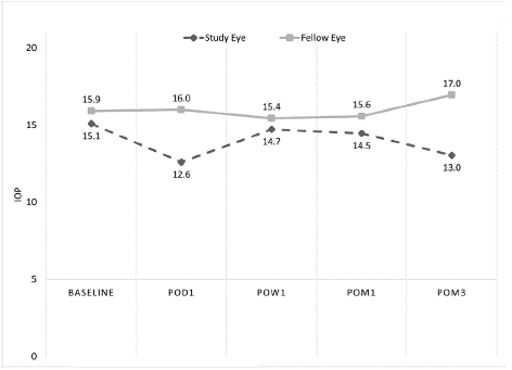
Median baseline IOP prior to insertion of silicone oil were 15.1 mmHg in the study eyes and 15.9 mmHg in the fellow eyes. There was no significant difference between the two groups (U=209.5, p=0.159). 64% of the silicone oil insertions and 76% of the removals were performed with 23-gauge system and the other 36% of the insertions and 24% of the removals were done with 25 gauge systems. The mean number of days between placement and removal of silicone oil was 122.9 (SD 88.2). No significant differences in IOP were found when comparing the study eyes to the fellow eyes at the last measurement prior to removal of silicone oil (U=230.5, p=0.829), at POD1 (U=48.5, p= 0.194), at POW1 (U=187.0, p=0.572), at POM1 (U=185, p=0.384), or at POM3 (U=168.5, p=0.086) after oil removal (Figure 1). No statistically significant differences were appreciated when comparing IOP changes from baseline to POM3 when factoring for the duration of silicone oil tamponade (less than 6 months versus longer than 6 months) (U=25, p=0.223)). Similarly, no significant difference was garnered from sub-group analysis by gauge size for silicone oil removal (U=31, p=0.411). Gauge size also did not lead to significant difference between the groups at POD1 (U=50.5, p=0.833). However, these subgroup analyses were severely limited by the small sample size (n=21 vs n=4 and n=19 vs n=6 respectively). Similarly, there were no significant differences in IOP change when accounting for a history of prior vitreoretinal surgeries (U=43, p=0.523). Only one patient underwent 5000 cs silicone oil placement and IOP changes could not be compared with 1000 cs silicone oil use.

Figure 1: IOP comparison in the study eye and fellow eye over the study period. IOP listed in mmHg.
Discussion
This study investigated use of small gauge instrumentation for silicone oil insertion and removal and its possible effects on IOP changes with the hypothesis that in the use of small gauge instruments, changes in IOP after oil removal would not be significant. Our study found that there were no significant differences in IOP after silicone oil removal compared to baseline when measured shortly after procedure (POD1) and at longer interval follow up (POM3). Three eyes required additional IOP lowering medications during the three-month postoperative period, but all of these eyes experienced complications that lead to IOP elevations including acute angle closure from pupil block (n=2) and hyphema (n=1). Hence, the need for more aggressive IOP control was due to these general surgical and pathological complications and not related to the actual silicone oil removal. Sub-group analyses exploring gauge size and duration of silicone oil retention also found no statistically significant differences but were limited due to small sample size which was exacerbated by the uneven sizes of these subgroups.
Prior studies have assessed IOP differences with larger gauge techniques, when use of 20G systems were more common. One such study by Zhang et al. compared changes in post-vitrectomy IOP between 20G and 23G groups at POD1, POW1, POM1, and POM6. Differences were only significant at POD1, in which the smaller 23G group (17.57±2.9 mmHg) had significantly higher IOP than the 20G group (10.1±5.6 mmHg) (p=0.000) [7]. While no prior studies have specifically examined IOP changes after silicone oil removal with small gauge instruments, prior studies have examined outcomes in small gauge instrument retina surgeries. One such study by Charles et al. was a multicenter clinical trial comparing multiple outcome measures for 27 vs 23-gauge PPV surgeries (n=68) [8]. They found that the smaller diameter 27-gauge vitrectomy instrument use led to smaller reductions in immediate postoperative IOP. Mean change from immediate preoperative to immediate postoperative IOP were -0.40 ± 6.60 mmHg in the 27-gauge group and -3.05±7.64 mmHg I the 23-gauge group (P=0.013). This contrasts with the findings of the discussed 20G versus 23G study. Charles et al postulated that large instruments caused more fluid and air egress during instrument removal. Long term IOP changes were not part of this study [8]. One systematic review and meta-analysis of 27-gague vs 25-gauge vitrectomy surgery by Jinlan Ma et al, included 11 studies, eight of which recorded IOP on POD1. These eight studies examined small gauge use for treatment of rhegmatogenous retinal detachments (n=4) and epiretinal membrane (n=4) and were not exclusive to use of silicone oil [9]. All eight studies found no significant differences between the two gauge sizes in controlling postoperative IOP at POD1 (MD=0.53; 95% CI: -1.49, 2.54; P=0.61) [9-17]. The meta-analysis only looked at IOP changes for POD1. Our study also did not find significant differences in POD1 IOP between 23G and 25G treated eyes as described above (U=50.5, p=0.833). One of these eight studies included only patients treated with silicone oil tamponade [10]. Li, Jie et al published a retrospective comparison of 25- and 27-gauge systems for treatment of rhegmatogenous retinal detachments with use of silicone oil tamponade [10]. Regarding IOP changes, they found that the post-operative IOP was significantly higher at each time point which included one day, one week, one month, and the last documented visit (p<0.05). However, there was no significant differences in IOP between the 25G and 27G groups. They found no change between the two groups for their other outcome measures, including VA improvement and transient ocular hypertension. Additionally, this study differs from ours in that the follow up data was acquired for time periods after injection of silicone oil into the eye, whereas the focus of our study was on IOP changes after silicone oil removal.
Our study has limitations that are inherent to every retrospective study, including many potentially confounding variables which prevent any firm conclusions. The main limitation of our study is the small sample size, which may have led to an inability to detect differences among our comparison groups.
In summary, mean IOP was not significantly affected after silicone oil removal with use of newer small gauge instruments when analyzed in the immediate (POD1) or 3 months post-operative period. This supports use of small gauge system in current and future PPV surgeries, though larger scale studies would be beneficial to further support this hypothesis.
References
- Lean JS, Boone DC, Azen SP, Lai MY, Linton KLP, McCuen II BW, et al. Vitrectomy with Silicone Oil or Sulfur Hexafluoride Gas in Eyes with Severe Proliferative Vitreoretinopathy: Results of a Randomized Clinical Trial: Silicone Study Report 1. Arch Ophthalmol 1992; 110: 770–779.
- Han DP, Lewis H, Lambrou FH Jr, Mieler WF, Hartz A. Mechanisms of Intraocular Pressure Elevation After Pars Plana Vitrectomy. Ophthalmol. 1989; 96: 1357-1362.
- Zhang X, Chen B, Yang HJ, Song Y, Zhang D, Soetikno BT. The Correlation of Pars Plana Incision and Transient Hypotony After Silicone Oil Removal. Ophthalmic Surg Lasers Imaging Retina. 2018; 49: e44–e51.
- Mohamed AA and Abdrabbo M. Outcome of 20-Gauge Transconjunctival Cannulated Sutureless Vitrectomy Using Silicone Oil or Air Tamponade. Clin Ophthalmol. 2013; 7: 379-384.
- Al-Wadani SF, Abouammoh MA, and Abu El-Asrar AM. Visual and Anatomical Outcomes After Silicone Oil Removal in Patients with Complex Retinal Detachment. Int Ophthalmol. 2014; 34: 549–556.
- Issa R, Xia T, Zarbin MA Bhagat N. Silicone Oil Removal: Post-Operative Complications. Eye. 2020; 34: 537-543.
- Zhang J, Li Y, Zhao X, Yu X, Lu L. Comparison of Clinical Features After 20-Gauge Vitrectomy Versus 23-Gauge Vitrectomy. Asia Pac J Ophthalmol. 2015; 4: 367-370.
- Charles S, Ho AC, Dugel PU, Riemann CD, Berrocal MH, Gupta S, et al. Clinical Comparison of 27-Gauge and 23-Gauge Instruments on the Outcomes of Pars Plana Vitrectomy Surgery for the Treatment of Vitreoretinal Diseases. Curr Opin Ophthalmol. 2020; 31: 185-191.
- Ma J, Wang Q, Niu H. Comparison of 27-Gauge and 25-Gauge Microincision Vitrectomy Surgery for the Treatment of Vitreouretinal Disease: a Systematic Review and Meta-Analysis. J Ophthalmology. 2020.
- Li J, Zhao B, Liu S, Li F, Dong W, Zhong J. Retrospective Comparison of 27-Gauge and 25-Gauge Microincision Vitrectomy Surgery with Silicone Oil for the Treatment of Primary Rhegmatogenous Retinal Detachment. J Ophthalmol. 2018.
- Otsuka K, Imai H, Fujii A, Miki A, Tagami M, Azumi A, et al. Comparison of 25- and 27-Gauge Pars Plana Vitrectomy in Repairing Primary Rhegmatogenous Retinal Detachment. J Ophthalmol. 2018.
- Romano MR, Cennamo G, Ferrara M, Cennamo M, Cennamo G. Seven-Gauge Versus 25-Gauge Vitrectomy for Primary Rhegmatogenous Retinal Detachment. Retina. 2017; 37: 637–642.
- Sborgia G, Niro A, Sborgia L, Grassi MO, Gigliola S, Romano MR, et al. One-Year Outcomes of 27-Gauge Versus 25-Gauge Pars Plana Vitrectomy for Uncomplicated Rhegmatogenous Retinal Detachment Repair. Int J Retina Vitreous. 2019; 5: 13.
- Lubinski W, Goslawski W, Podboraczynska-Jodko K, Mularczyk M, Post M. Comparison of 27-Gauge Versus 25-Gauge Vitrectomy Results in Patients with Epiretinal Membrane: 6-Month Follow-Up. Int Ophthalmol. 2020; 40: 867–875.
- Mitsui K, Kogo J, Takeda H, Shiono A, Sasaki H, Munemasa Y, et al. Comparative Study of 27-Gauge vs 25-Gauge Vitrectomy for Epiretinal Membrane. Eye. 2016; 30: 538–544.
- Naruse S, Shimada H, and Mori R. 27-Gauge and 25-Gauge Vitrectomy Day Surgery for Idiopathic Epiretinal Membrane. BMC Ophthalmol. 2017; 17: 188.
- Takashina H, Watanabe A, and Tsuneoka H. Perioperative Changes of the Intraocular Pressure During the Treatment of Epiretinal Membrane by Using 25- or 27-Gauge Sutureless Vitrectomy Without Gas Tamponade. Clin Ophthalmol. 2017; 11: 739–743.