Case Report
J Ophthalmol & Vis Sci. 2024; 9(2): 1096.
Morphological Aspect and Management of Pediatric Cataract in Treacher Collins Syndrome
Tartarella MB¹; Verçosa IM²; Caliope M²; Botelho C²; Ellery M²; Fortes Filho JB³*
1Department of Ophthalmology, Centro Integrado de Oftalmologia Tartarella, São Paulo SP, Brazil
2Department of Ophthalmology, CAVIVER Institute, Fortaleza CE, Brazil
3Department of Ophthalmology, Federal University of Rio Grande do Sul, Porto Alegre RS, Brazil
*Corresponding author: Fortes Filho JB, Department of Ophthalmology, Federal University of Rio Grande do Sul, Porto Alegre RS, 2350 Ramiro Barcelos CEP 900035- 903, Brazil. Tel: +5551999698081 Email: joaoborgesfortes@gmail.com
Received: October 09, 2024; Accepted: October 29, 2024 Published: November 05, 2024
Abstract
The morphological aspect of lens opacification in a 13-year-old patient with Treacher Collins Syndrome was discussed. The outcome of the cataract surgical treatment was presented. Initial visual acuity was 20/200 at the right eye and 20/100 at the left eye. A lamellar opacification of the lens, as bananatree- leaves-shaped cataracts, were observed in both eyes. Phacoemulsification with intraocular lens implantation was performed. Final visual acuity was 20/30 in both eyes. Cataract is a rare feature in Treacher Collins Syndrome and the lens opacity morphology detected in this patient was very unique.
Keywords: Cataract; Pediatric cataract; Treacher Collins Syndrome; Mandibulofacial dysostosis
Abbreviations: TCS: Treacher Collins Syndrome
Introduction
Treacher Collins Syndrome (TCS) or mandibulofacial dysostosis is a genetic disorder characterized by typical facies with malar and mandibular hypoplasia, antimongoloid eyelid position, auricular pavilion malformations, conductive deafness, cleft palate, and a bird's beak shaped nose. Coloboma of the lower eyelid and microphthalmia are often seen in TCS [1,2].
Cataract in patients with TCS is not a common occurrence and has been rarely described in previous literature. We report a case of a patient with bilateral cataract associated with TCS.
Case Report
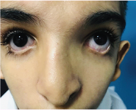
A 13-year-old male diagnosed with TCS that presented with mild hearing loss, low ear implantation, and bird´s beak shaped nose (Figure 1), was referred to CAVIVER Eye Clinic, in the city of Fortaleza, CE, Brazil, for ophthalmological evaluation. The patient´s parents were first cousins.

    Figure 1: Facial features: partial bilateral inferior eyelid coloboma, nose
and ear malformations.
The patient complained of progressive bilateral vision loss. A comprehensive ophthalmic evaluation disclosed visual acuity of 20/200 and 20/100 in the right eye (RE) and left eye (LE), respectively. Indirect binocular ophthalmoscopy revealed normal eye fundus. Refraction was not feasible in RE due to dense lens opacity. Refraction of the LE was - 2,25 sph = - 4,50cyl (135°). Neither nystagmus nor strabismus was observed. A partial bilateral inferior eyelid coloboma was present.
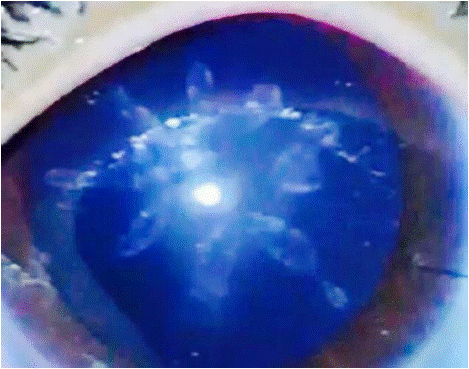
A bilateral lamellar lens opacification resembling banana-treeleaves was observed through slit-lamp examination. Peripheral lens cortex was clear in both eyes (Figure 2). Axial length was 26.7mm in RE and 25.5mm in LE. Keratometric readings were 42.25 x 45.75 diopters (120°) in RE and 42.00 x 46.50 diopters (145°) in LE. Intraocular lens power calculation was +11.0 and +13.0 diopters, respectively in the RE and LE.

Figure 2: Morphology: banana-tree-leaves-shaped cataract.
A phacoemulsification with an anterior continuous circular capsulorhexis was performed in both eyes. The procedure was uneventful. Aspiration of the soft nucleus needed low ultrasound power. A hydrophobic monofocal foldable acrylic intraocular lens was implanted in the bag. Posterior capsule was clear and was left intact bilaterally. Post-operative refraction was + 0.75 sph = - 3.00 cyl (40°) and - 1.00 sph = - 4.00 cyl (145°), respectively in RE and LE. Final best corrected visual acuity achieved 20/30 in both eyes.
Discussion
Treacher Collins syndrome is a genetic condition with a marked craniofacial abnormality. The gene responsible for TCS at chromosome 5 (5q31.3- q33.3) was studied and genetic testing for prenatal diagnosis is available for affected families [2]. Consanguinity was present in the reported case.
Rooijers et al. studied the prevalence of ocular and adnexal anomalies in 194 patients with TCS. Primary ocular anomaly, as inferior lid coloboma, was reported in 98.5% of cases, and secondary anomalies in 34.5%, strabismus in 27.3%, refractive errors in 49.5%, and visual impairment in 4.6%. No cataract was reported in this multicenter study with a large number of affected patients [1].
The occurrence of lens opacification in TCS was previously reported in only one published paper. Authors reported bilateral extracapsular cataract extraction with posterior chamber lens implantation in a 13-month-old patient with delayed-onset nuclear cataract [3].
The pediatric cataract morphology may be associated with specific genetic disorders suggesting the possible etiology of the lens opacification and suggesting heritability [4]. Cataract morphology can be a visual prognosis indicator. Different morphological types may have a better visual prognosis than others, with lamellar cataracts doing well and total cataracts relatively poorly. Lamellar cataracts frequently develop at a later stage and can be progressive. In a published study of children with congenital cataracts, the results showed that visual prognosis may depend on the morphological type, with less favorable outcomes in cases of total cataracts [5,6].
Pediatric cataracts can exhibit phenotypical heterogeneity. In a previous study, 207 patients with pediatric cataract were studied. Age of the patients ranged from 19 days to 12 years. Zonular cataracts occurred with the highest frequency, in 72 (33.8%) patients. Among the zonular cases, the lamellar subtype was the most common (66.7%). Total cataract occurred in 31.9% of the patients. Among the patients with genetic disorders or syndromes, 87,5% had bilateral cataract. Morphology or laterality of pediatric cataract may be indicative of its etiology [7].
The banana-tree-leaves-shaped cataract morphology was a very unique finding in the case of TCS here reported. This cataract morphology was not previously described in the literature and it could be associated with early stages of the lamellar lens opacification in TCS patients. The morphological heterogeneity of pediatric cataracts makes the morphological classification challenging, but morphology is of great importance to guide to etiology diagnosis, and treatment options.
Within a couple of months, progressive lens opacification resulted in vision loss due to the development of a total cataract in both eyes. Phacoemulsification with intraocular lens implanted in the bag was performed in both eyes with no complications [5]. Due to the late onset of the lens opacification, which occurred subsequent to the amblyogenic period, a good visual acuity was restored after cataract surgery.
Accurate diagnosis of the etiology of pediatric cataract is important for epidemiological studies and to promote ocular health preventive programs with the goal to prevent childhood blindness.
Conclusion
The banana-tree-leaves-shaped cataract morphology in TCS is a very unique finding in the case described here.
Patients with TCS need a multidisciplinary approach to achieve a better quality of life. Ophthalmological evaluation is recommended in order to avoid visual impairment.
References
- Rooijers W, Schreuder MJ, Loudon SE, Forrest CR, Wan MJ, Dunaway DJ, et al. Ocular and adnexal anomalies in Treacher Collins syndrome: a retrospective multicenter study. J AAPOS. 2022; 26: 10-12.
- Hertle R, Ziylan S, Katowitz JA. Ophthalmic features and visual prognosis in the Treacher-Collins syndrome. Br J Ophthalmol. 1993; 77: 642-645.
- Biebesheimer JB, Fredrick DR. Delayed-Onset Infantile Cataracts in a Case of Treacher Collins Syndrome. Arch Ophthalmol. 2004; 122: 1721-1722.
- Taylor D. The Doyne Lecture Congenital cataract: the history, the nature and the practice. Eye Royal College of Ophthalmologists. 1998; 12: 9-36.
- Tartarella MB, Verçosa ICV. Surgical techniques in Pediatric Cataracts. In: Verçosa I, Tartarella MB editors: Catarata na Criança [Cataract in Children], Fortaleza, CE, Brazil. Editora Celigrafica. 2008: 105-118.
- Ejzenbaum F, Salomão SR, Berezovsky A, Waiswol M, Tartarella MB, Sacai PY, et al. Amblyopia after unilateral infantile cataract extraction after six weeks of age. Arq Bras Oftalmol. 2009; 72: 645-649.
- Tartarella MB, Britez-Colombi GF, Milhomem S, Cordeiro M, Lopes E, Fortes Filho JB. Pediatric cataracts: clinical aspects, frequency of strabismus and chronological, etiological, and morphological features. Arq Bras Oftalmol. 2014; 77: 143-147.