Case Report
Austin J Orthopade & Rheumatol. 2014;1(1): 2.
The Largest Osteochondral Fracture of Patella, Case Report
Meric Unal1*and Hasan Tatari2
1Isparta Sifa Hospital Department of Orthopaedics and Traumatology, Turkey
2Dokuz Eylül University Faculty of Medicine Department of Orthopaedics and Traumatology, Turkey
*Corresponding author: Meric Unal, Department of Orthopedics, Dokuz Eylül University, Izmir, Turkey. Email: abdmunal@yahoo.com
Received: September 29, 2014; Accepted: October 24, 2014; Published: October 30, 2014
Abstract
Fractures of the patella, generally occuring by direct trauma, constitute 1% of all fractures. The most comprehensive classification of the patella fractures is the Orthopaedic Trauma Association (OTA) classification. This is a report of a 15-year-old male patient examined after falling down and sufferred from a patella fracture. The fracture could not be classified by the current classifications. At the operation, the extensor mechanism was normal and the medial joint surface of the patella was displaced superiorly. The fragment was considered as a very big osteochondral fragment and two headless full-threaded compression screws were used for fixation.
Introduction
Patella is the biggest sesamoid bone in the body [1] that is necessary for the extensor mechanism of the knee. Fractures of the patella constitute approximately 1% of all fractures [1,2] generally occuring after direct trauma. Indirect trauma by pulling the tendon is another mechanism. Most prominent effects of patellar fracture are the loss of extensor mchanism and irregularity of the patellofemoral joint [3,8]. Osteochondral fractures are generally seen after patellar dislocation [4].
Patellar fractures are basicly classified as displaced and nondisplaced [5]. Displaced fractures have some subtypes like; transverse, vertical, lower and upper pole, comminuted and osteochondral [5]. The most comprehensive classification for patellar fractures is OTA classification [1]. In this classification there are three types basicly distinguished with joint involvement. Type-1 is extra-articular, type-2 is particularly articular and type-3 is completely articular [6]. Type 1 and 2 have two subtypes and type-3 has three subtypes [6]. The fractures, those are just intraarticular and do not destroy the extensor mechanism, are very rare and can not be classified with current classifications. These types of fractures may be recognized as osteochondral fractures of the patella.
In this article, a different type of patella fracture case, that is not classified by current classifications, is presented.
Case
A 15-year-old male patient was consulted to us with a pain in his left knee after falling down during running. Physical examination demonstrated tenderness over the patella, minimal effusion and locking. Neurovascular examination was normal.
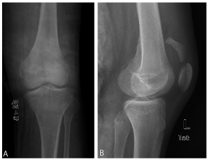
For radiological examination, direct anteroposterior (AP) and lateral views of the knee were taken and the fracture of the patella was diagnosed (Figure 1A,1B). Open reduction and internal fixation was planned.
A: Preoperative anteroposterior radiography of the left knee.
B: Preoperative lateral radiography of the left knee.Figure 1A and 1B:A: Preoperative anteroposterior radiography of the left knee.
B: Preoperative lateral radiography of the left knee.
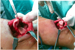
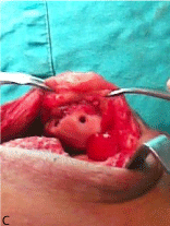
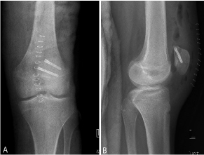
Under general anesthesia, the operation was made through an anterior longitudinal incision. The extensor mechanism was totally normal. The joint was opened with median parapatellar incision. At the patellar joint surface, a longitudinal fracture line containing only the posterior cortex was demonstrated and the medial joint surface of the patella was displaced superiorly like a big osteochondral fragment. (Figure 2A, 2B). After the reduction of the fragment, fixation was performed with two full-threaded headless compression screws (Figure 2C). Reduction and fixation were controlled with postoperative direct radiographies (Figure 3A, 3B).
A: Intraoperative view of the displaced fragment.
B: The fragment before reduction.Figure 2A and 2B:A: Intraoperative view of the displaced fragment.
B: The fragment before reduction.
Figure 2C: Intraoperative view after fixation of the fragment.
A: Postoperative anteroposterior radiography.
B: Postoperative lateral radiography.Figure 3A and 3B:A: Postoperative anteroposterior radiography.
B: Postoperative lateral radiography.
Discussion
Direct anteroposterior and lateral views of the knee are generally sufficient for radiological examination. Skyline views may be needed to establish the longitudinal and osteochondral fractures [1]. In the present case, the fracture was diagnosed only with anteroposterior and lateral radiographies.
The purpose of treatment in patellar fractures is to restore the extensor mechanism and the joint surface. Tension band technique is generally used for restoring the extensor mechanism. Interfragmentary screws and the combinations of these are also used [2,3]. For the joint surface fractures, like osteochondral fractures, the use of intefragmentary headless compression screws may be a suitable technique for treatment [7].
To our knowledge, the present case could not be classified with current classifications. This type of fracture may be considered as an unicortical (osteochondral) fracture pattern with a very big fragment. When the literature was reviewed; we could not find any patellar fracture that could not be classified with OTA classification and also did not destroy the extensor mechanism containing only the joint surface. If it is considered as an osteochondral fracture, there is no case in the literature having such a big fragment as in our case. Osteochondral fractures may be sonsidered more in comprehensive fracture classifiying systems.
References
- Rüedi TP, Murphy WM. AO principles of fracture management. 2001: 483-497.
- Galla M, Lobenhoffer P . [Patella fractures]. Chirurg. 2005; 76: 987-997.
- Whittle AP. Fractures of the lower extremity. Canale ST, Beaty JH (ed) Campbell’s Operative Orthopaedics. 2008; 3: 3085-3236.
- Kanamiya T, Naito M, Cho K, Kitamura T, Takeda T, Goto E, et al. Unicortical transverse osteochondral fracture of the patella: a case report. Knee. 2006; 13: 167-169.
- Harris RM. Fractures of patella. In: Rockwood and Green’s fractures of adults, Buchols RW ,Heckman JD (ed) 2001; 2: 1775-1800.
- Marsh JL, Slongo TF, Agel J, Broderick JS, Creevey W, DeCoster TA, et al. Fracture and dislocation classification compendium - 2007: Orthopaedic Trauma Association classification, database and outcomes committee. J Orthop Trauma. 2007; 21: 1-133.
- Marsh JL, Slongo TF, Agel J, Broderick JS, Creevey W, DeCoster TA, et al. Fracture and dislocation classification compendium -2007: Orthopaedic Trauma Association classification, database and outcomes committee. J Orthop Trauma. 2007; 21: 1-133.
- Cekin T, Tükenmez M, Tezeren G . [Comparison of three fixation methods in transverse fractures of the patella in a calf model]. Acta Orthop Traumatol Turc. 2006; 40: 248-251.
- Tandogan R, Karaman A, Ersözlü S. Turkiye Klinikleri. J Surg Med Sci. 2006; 2: 123-127.


