
Research Aricle
Austin J Orthopade & Rheumatol. 2023; 10(1): 1114.
The Optimum Size of K-Wires for Fifth Metacarpal Neck Fractures: Double 1.6 mm K-Wires Antegrade Intramedullary Nailing Technique
Kaymin W¹, Rong C¹, Youwu H¹, Gang L¹, Xingqun Z¹, Yi Y¹, Zipu W², Lijiang H³ and Liang W¹*
¹Department of Hand and Plastic Surgery, The firstpeople’s Hospital of Lingping District, China
²Department of Surgery; Lu’an Hospital of Anhui Medical University, China
³Department of Orthopedic Surgery; Second People’s Hospital of Yuhang District, China
*Corresponding author: Liang WuDepartment of Hand and Plastic Surgery; The first people’s Hospital of Lingping District; No. 369, Rd. Ying Bin, City Hangzhou, Province Zhejiang, China
Received: January 17, 2023; Accepted: February 22, 2023; Published: February 28, 2023
Abstract
Objective: Previous researches did not pay significant attention to the 5th metacarpal’s intramedullary diameter. This research aims to determine the optimal K-wires for 5th metacarpal neck fractures based on the narrowest part of 5th metacarpal.
Methods: We retrospectively studied 31 patients with fifth metacarpal neck fractures. All patients underwent Intramedullary (IM) nailing surgery with two 1.6 mm K-wires. The narrowest part of the fifth metacarpal was measured preoperatively based on CT scan. Quick-DASH was assigned to be the primary outcome, pain, satisfaction, motor function and complications were also recorded.
Results: Well reductions were observed in all patients. QuickDASH sore was median 0 with the range of 0-32, pain score was median 0 with the range of 0-2, VAS satisfaction was median 5 with the range of 3-5 at the endpoint. Grip strength, metacarpophalangeal joint and total active motion were improved immediately after operation. Two patients remained persistent paresthesia, and three patients experienced persistent pain at the endpoint with the VAS score of 1, 2, 2.
Conclusion: The use of double 1.6 mm K-wires antegrade intramedullary nailing could be an optimum option for patients with fifth metacarpal neck fractures.
Keywords: Metacarpal neck fracture; K-wire; Marrow cavity; Antegrade intramedullary nailing; Surgery
Abbreviations: MCP: Metacarpophalangeal; AIMN: Antegrade Intramedullary Nailing; IM: Intramedullary; CT: Computed Tomography; 3D: 3-Dimensional; ROM: Range of Motion; TAM: Total Active Motion
Introduction
Fifth metacarpal neck fractures are the most commonly injured metacarpal, representing around 20% of all hand fractures and usually occurring in the young, working population. It is also known as the boxer's fracture because it is most often caused by an axial impact, most commonly a direct punch to the knuckles [1,2].
Conservative treatment and surgery are both suitable for patients with closed 5th metacarpal neck fracture [3]. However, inappropriate treatment of these fractures might result in a decrease in the range of motion of the Metacarpophalangeal (MCP) joint, distal aspect deformity of the fifth metacarpal, and a decrease in the grip strength [4,5]. There is no consensus on the indications for surgery or best operative management for extra-articular 5th metacarpal neck fractures [3]. An excessive dorsal angulation and malrotation are usually indications for surgery, while the tolerable limit of dorsal angulation still remains controversial, above 30° to above 45° [6-8]. Lots of surgical techniques have been described for the treatment of these fractures, such as transverse k-wires pinning [9,10], Antegrade Intramedullary Nailing (AIMN) [9,11-15], low profile plates [14] and external fixation [16]. In recent years, the use of AIMN has won attractiveness owing to being relatively simple, minimally invasive and cost-effective with reports of excellent clinical outcomes [17,18]. Yammine et al. had reported significantly better clinical and radiological outcomes when using the double K-wires AIMN with the advantage of less complications [18]. Some researchers also reported using single K-wire to treat these fractures with favourable outcomes [19-25].c
However, previous studies did not pay close attention to the intramedullary diameter of 5th metacarpal. We aim to study the narrowest part of 5th metacarpal to find the optimum K-wires for 5th metacarpal neck fractures.
Materials and Methods
Patients and Methods
31 patients treated for the 5th metacarpal neck fracture between January 2018 and December 2020 was retrospectively reviewed in our department. Open fractures and fractures extending to the articular surface or the metacarpal shaft were excluded from the study. Inclusion criteria were as follows: (I) patients with unstable fractures with a dorsal angulation of more than 30°or (II) with a shortening more than 3 mm.
Pre- and post-operative radiographs were obtained by using a standardized protocol. All patients underwent pre-operative Computed Tomography (CT) scan. Image data were post-processed using Mimics 10.0/15.0 to rebuild 3-Dimensional (3D) bone models. The narrowest part of the marrow cavity was measured to calculate the optimised diameter of K-wires and the length of K-wire depends on the length of 5th metacarpal.
Sagittal angulation was measured by using the angle between the axis line through the neck and the center of the head and the line through the shaft axis.
Ethical approval was granted from the institutional board prior to the conduct of the study.
Surgical Technique
All surgeries were performed underaxillary plexus anaesthesia by one senior surgeon. A tourniquet was applied over the upper arm and inflated after elevation of the arm. Jahss technique was used to control fracture reduction under fluoroscopy. Not all the fractures were anatomically reduced to fully considering the accuracy of reduction, but all patients gained much better positions.
A short skin incision vertical to the metacarpal shaft was made over the dorsoulnar aspect of the metacarpal base to avoid scar contracture, the medullary cavity was entered through a small drill hole and the first K-wire (1.6 mm) was drilled smoothly to extend as far as the subchondral bone of the metacarpal head. The second K-wire (1.6 mm) was inserted 0.5 cm distal to the first and terminated at the same level. the K-wires were cut beneath the skin.
All patients underwent a standard post-operative rehabilitation protocol. Gentle mobilization exercise started 3 days after the operation with the guide of exceptional physiotherapists. Patients were recommended to avoid heavy exertion at least 6 weeks after surgery. K-wires were removed at 4 to 6 weeks under local anaesthesia.
Outcome Evaluation
Each patient was routinely evaluated in our clinic three times after surgery: 4 weeks, 3 months and 1 year. The primary outcome measure was selected to be the QuickDASH at the endpoint. It was also obtained at the time of inclusion (recall baseline function), 4 weeks later, and 3 months later.
The secondary subjective outcome measures were pain and Visual Analogue Scale (VAS) satisfaction at all follow-up points. Pain was recorded both at rest and activity based on VAS (0-10, 0 best); VAS satisfaction was scaled by a five-level scale Likert satisfaction score (0-5, 5 best). The Range of Motion (ROM) in little fingers was measured by a goniometer to compare with the normal side, as passive and active extension and flexion of theMP joint and total passive motion and Total Active Motion (TAM) the little fingers. Grip strength was also recorded in both hands as the best of five attempts.
Complications were recorded at all follow-up points, as well as the length of patients recovered. Radio imaging examinations were reported by an experienced radiologist pre- and postoperatively.
Results
All patients successfully implanted two 1.6mm K-wires. Of the 31 patients (22 are workers) included in the study (Table 1), the sex ratio showed a 26/5 male predominance, and the mean age was 29.9 ± 10.7 years (range 14-57 years). Mean operation time was 19.4 minutes (range 14-31 minutes). Mean clinical follow-up period was 16.3 ± 3.3 months (range 12-25 months). Mean c-arm usage was 6.3 times with range of 4-12 (number of clicks). Mean diameter of the narrowest part of the marrow cavity was 4.3 ± 0.5 mm (range 3.6-5.4 mm). Mean angle before surgery was 47.5° ± 9.3°, and 2.5° ± 1.2° post-operatively.
![]()
    Male/Female

    26/5


    Age (years)

    29.9±10.7 (14-57)


    Left Hand/Right Hand

    12/19


    Narrowest Part of    Marrow Cavity (mm)

    4.3±0.5 (3.6-5.4)


    Operation Time (min)

    19.4 (14-31)


    Follow Up Time    (month)

    16.3±3.3 (12-25)


    Pre-operative Dorsal    Angulation (°)

    47.5±9.3 (32.1-65.0)


    Post-operative    Dorsal Angulation (°)

    2.5±1.2 (0.1-4.4)


    Continuous    data are given as median (range)


      Discrete    data are given as average ± standard deviation (range)
Table 1: Baseline characteristics.
A 29 years old male had the most minor intramedullary diameter of the fifth metacarpal at about 3.6mm. The dorsal angulation of this patient was about 45°. K-wires were smoothly implanted in this patient, and the fifth metacarpal achieved a good reduction.
The youngest patient was a 14-year-old boy, his narrowest part of the marrow cavity of the fifth metacarpal was about 4.0 mm. He also achieved a good reduction (Figure 1). The QuickDASH sore at the endpoint was median 0 with the range of 0-32. Pain score was median 0 with the range of 0-2. VAS satisfaction was median 5 with the range of 3-5. TAM, MP joint motion and grip strength were improved immediately after surgery. Mean grip strength was a quarter of the normal side before operation (Figure 2), mean TAM and mean MP joint motion were about 50% (Figures 3 & 4). All of them were approximately equal to the normal side at the endpoint.
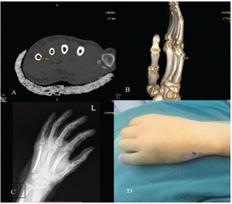
Figure 1: A 29 years old male. (A) The most minor intramedullary diameter of the fifth metacarpal was about 3.6mm in our study. (B) The metacarpal neck angulation of this patient was about 45° before surgery. (C) His fracture achieved a well reduction. (D) The skin incision was about 5mm and vertical to the metacarpal shaft.
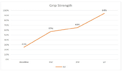
Figure 2: Grip Strength.
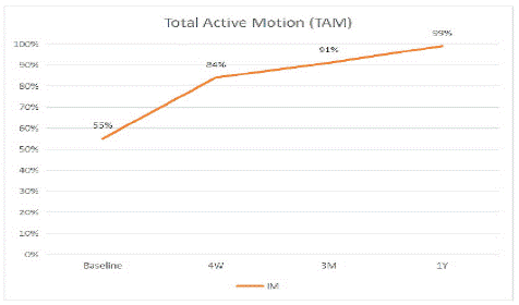
Figure 3: Total Active Motion.
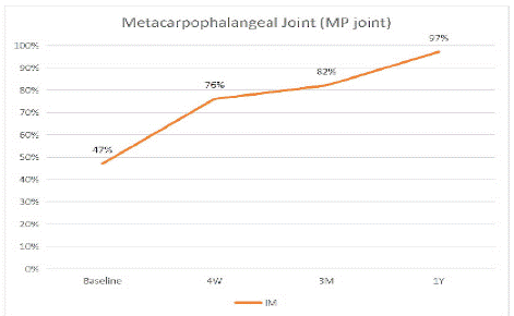
Figure 4: Metacarpophalangeal Joint.
Eight patients suffered local paresthesia during the follow-up period, and only two remained persistent paresthesia at the endpoint. Six of them recovered six months later after surgery. Three patients experienced persistent pain with the VAS score of 1, 2, 2. No infection, pin migration and pin bending were observed among these patients.
Discussion
The fifth metacarpal neck fractures mainly occur in young male population. Most of the fractures coursed by aggression [26]. The management of the fifth metacarpal neck fracture is still controversial. Fractures presenting with small displacement will have good results with conservative treatment. Some researchers suggested that the upper limitation of acceptable angulation of palmar displacement of these fractures is 30° [1,4]. On the other hand, some clinical studies indicate that the small finger metacarpal neck may tolerate up to 70° of angulation in the sagittal plane due to the compensatory movement of the little finger carpometacarpal joint of 20° to 30° [2,27,28]. Van Aaken et al. treated 25 patients with palmar angulations ranging from 30° to 75° conservatively; clinical outcome was evaluated at 5 months showed all patients were satisfied [29]. Moreover, Westbrook et al. studied 105 patients and found that palmar deformity severity did not affect the outcome [30]. Low et al. revealed that angulation (>30°) and shortening (>3 mm) of extrinsic tendons resulted in a reduction in flexion force [31]. In a cadaver model, Meunier et al. assessed intrinsic muscle fiber length after metacarpal shortening. The findings indicated that a 2 mm shortening resulted in an 8% loss of muscular power, while an 8 mm shortening resulted in a 45 percent drop in optimal power [5]. With respect to these studies, we included all patients who had an angulation at least 30° or metacarpal shorten than 3mm.
Many surgical techniques have been described for the osteosynthesis of fifth metacarpal neck fractures, such as locked nailing, perpendicular nailing, non-locked plate fixation, external fixation, intramedullary K-wires, and single antegrade intramedullary K-wire technique [32-35]. The principle of intramedullary nailing using double K-wires for 5th metacarpal neck fractures has not been well established. Our study demonstrated excellent clinical outcomes using optimized K-wires with the diameter of 1.6 mm for patients. Intramedullary multiple K-wiring was first described by Foucher and had became increasingly popular among surgeons owing to its simplicity and minimal invasiveness [36]. A meta-analysis had proved double Intramedullary nailing achieved better short- and mid-term clinical and radiological outcomes compared with other techniques [18]. Moreover, Mohammed et al. made the first attempt to use a single K-wire for fifth metacarpal neck fractures and achieved an excellent outcome [19]. However, the mean tensile strength of a 1.6mm K-wire is about 134.6N, making rotational deformities difficult to correct. Double 1.6 mm K-wires intramedullary nailing technique provides desirable strength similar to I plate [37]. In our study, the inside diameter of the narrowest part of the metacarpal ranged from 3.6 mm to 5.4 mm. Even in the youngest patient (14 y), his metacarpal inside diameter was greater than 3.2 mm, which was able to contain two 1.6 mm K-wires. We believe that two 1.6 mm K-wires intramedullary nailing is firm enough for most patients with metacarpal neck fractures.
The overall outcome was excellent. Intramedullary nail fixation with two 1.6 mm K-wires proved quite efficient in stabilizing metacarpal neck fractures anatomically. In our study, mean grip strength, mean TAM and mean MP joint motion were improved immediately after surgery. After one year of follow-up, patients achieved similar motor functions compared with the normal sides. In the past, numerous researchers had demonstrated that plate fixation results in a more rigid mechanical strength than intramedullary nail fixation in hand fracture models [38,39]. Fujitani et al. compared patients underwent intramedullary nail fixation with plate fixation. Intramedullary nail fixation showed a better outcome in the range of finger motion while plate fixation achieved a greater result in grip strength. The author recommended that plate fixation is suitable for patients who reject braces or hardware removal and need rapid return of robust hand function. Intramedullary fixation is advised for patients who want less invasive surgery or choose adequate finger range of motion over-vigorous hand function [14]. In our study, regretfully, we did not set a group with plate fixation to make a comprehensive comparison. This is one of the limitations of this study. Another limitation was the lack of data on children and adolescents who may have smaller marrow cavities in this study. Further studies are needed to confirm the optimal population.
Conclusion
According to the findings of this study, double 1.6 mm K-wires intramedullary pinning is a highly effective therapy for treating fifth metacarpal neck fractures. This approach produced excellent results in terms of bone union, post-operative dorsal angle, QuickDASH sore, satisfaction score, and functional outcomes. With potential benefits such as well surgical outcome and desirable strength, the use of double 1.6 mm K-wires AIMN could be an optimum option for patients with fifth metacarpal neck fractures.
References
- Ali A, Hamman J, Mass DP. The biomechanical effects of angulated boxer’s fractures. J Hand Surg Am. 1999; 24: 835-44.
- Hunter J, Cowen N. Fifth metacarpal fractures in a compensation clinic population. A report on one hundred and thirty-three cases. The Journal of bone and joint surgery American volume. 1970; 52: 1159-65.
- Giddins GE. The non-operative management of hand fractures. J Hand Surg Eur Vol. 2015; 40: 33-41.
- Birndorf MS, Daley R, Greenwald DP. Metacarpal fracture angulation decreases flexor mechanical efficiency in human hands. Plast Reconstr Surg. 1997; 99: 1079-83.
- Meunier MJ, Hentzen E, Ryan M, Shin AY, Lieber RL. Predicted effects of metacarpal shortening on interosseous muscle function. J Hand Surg Am. 2004; 29: 689-93.
- Kollitz KM, Hammert WC, Vedder NB, Huang JI. Metacarpal fractures: treatment and complications. Hand (N Y). 2014; 9: 16-23.
- Theeuwen GA, Lemmens JA, van Niekerk JL. Conservative treatment of boxer’s fracture: a retrospective analysis. Injury. 1991; 22: 394-6.
- Kim JK, Kim DJ. Antegrade intramedullary pinning versus retrograde intramedullary pinning for displaced fifth metacarpal neck fractures. Clin Orthop Relat Res. 2015; 473: 1747-54.
- Wong T, Ip F, Yeung S. Comparison between percutaneous transverse fixation and intramedullary K-wires in treating closed fractures of the metacarpal neck of the little finger. Journal of hand surgery (Edinburgh, Scotland). 2006; 31: 61-5.
- Zhang X, Huang X, Shao X. Reduction of fifth metacarpal neck fractures with a Kirschner wire. J Hand Surg Am. 2015; 40: 1225-30.
- Winter M, Balaguer T, Bessiere C, Carles M, Lebreton E. Surgical treatment of the boxer’s fracture: transverse pinning versus intramedullary pinning. J Hand Surg Eur Vol. 2007; 32: 709-13.
- Facca S, Ramdhian R, Pelissier A, Diaconu M, Liverneaux P. Fifth metacarpal neck fracture fixation: Locking plate versus K-wire? Orthop Traumatol Surg Res. 2010; 96: 506-12.
- Strub B, Schindele S, Sonderegger J, Sproedt J, von Campe A, et al. Intramedullary splinting or conservative treatment for displaced fractures of the little finger metacarpal neck? A prospective study. J Hand Surg Eur Vol. 2010; 35: 725-9.
- Fujitani R, Omokawa S, Shigematsu K, Tanaka Y. Comparison of the intramedullary nail and low-profile plate for unstable metacarpal neck fractures. J Orthop Sci. 2012; 17: 450-6.
- Sletten IN, Hellund JC, Olsen B, Clementsen S, Kvernmo HD, et al. Conservative treatment has comparable outcome with bouquet pinning of little finger metacarpal neck fractures: a multicentre randomized controlled study of 85 patients. J Hand Surg Eur Vol. 2015; 40: 76-83.
- Margic K. External fixation of closed metacarpal and phalangeal fractures of digits. A prospective study of one hundred consecutive patients. Journal of hand surgery (Edinburgh, Scotland). 2006; 31: 30-40.
- Manueddu CA, Della Santa D. Fasciculated intramedullary pinning of metacarpal fractures. J Hand Surg Br. 1996; 21: 230-6.
- Yammine K, Harvey A. Antegrade intramedullary nailing for fifth metacarpal neck fractures: a systematic review and meta-analysis. Eur J Orthop Surg Traumatol. 2014; 24: 273-8.
- Mohammed R, Farook MZ, Newman K. Percutaneous elastic intramedullary nailing of metacarpal fractures: surgical technique and clinical results study. J Orthop Surg Res. 2011; 6: 37.
- Lieber J, Harter B, Schmid E, Kirschner HJ, Schmittenbecher PP. Elastic stable intramedullary nailing (ESIN) of pediatric metacarpal fractures: experiences with 66 cases. Eur J Pediatr Surg. 2012; 22: 305-10.
- Boussakri H, Elidrissi M, Azarkane M, Bensaad S, Bachiri M, Shimi M, et al. Fractures of the neck of the fifth metacarpal bone, treated by percutaneous intramedullary nailing: surgical technique, radiological and clinical results study (28 cases). Pan Afr Med J. 2014; 18: 187.
- Shen K, Xu Y, Cao D, Wang Z, Cai H. Outcome of antegrade intramedullary fixation for juvenile fifth metacarpal neck fracture with titanium elastic nail. Exp Ther Med. 2017; 13: 2997-3002.
- She Y, Xu Y. Treatment of fifth metacarpal neck fractures with antegrade single elastic intramedullary nailing. BMC Musculoskelet Disord. 2017; 18: 238.
- Amsallem L, Pierrart J, Bihel T, Sekri J, Lafosse T, et al. Simplified internal fixation of fifth metacarpal neck fractures. Orthop Traumatol Surg Res. 2018; 104: 257-60.
- Assi C, Mansour J, Samaha C, Ajjoub S, Yammine K. A single antegrade intramedullary k-wire for fifth metacarpal neck fractures. Eur J Trauma Emerg Surg. 2020; 46: 389-95.
- Gudmundsen TE, Borgen L. Fractures of the fifth metacarpal. Acta Radiol. 2009; 50: 296-300.
- Ford DJ, Ali MS, Steel WM. Fractures of the fifth metacarpal neck: is reduction or immobilisation necessary? J Hand Surg Br. 1989; 14: 165-7.
- Statius Muller MG, Poolman RW, van Hoogstraten MJ, Steller EP. Immediate mobilization gives good results in boxer’s fractures with volar angulation up to 70 degrees: a prospective randomized trial comparing immediate mobilization with cast immobilization. Arch Orthop Trauma Surg. 2003; 123: 534-7.
- van Aaken J, Kampfen S, Berli M, Fritschy D, Della Santa D, Fusetti C. Outcome of boxer’s fractures treated by a soft wrap and buddy taping: a prospective study. Hand (N Y). 2007; 2: 212-7.
- Westbrook AP, Davis TR, Armstrong D, Burke FD. The clinical significance of malunion of fractures of the neck and shaft of the little finger metacarpal. J Hand Surg Eur Vol. 2008; 33: 732-9.
- Low CK, Wong HC, Low YP, Wong HP. A cadaver study of the effects of dorsal angulation and shortening of the metacarpal shaft on the extension and flexion force ratios of the index and little fingers. J Hand Surg Br. 1995; 20: 609-13.
- Margic K. External fixation of closed metacarpal and phalangeal fractures of digits. A prospective study of one hundred consecutive patients. J Hand Surg Br. 2006; 31: 30-40.
- Wong TC, Ip FK, Yeung SH. Comparison between percutaneous transverse fixation and intramedullary K-wires in treating closed fractures of the metacarpal neck of the little finger. J Hand Surg Br. 2006; 31: 61-5.
- Page SM, Stern PJ. Complications and range of motion following plate fixation of metacarpal and phalangeal fractures. J Hand Surg Am. 1998; 23: 827-32.
- Orbay JL, Touhami A. The treatment of unstable metacarpal and phalangeal shaft fractures with flexible nonlocking and locking intramedullary nails. Hand Clin. 2006; 22: 279-86.
- Foucher G, Chemorin C, Sibilly A. A new technic of osteosynthesis in fractures of the distal 3d of the 5th metacarpus. Nouv Presse Med. 1976; 5: 1139-40.
- Oh JR, Kim DS, Yeom JS, Kang SK, Kim YT. A Comparative Study of Tensile Strength of Three Operative Fixation Techniques for Metacarpal Shaft Fractures in Adults: A Cadaver Study. Clin Orthop Surg. 2019; 11: 120-5.
- Black D, Mann RJ, Constine R, Daniels AU. Comparison of internal fixation techniques in metacarpal fractures. J Hand Surg Am. 1985; 10: 466-72.
- Firoozbakhsh KK, Moneim MS, Howey T, Castaneda E, Pirela-Cruz MA. Comparative fatigue strengths and stabilities of metacarpal internal fixation techniques. J Hand Surg Am. 1993; 18: 1059-68.