
Case Report
Austin J Orthopade & Rheumatol. 2023; 10(1): 1118.
Intra-Articular Osteoid Osteoma of the Hip: An Observation and the Review of the Literature
El Maqrout Amine, MD¹*; Ghannam Abdelaziz, MD¹; Mekkaoui Jalal, PhD¹; Moncef Boufettal, PhD²; Bassir Reda Allah, PhD²; Kharmaz Mohammed, PhD¹; Lamrani Moulay Omar, PhD¹; Berrada Mohammed Saleh, PhD¹
¹Department of Trauma and Orthopedic Surgery, Ibn Sina University Hospital, Faculty of Medicine, Mohamed V University of Rabat, Rabat, Morocco
²Department of Anatomy, Faculty of Medicine, Mohamed V University of Rabat, Rabat, Morocco
*Corresponding author: El Maqrout Amine Department of Trauma and Orthopedic Surgery, Ibn Sina University Hospital, Faculty of Medicine, Mohamed V University of Rabat, Rabat, Morocco. Email: amineelmaqrout96@gmail.com
Received: March 04, 2023 Accepted: May 04, 2023 Published: May 11, 2023
Abstract
Osteoid osteoma is a common benign bone tumor that affects young adults and has a typical clinical and radiographic presentation in long bones. However, when it appears in unusual intra-articular locations, the clinical symptomatology is then atypical and can lead to misdiagnosis constituting a diagnostic challenge for clinicians and causing delay in management. We present the case of a girl of 16 years with an osteoma osteoid intra-articular right hip involving the femoral head. This unusual location was the cause of unexplained pain and a delay in diagnosis of 20 months after the onset of symptoms, as the initial Magnetic Resonance Imaging (MRI) examination could not identify the lesion. The tomodensitometry is in this indication the most specific examination which made it possible to evoke the diagnosis of osteoid osteoma. Once the diagnosis is made, given the complexity of the location, it makes it difficult to access radio-guided techniques. The open surgical excision allowed healing with complete disappearance of pain. After complete surgical excision of the tumor, histopathological examination confirmed the final diagnosis of intra-articular osteoid osteoma. No recurrence was observed.
Keywords: Osteoid osteoma; CT; Bone tumor
Introduction
Osteoid Osteoma (OO) is a relatively common benign osteoblastic tumor (2-3% of all bone tumors). It was described by Jaffé in 1935 [1] which mainly affects children [2] and young adults, affects men twice as often as women and generally occurs in the cortico-diaphyseal or metaphyseal region of the long bones (femur, tibia) [3,4]. Intra-articular localization is less frequent representing around 10% of cases [5,6], resulting in a more confused diagnosis [7,8] and producing nonspecific clinical symptoms which may mimic inflammatory monoarthritis [9,10]. Intra-articular OO is generally an atypical clinical condition that can delay diagnosis and treatment [7,11]. We report the case of an e daughter of 16 years presentant osteoma osteoid intra- articular and we try to describe the peculiarities clinical-radiologiques and terms Therapeutic this location knowing that this location is anatomically unreachable for treatment
Observation
A young girl of 16 years with no notable history was presented to our formation with intermittent right hip pain, with a nocturnal recurrence, which suggests cruralgia that appeared without triggers. These pains are slightly calmed by analgesics and non-steroidal anti-inflammatory drugs. It causes limping and limitation of the range of motion of the right hip. The clinical examination finds a significant and painful limitation of the movements of the right hip. This pain radiating along the front of the thigh. Lasegue's sign was negative and examination of the dorsolumbar spine and sacroiliac joints was normal.
Biological examinations showed: the sedimentation rate is normal and CRP<4mg/l, normal white blood cells. The white line and the immunological examinations were negative. The pelvic and lumbar spine x-rays were normal. The first radiographic examinations (x-rays of the pelvis and the right hip (Figure 1), Computed Tomography (CT-scan) of the dorsolumbar spine, MRI of the hip and thigh (Figure 2) did not reveal any abnormalities, with the exception of the presence of a misdirected benign osteocondensation located in the right femoral head, which could not be correlated with the patient's clinical picture as well as a gap with posterior hyper signal of the femoral head and synovial reaction has MRI.
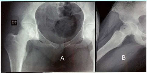
    Figure 1: X-ray of the left hip. Osteocondensation of the superior edge of the right femoral head.

A: strict face; B: internal rotation side
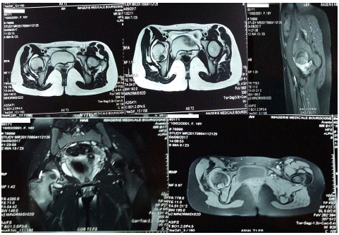
Figure 2: MRI of the hip right that visualizes gap after the head femoral with synovial reaction.
As the location of the lesion did not allow safe access for standard X-ray guided ablation, surgical resection of the tumor by a minimally invasive posterolateral approach was planned. The posterior femoral circumflex vessels were identified and preserved, and then a T-shaped arthrotomy (designed on a trochanteric basis) was performed after passing through the piriformis and upper twin muscles. The hip was then placed in flexion, adduction and maximum internal rotation for good visualization of the tumor region, which was exposed macroscopically outside the acetabulum coverage area. Abundant effusion, inflammatory synovitis, the superficial area of tumor intrusion appeared in stellate depression of the posterior face of the femoral head under the rim of the posterior face, which facilitated its identification. A block resection, after perforation of this area and the discovery of a gap of one centimeter containing a granular tissue (nidus), with additional curettage of the perilesional bone without filling was performed then the entire excised specimen (Figure 3). Was sent to the laboratory for pathological analysis. Histopathological examination confirmed the diagnosis of osteoid osteoma due to the development of a bone substance calcified by osteoblast cells. The osteoblasts were devoid of atypia and showed no mitotic activity (Figure 4), thus confirming the benignity of the lesion. The postoperative follow-up was simple (Figure 5). Support is prohibited for a period of 45 days. The operative consequences are simple with immediate disappearance of pain and complete recovery. Two months after the operation, the patient did not complain of any pain. The range of motion was normal and symmetrical. The patient had resumed his professional activities. After five years of follow-up, the clinical examination was strictly normal and the patient had resumed his sports activities without complaining of pain.
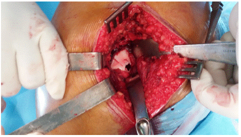
Figure 3: Operative view. Arthrotomy of the hip through posterolateral side - excision of the nidus.
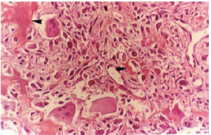
Figure 4: Pathological examination of the excision piece. Richly vascularized proliferation of the nidus surrounded by osteoid trabeculae rich in osteoblastic cells.
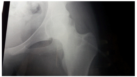
Figure 5: Postoperative control radiography.
Discussion
Osteoid Osteoma (OO) is a common benign primary bone tumor. It represents 2 to 3% of all bone tumors and 10 to 20% of all benign bone tumors [4-6]. It is preferentially located in the long bones [6,12] with a predilection for the lower limbs [13], in particular the tibia and the femur. Intra-articular localization is rare and its frequency is difficult to assess about 10% of all osteoid osteomas and occurs mainly in the elbow, carpus, ankle or hip joints [7,9].
The clinical manifestations of OO are most often made by nighttime pain, insomnia, calmed by taking salicylates [13,14].
However, Intra-Articular OO (OOIA) frequently evolves in a misleading context [15] delaying diagnosis and adequate management [15]. Due to the rarity and non-specificity of its clinical manifestations, such as pain or even monoarthritis [7,8,16], diagnosis is difficult. Osteoid osteoma can be confused with other causes, such as inflammatory or infectious arthritis, aseptic osteonecrosis of the femoral head, stress fracture, radicular syndrome and even pigmented villonodullary synovitis [17].
In the literature, the diagnostic times for intra-articular OO range from 4 months to 5 years [5,9,15,18], longer than in other locations. In our observation, the diagnostic delay was almost 2 years.
As there is no sclerosis or periosteal reaction around the lesion, standard radiographs can only give subtle results, different from extra-articular locations [8].
In most cases, there is no typical image of the nidus bordered by surrounding bone sclerosis in its intra-articular location [18]. X-rays are either normal or show osteopenia around the joints [5,9]. Reactive sclerosis may mask the radiolucent nidus and may be associated with demineralization which may be necessary for algodystrophy [19]. In the hip, this regional osteoporosis is frequent [20], and in patients whose pain lasts at least 3 months, an enlarged femoral neck may be observed [20].
All these clinical and radiological characteristics of intra-articular OO require the frequent use of several imaging means [9].
According to many authors, bone scintigraphy and computed tomography are the first examinations of choice for a correct diagnosis [16,21]. Bone scintigraphy is very sensitive reaching 100% but has a lower specificity than the CT scan, in particular in the case of intra-articular localization, reveals an early and intense localized fixation "in spot" [3], because the bone sclerosis around the nidus cannot be detected early because it is less intense due to the associated synovial reaction [22]. As the osteoid osteoma progresses, the signal strength of peripheral bone sclerosis decreases, while the nest can be more easily detected on scintigraphy. But its interest lies in looking for other locations that are generally exceptional [14,23] and is used to precisely target the rest of the imaging work-up (CT and MRI) [12]. Computed tomography remains the examination of choice when it uses contiguous fine millimeter slices at high resolution, thus providing precise data on the size and location of the lesion [5]. It makes it possible to locate the lesion with precision, to measure the exact dimension of the nidus and to evaluate its local extension, which makes it possible to orient the therapeutic strategy [13,23]. Consequently, according to some authors, MRI remains the preferred means to explore bone tumors [5]. On magnetic resonance imaging, the osteoid osteoma usually shows lower signal intensity on T1 and T2-weighted images, edema of the bone marrow around the nest, and a sharp increase in contrast after administration of gadolinium Lesions intra-articular can be accompanied by a thickening of the synovial membrane, visible on MRI and diagnosed after injection of contrast product. However, the precise location of the nidus may not be easy. In 35% of cases, the nidus cannot be detected due to the associated peri-lesional swelling around the lesion, while in 50% of cases; the nidus is atypical and may lead to a misdiagnosis. [21]. in our case, the MRI could not provide an early diagnosis of osteoid osteoma because of the small size of the lesion and to even misleading twice with the presence of an énostose.
In the literature, the treatment of choice for this benign tumor, although it can spontaneously involute after years, is surgical excision, which must be complete to avoid recurrence. The en bloc resection is the technique, which allows the entire resection of the nidus [13] and therefore healing. This approach can only be considered in the event of a confused diagnosis before the pathological examination or when the location of the tumor does not allow easy access to radio-guided techniques [3]. Since the 1990s, minimally invasive techniques have taken an important place in the therapeutic arsenal of OOs, particularly such as percutaneous surgery for drilling and resection procedures under CT guidance [24], laser photocoagulation [25], Radiofrequency coagulation [26] or even arthroscopy of the hip [27] could not be offered, despite their proven reliability and effectiveness and the limited number of complications and recurrences.
The drawback of these nidus destruction techniques is the lack of possibility of pathological examination. Due to the localization of the osteoid osteoma next to the femoral cartilage, the use of thermocoagulation, laser photocoagulation or even radiofrequency seemed difficult [28] while the postero-inferior localization of the lesion did not allow not safe access for arthroscopic excision. Percutaneous techniques should be used in the event of a reliable diagnosis and easy access to the lesion without associated iatrogenic risk. Otherwise, conventional surgery by the minimally invasive approach should be performed for effective curative treatment with precise histopathological examination. Open surgery therefore appeared to be the best management option and was performed by a limited posterior approach with dissection of the circumflex pedicle to preserve the cephalic vascularization [29] while providing sufficient exposure without an intraoperative referencing technique and allowing a Complete excision of the lesion which was subsequently confirmed by histopathological analysis and demonstrating the absence of recurrence. Our patient underwent a one-piece open excision, the consequences of which were marked by a good clinical course. This surgery also made it possible to obtain a histological confirmation often necessary for the diagnosis and to confirm the completeness of the resection.
Conclusion
Intra-articular locations of osteoid osteomas are rare. The clinical presentation is most often atypical, their diagnosis is usually difficult, and it is facilitated by the contribution of medical imaging techniques. If there is any doubt about the diagnosis, the tomodensitometry represents the most specific examination allowing a positive diagnosis. Complete surgical excision of the lesion usually allows total healing and prevents recurrence.
Author Statements
Conflicts of Interest
The authors declare no conflict of interest.
Authors' Contribution
All the authors contributed to the care of the patient and the writing of the manuscript.
References
- Jaffe HL. Osteoid osteoma. Arch Surg. 1935; 31: 709-28.
- Jacopin S, Launay F, Viehweger E, Glard Y, Jouve JL, et al. Hip subluxation and coxa valga secondary to an osteoid osteoma. Rev Chir Orthop. 2008; 94: 758-62.
- Bonnevialle P, Railhac JJ. Osteoma Osteoid, Oosteoblastoma. Encycl Med Chir (Scientific and medical editions Elsevier SAS, Paris), Musculoskeletal system. 2001; 14-712.
- Lee EH, Shafi M, Hui JH. Osteoid osteoma: a current review. J Pediatr Orthop. 2006; 26: 695-700.
- Allen SD, Saifuddin A. Imaging of intra-articular osteoid osteoma. Clin Radiol. 2003; 58: 845-52.
- Clavert JM. Osteoid osteoma and osteoblastoma. In: SOFC OT teaching notebooks. Teaching conferences.
- Franceschi F, Marinozzi A, Papalia R, Longo UG, Gualdi G, et al. Intra- and juxta-articular osteoid osteoma: a diagnostic challenge: misdiagnosis and successful treatment: a report of four cases. Arch Orthop Trauma Surg. 2006; 126: 660-7.
- Cassar-Pullicino VN, McCall IW, Wan S. Intra-articular osteoid os teoma. Clin Radiol. 1992; 45: 153-60.
- Szendroi M, Kollo K, Antal I, Lakatos J, Szoke G. Intra-articular osteoid osteoma: clinical features, imaging results, and comparison with extra-articular localization. J Rheumatol. 2004; 31: 957-64.
- Kellner H, Spathling S, Kuffer G, Herzer P. Intra-articular osteoid osteoma: a rare cause of coxitis. Z Rheumatol. 1991; 50: 114-6.
- Davies M, Cassar-Pullicino VN, Davies AM. The diagnostic accuracy of MR imaging in osteoid osteoma. Skeletal Radiol. 2002; 31: 559-69.
- Girard J, Bequet E, Limousin M, Chantelot C, Fontaine C. Osteoma osteoid of trapezoid bone: a case-report and review of the literature. Chir Main. 2005; 24: 35-8.
- Saidi H, El Bouanani A, Ayash A Fikry T. Osteoma osteoid of the lunate: about one case. Chir Main. 2007; 26: 173-5.
- Kalb K, Schlör U, Meier M, Schmitt R, Lanz U. Osteoid osteoma of hand and wrist. Handchir Mikrochir. Plast Chir. 2004; 36: 405-10.
- Efstathopoulos N, Sapkas G, N Xypnitos F, Lazarettos I, Korres D, et al. Recurrent intra- articular osteoid osteoma of the hip after radiofrequency ablation: a case report and review of the literature. Journal boxes. 2009; 2: 6439.
- Caillieret J, Fontaine C, Ducloux M, Letendart J, Duquennoy A. Osteoid osteoma of the upper extremity of the femur. Rev Chir Orthop. 1986; 72: 101-3.
- Bauer TW, Zehr RJ, Belhobek GH, Marks KE. Juxta-articular osteoid osteoma. Am J Surg Pathol. 1991; 15: 381-7.
- Eggel Y, Theumann N, Lüthi F. Intra-articular osteoid osteoma of the knee: clinical and therapeutical particularities. Joint Bone Spine. 2007; 74: 379-81.
- Laffosse JM, Tricoire JL, Cantagrel A, Wagner A, Puget J. Osteoid osteoma of the carpal bones - Two case reports. Joint Bone Spine. 2006; 73: 560-3.
- Kumar SJ, Harcke HT, MacEwen GD, Ger E. Osteoid osteoma of the proximal femur: new techniques in diagnosis and treatment. J Pediatr Orthop. 1984; 4: 669-672.
- Davies M, Cassar-Pullicino VN, Davies AM. The diagnostic accuracy of MR imaging in osteoid osteoma. Skeletal Radiol. 2002; 31: 559-69.
- Koliakos G, Katsiki N. Osteoid osteoma mimicking chronic arthritis. Diagnosis by bone scintigraphy. Hell J Nucl Med. 2005; 8: 171-3.
- Haddam A, Bsiss A, Ech charraq I, BenRaïs N, et al. Optimization of the treatment of osteoid osteoma by intraoperative isotopic detection: report on a case. Nuclear medicine. 2009; 33: 375–379.
- Guyot-Drouot MH, Migaud H, Cotten A, Cortet B, Delezenn e A, et al. Long-term efficacy of percutaneous drill-biopsy under computed tomography guidance of osteoid osteomas of the hip and femur. A review of seven cases. Joint Bone Spine. 2000; 67: 204-9.
- Gangi A, Dietemann JL, Clavert JM, Dodelin A, Mortazavi R, et al. Treatment of osteoid osteoma using laser photocoagulation . About 28 cases. Rev Chir Orthop. 1998; 84: 676-84.
- Ghanem I, Collet LM, Kharrat K, Samaha A, Deramon H, et al. Percutaneous radiofrequency coagulation of osteoid osteoma in children and adolescents. J Pediatr Orthop. 2003; 12: 244-52 .
- Lee DH, Jeong WK, Lee SH. Arthroscopic excision of osteoid osteomas of the hip in children. J Pediatr Orthop. 2009; 29: 547-51.
- Bosschaert PP, Deprez FC. Acetabular osteoid osteoma treated by percutaneous radiofrequency ablation: delayed articular cartilage damage. JBR-BRT. 2010; 93: 204-6.
- Goldstein WM, Branson JJ, Berland KA, Gordon AC. Minimal- incision for total hip arthroplasty. J Bone Joint Surg (Am). 2003; 85: 33-8.