Clinical Image
Austin J Orthopade & Rheumatol. 2023; 10(2): 1119.
Tumor of Hallux: A Case Report and Review of Literature
MR El Galiou*; Y Houass; J Mekkaoui; M Boufettal; RA Bassir; MO Lamrani; M Kharmaz; MS Berrada
Orthopedic Surgery Department of Ibn Sina Hospital, University Mohamed V, Rabat, Morocco
*Corresponding author: Mohamed Rida El Galiou Orthopedic Surgery Department of Ibn Sina Hospital, University Mohamed V, Rabat, Morocco. Email: medrida.elgaliou@gmail.com
Received: June 26, 2023 Accepted: July 28, 2023 Published: August 05, 2023
Abstract
Glomus tumors are rare and benign, arising from a neuromyoarteriel glomus body. If the digital location is well known in surgery of the hand, the extradigitales locations suffer from a misunderstanding ending in diagnostic and therapeutic errors. We report a new case of tumor glomique of the hallux, and through a review of the literature, we would try to draw the attention towards these atypical location.
Keywords: Glomus; Tumor; Hallux
Introduction
Glomus tumors are benign and rare tumors that develop from the neuromyoarterial glomus of arteriovenous anastomoses. They mainly affect the digital extremities, but extra-digital localizations are not rare but above all unknown, which is responsible for the delay in their diagnosis and their management. The aim of our work is to report a new case of hallux glomus tumor and to draw attention to this unusual location.
Clinical Case
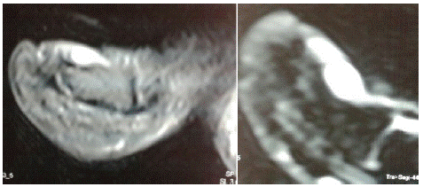
This is a 41 year-old patient, with no particular pathological history, who has been presenting for 3 years with intermittent pain at the base of the left big toe, coinciding with trauma to the same toe. We received the patient who consulted several times given the increase in pain intensity and the lack of improvement under symptomatic treatment. The clinical examination of this patient showed a discoloration of the base of the nail which was very painful on palpation. The osteoarticular examination of the left hallux was unremarkable. The standard X-ray and the biological assessment were normal. The MRI objectified a hyper signal around the nail bed suggesting the possibility of a glomus tumor (Figure 1). A lateral subungual excision was performed and the pathological examination confirmed the diagnosis of glomus tumor. The postoperative course was simple, the patient had immediate pain relief and the clinical examination of the patient after a follow-up 2 years did not show any recurrence of symptoms.

Figure 1: MRI aspect showing a hyper signal around the nail bed.
Discussion
Glomus tumors are rare and benign, representing about 1 to 5% of all soft tissue tumors [1]. These are tumors that affect the adult subject, the average age is 40 years, it is rare before 20 years [2]. While digital localization is well known in hand surgery [3], atypical localizations [4-8], like our case in the hallux, are often overlooked, leading to diagnostic and therapeutic errors. The clinical symptomatology associates a classic triad with severe pain, sore spot and intolerance to cold. What also characterizes the symptomatology is the contrast between the intensity of the subjective signs and the poverty of the objective signs. Rarely are the cases where there is a traumatic history as in our case [9]. There is no specific imaging allowing diagnostic confirmation, however ultrasound, despite its low specificity, helps to locate the lesion [10]. MRI remains the gold standard in the diagnosis of glomus tumours, it specifies the exact site of the lesion and its relationship with the neighboring structures [11-13]. Its treatment is always surgical, it consists of surgical excision which leads to a rapid disappearance of the pain. Recurrences are rare but possible, they are always early and the fact of incomplete excision, hence the recommendation of some authors [13-15] to excise more than the apparent limits of the tumour.
Conclusion
Glomus tumors are rare but not exceptional. They can sit wherever the glomus exists. Faced with any pain with or without a palpable mass, without obvious etiology, the diagnosis of glomus tumor should be considered.
References
- Akgün RC, Güler UÖ, Onay U. A glomus tumor anterior to the patellar tendon: a case report. Acta Orthop Traumatol Turc. 2010; 44: 250-3.
- Johnson DL, Kuschner SH, Lane CS. Intraosseous glomus tumor of the phalanx: a case report. J Hand Surg Am. 1993; 18: 1026-8.
- Carroll RE, Berman AT. Glomus tumors of the hand: review of the literature and report on twenty-eight cases. J Bone Joint Surg Am. 1972; 54: 691-703.
- Proietti A, Alì G, Quilici F, Bertoglio P, Mussi A, Fontanini G. Glomus tumor of the shoulder: A case report and review of the literature. Oncol Lett. 2013; 6: 1021-4.
- Chun JS, Hong R, Kim JA. Extradigital glomus tumor: A case report. Mol Clin Oncol. 2014; 2: 237-9.
- Balaram AK, Hsu AR, Rapp TB, Mehta V, Bindra RR. Large solitary glomus tumor of the wrist involving the radial artery. Am J Orthop (Belle Mead NJ). 2014; 43: 567-70.
- Amillo S, Arriola FJ, Muñoz G. Extradigital glomus tumor causing thigh pain:a case report. J Bone Joint Surg Br. 1997; 79: 104-6.
- Beksaç K, Dogan L, Bozdogan N, Dilek G, Akgul GG, Ozaslan C. Extradigital glomus tumor of thigh. Case Rep Surg. 2015; 2015: 638283.
- Koti M, Bhattacharryya R, Ewen SW, Maffulli N. Subungual glomus tumor of the hallux. A case report. Acta Orthop Belg. 2001; 67: 297-9.
- Smith KA, Mackinnon SE, Macauley RJ, Mailis A. Glomus tumor originating in the radial nerve:a case report. J Hand Surg Am. 1992; 17: 665-7.
- Shih TT, Sun JS, Hou SM, Huang KM, Su TT. Magnetic resonance imaging of glomus tumor in the hand. Int Orthop. 1996; 20: 342-5.
- Dupuis P, Pigeau I, Ebelin M, Barbato B, Lemerle JP. The contribution of MRN in the study of glomus tumors. Ann Chir Main Memb Super. 1994; 13: 358-62.
- Matloub HS, Muoneke VN, Prevel CD, Sanger JR, Yousif NJ. Glomus tumor imaging:use of MRI for localization of occult lesions. J Hand Surg Am. 1992; 17: 472-5.
- Varian JP, Cleak DK. Glomus tumors in the hand. Hand. 1980; 12: 293-9.
- Rettig AC, Strickland JW. Glomus tumor of the digits. J Hand Surg Am. 1977; 2: 261-5.