Case Report
Austin J Orthopade & Rheumatol. 2023 ; 10(2) : 1120.
Hoffa’s Fracture Case Report
Abdelhay Rabah*
Department of Orthopedic A Traumatological Surgery, Mohamed V Military Hospital, Rabat-Morocco
*Corresponding author: Abdelhay Rabah Department of Orthopedic A Traumatological Surgery, Mohamed V Military Hospital, Rabat-Morocco. Email: rabahabdelhay7@gmail.com
Received: September 27, 2023 Accepted: November 02, 2023 Published: November 09, 2023
Abstract
Unicondylar fractures of the distal third of the femur in the coronal plane are known as Hoffa’s fractures, they are rare and very rare. Few cases have been reported of this type of fracture, by definition they are unstable fractures and therefore require surgical treatment. The mechanism of trauma in this type of fracture is direct with the knee in flexion. Often they can go unnoticed in the Anteroposterior (AP) X-ray projection, which is why it is important to assess the lateral projection. When they are suspected or highlighted, it is necessary to pass a Computed Tomography (CT) in order to clearly define its surgical resolution, since the approach and method of fixation are controversial. Although today the availability and characteristics of different types of implants have increased, there is no consensus in the literature mainly due to the lack of expertise in these rare cases.
Introduction
Hoffa's fracture, first described in 1904 [1], is a rare type of fracture of the femoral condyle with its unique coronal cut. Lateral Hoffa fractures are more common, but medial Hoffa fractures have been described and are extremely rare. This type of fracture is intra-articular by definition and the principles of treatment are generally similar to those of typical intra-articular fractures. Moreover, this type of injury often goes unnoticed due to lack of clinical suspicion and inadequate radiographic imaging [2]. The mechanism of injury is usually high velocity energy [3–6]. Here we present a case of a 25-year-old patient with a rare type of injury involving the lateral femoral condyle; surgically treated by open reduction and internal fixation.
Clinical Observation
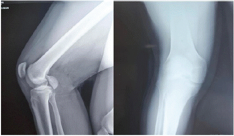
This is a young soldier who presented to our emergency services with right knee pain and total functional impotence of the right lower limb following a road accident collision between 2 motorcycles with landing on the knee right. Clinical examination revealed that his knee was enlarged with tight skin and peri-patellar bruising with no skin opening and range of motion was limited due to pain. Neurovascular examination was normal with no signs of compartment syndrome. Knee x-rays revealed a lateral Hoffa fracture (Figure 1).

Figure 1: AP and profile X-ray of the knee showing Hoffa’s fracture.
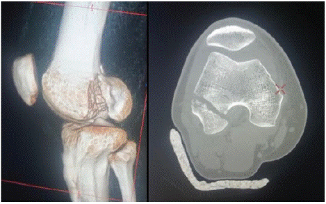
Computed tomography with three-dimensional reconstruction showed a displaced lateral coronal condylar fracture (Figure 2).

Figure 2: CT scan of the knee showing Hoffa’s fracture.
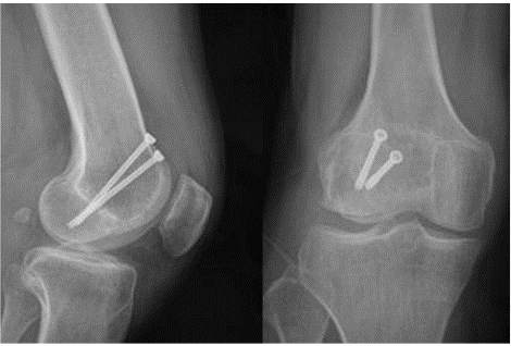
The patient underwent surgical treatment with means of open reduction and internal fixation: The fracture was exposed by a lateral parapatellar approach Under image intensifier control, the fracture was reduced using bone clamp and secured with two 4.5 mm cancellous screws, placed perpendicular to the fracture line. The screw heads were placed extra-articularly. After open reduction and internal fixation, the assembly was protected by a Zimmer knee splint in extension for 6 weeks, then functional knee rehabilitation sessions were initiated for gradual recovery of joint amplitude and muscle strengthening. Strict instructions were given to avoid any weight-bearing flexion during this six-week period to minimize the shear force on this type of coronal fracture. Partial weight bearing was authorized after 6 weeks postoperatively. At 10 weeks after the surgical procedure, he gradually progressed towards full weight bearing. At the six-month follow-up, he was walking unsupported and pain-free and the knee range of motion was 15 to 130° with good right lower limb musculature. Additionally, there was no angular deformity or limb length difference. Control radiographs showed a well-consolidated fracture with no signs of femoral condyle collapse or pseudarthrosis (Figure 3).

Figure 3: AP and profile X-ray showing consolidation of Hoffa’s fracture.
Discussion
The so-called “Hoffa” fracture refers to an isolated fracture, a coronal fracture of one or the other of the femoral condyles, with intra-articular extension [5]. This rare injury corresponds to orthopedic trauma association type 33-B3 fracture (partial frontal articular fracture of the distal femur) [6]. These injuries have already been classified. Type 1 fractures extend from an extra-articular region located between the junction of the posterior femoral shaft and the proximal aspect of the femoral condyle at the top and the lower surface of the posterior aspect of the condylar joint, so that the The insertion of the popliteus tendon and the lateral head of gastrocnemius remain attached to the condylar fragment. The insertions of the anterior cruciate ligament and the lateral ligament can be attached to either the condylar or diaphyseal fragment. They have type II origins posterior to the posterior femoral shaft-condylar junction, and are therefore entirely intra-articular.
Compared to Type I fractures, the aforementioned ligamentous insertions are less likely to be attached to the condylar fragment. In Type III fractures, all lateral ligament insertions remain attached to the condylar fragment. No relationship between the incidence of avascular necrosis and the type of fracture has been conclusively demonstrated [5].
Hoffa fractures are seen in high energy trauma. Four out of five patients with isolated Hoffa fractures in a series were caused by a road accident [7]. In another series, five of seven fractures were sustained in road accidents, while a sixth involved a pedestrian struck by a car [5]. In a large retrospective review of intra-articular distal femoral fractures performed at a large level 1 trauma center, Hoffa fracture was an associated injury in 77 cases out of 202 supracondylar-intracondylar distal femoral fractures. Of these 77 fractures, 62 were seen in the context of trauma from road accidents [9]. A fall from a height seems to be the second most common scenario [5,7,9].
While some authors have considered direct impact with the knee in a flexed position as the mechanism of injury, others have attributed the fracture to simultaneous vertical shear and twisting forces [5,8]. The Hoffa fracture pattern has been described in the medial and lateral femoral condyle [5,7,9]. Bi-condylar involvement has also been previously reported [8,9,10]. As already mentioned, Hoffa fractures are not uncommonly seen in association with other fractures of the femur, both distal and more proximal [9,10,11]. Associated lesions of the patellar and/or quadriceps tendons have also been reported [10,12]. In one case, the avulsed patellar tendon became incarcerated between the fragments of a Hoffa fracture of the lateral femoral condyle, precluding closed reduction [12].
The oblique coronal orientation of the Hoffa fracture makes this injury difficult to detect on standard AP and lateral radiographs, especially if the fracture is nondisplaced (5,7,9). In the AP projection, the X-ray beam is not tangent to the edges of the fracture. The intact anterior surface of the fractured condyle further conceals the fracture [5]. Moreover, the fractured condyle with shortened limb can be neglected as simple varus or valgus. If not displaced, incomplete overlapping of the condyles may be indicative of a fracture; however, this a subtle finding may be misinterpreted as a slight obliquity of a contemplated lateral radiograph [7]. In large series evaluating Hoffa's fractures in the context of intra-articular distal femoral fractures, initial AP and lateral radiographs diagnosed only 66 of the 95 cases of Hoffa's unicondylar fracture. In 10 cases without preoperative computed tomography (CT) evaluation, Hoffa's fracture was only discovered intraoperatively [9]. In the presented case, the fracture was obvious and displaced.
Although the patient in our presented case was successfully treated conservatively with a knee brace and protected support, noted loss of anatomical alignment of undisplaced Hoffa fractures treated exclusively by nonoperative management is well described. This was the case in 3 of 7 patients in one series, and in 1 of 5 patients in another series. The rest of the fractures in each of these two series were treated surgically [5,7]. Hoffa's fractures are generally reduced and fixed with cancellous screws oriented anterior-posterior [5,7,8,13,114,15]. When there is metaphyseal extension of the fracture, the fixation is supplemented by a lateral support plate [14,15]. Lateral condylar surgical repair of Hoffa fractures can be performed without arthrotomy by performing a Gerdy tubercle osteotomy and raising the iliotibial band for better visibility [16]. Recently, the durability of postero-anterior screw fixation was compared with antero-posterior fixation in cadaveric femurs having undergone osteotomies to simulate Hoffa's fractures. The authors showed that those condyles fixed with posterior-anterior directed screws showed less displacement when subjected to a vertical force compared to an antero-posterior screw fixation [15].
Conclusion
To conclude, we believe that the diagnosis of this fracture can easily go unnoticed, so keep this in mind when dealing with high-energy knee trauma with total functional impairment and shortening of the traumatized limb. If necessary, complete the assessment with a CT scan with 3D reconstruction.
This fracture is best treated with open reduction and internal fixation allowing for quick and complete recovery while avoiding further long-term complications.
References
- Hoffa A. Lehrbuch der Frakturen und Luxationen, Stuttgart: V erlag von Ferdinand Enke. 1904; 451.
- Salunke A, Nambi GI, Singh S, Menon P, Girish GN, Vachalam D. Hoffa's fracture with ipsilateral fibular fracture in a 16-year-old girl: an approach to a rare injury. Chin J Traumatol. 2015; 18: 178-80.
- Lewis SL, Pozo JL, Muirhead-Allwood WFG. Coronal fractures of the lateral femoral condyle. J Bone Joint Surg Br. 1989; 71: 118-20.
- Flanagin BA, Cruz AI, Medvecky MJ. Hoffa fracture in a 14-year-old. Orthopedics. 2011; 34: 138.
- Lewis SL, Pozo JL, Muirhead-Allwood WFG. Coronal fractures of the lateral femoral condyle. J Bone Joint Surg Br. 1989; 71: 118-20.
- Marsh JL, Slongo TF, Agel J, Broderick JS, Creevey W, DeCoster TA, et al. Fracture and dislocation classification compendium – 2007. J Orthop Trauma. 2007; 21: S1-S133.
- Holmes SM, Bomback D, Baumgaertner MR. Coronal fractures of the femoral condyle: a brief report of five cases. J Orthop Trauma. 2004; 18: 316-9.
- Papadopoulos AX, Panagopoulos A, Karageorgos A, Tyllianakis M. Operative treatment of unilateral bicondylar Hoffa fractures. J Orthop Trauma. 2004; 18: 119-22.
- Nork SE, Segina DN, Aflatoon K, Barei DP, Henley MB, Holt S, et al. The association between supracondylar-intercondylar distal femoral fractures and coronal plane fractures. J Bone Joint Surg Am. 2005; 87: 564-9.
- Calmet J, Mellado JM, García Forcada IL, Giné J. Open bicondylar Hoffa fracture associated with extensor mechanism injury. J Orthop Trauma. 2004; 18: 323-5.
- Miyamoto R, Fornari E, Tejwani NC. Hoffa fragment associated with a femoral shaft fracture. A case report. J Bone Joint Surg Am. 2006; 88: 2270-4.
- Shetty GM, Wang JH, Kim SK, Park JH, Park JW, Kim JG, et al. Incarcerated patellar tendon in Hoffa fracture: an unusual cause of irreducible knee dislocation. Knee Surg Sports Traumatol Arthrosc. 2008; 16: 378-81.
- Kumar R, Malhotra R. The Hoffa fracture: three case reports. J Orthop Surg (Hong Kong). 2001; 9: 47-51.
- Agarwal S, Giannoudis PV, Smith RM. Cruciate fracture of the distal femur: the double Hoffa fracture. Injury. 2004; 35: 828-30.
- Jarit GJ, Kummer FJ, Gibber MJ, Egol KA. A mechanical evaluation of two fixation methods using cancellous screws for coronal fractures of the lateral condyle of the distal femur (OTA type 33B). J Orthop Trauma. 2006; 20: 273-6.
- Liebergall M, Wilber JH, Mosheiff R, Segal D. Gerdy’s tubercle osteotomy for the treatment of coronal fractures of the lateral femoral condyle. J Orthop Trauma. 2000; 14: 214-5.