
Case Report
Austin J Orthopade & Rheumatol. 2023; 10(2): 1122.
End Stage Hoffa’s Disease Presented as a Giant Osteochondroma: Case Report and Literature Review
El Mokhtari Kamal*
Department of Orthopedic Surgery and Traumatology, Military Hospital Mohammed V, Rabat, Morocco
*Corresponding author: El Mokhtari Kamal Department of Orthopedic Surgery and Traumatology, Military Hospital Mohammed V, Rabat, Morocco. Email: kamalelmokhtari@yahoo.com
Received: October 30, 2023 Accepted: November 30, 2023 Published: December 07, 2023
Abstract
Introduction: Hoffa’s disease appears to be an under-recognized ailment according to an extensive comprehensive literature assessment. Predisposing factors in sports training include torsion and hyperextexion at the knee should be taken into consideration when evaluating patients with chronic knee pain. We present the case of a young soldier suffering from left knee pain as a result of Hoffa’s disease
Case report: A 29-year-old male patient admitted to orthopedic department to assess an anterior mechanical pain on the left knee. He reported that he had a sport accident with notion of hyperextension and anterior direct trauma to the left knee. On physical examination we found a painful mass that seems located under the patellar tendon, passive flexion of the left knee triggered a sharp pain from 100°. On X ray we found bone-like mass located behind the silhouette of the patellar tendon without attachment to the bones of the leg. MRI identified a 4.2 osseous tumor with no attachment to the patellar tendon. A surgical excision was preformed
Conclusion: Hoffa disease is relatively a rare illness, its physiopathology is not yet completely clear. But many cases have been reported in literature. Its clinical presentation is not very specific, and its surgical excision which remains unavoidable, guarantees satisfying clinical and functional results. However, it remains to be determined whether it is a definitive solution in the context of “Hoffa’s disease”.
Keywords: Hoffa; Knee; Patella; Anterior; Pain
Introduction
Even though the Hoffa infrapatellar fat pad is usually injured, it is rarely described in the orthopedic literature. The infrapatellar fat pad (Hoffa) is an intracapsular extra synovial structure. Although the exact function of the Hoffa fat is unknown, it is clinically significant because it is the source of a variety of tumors and tumor-like abnormalities like osteochondroma and Hoffa's Disease.
We report the case of a patient who had a giant osteochondral tumor in association with Hoffa’s disease. A review of the literature reveals one case report which questions whether or not the latter was the outcome of end-stage Hoffa’s disease [1]. Our patient was informed that data concerning this case would be submitted for publication.
Case Report: Data and Findings
A 29-year-old patient, with no particular pathological history, admitted to our department for therapeutical management of mechanical gonalgia becoming resistant to the usual analgesic and anti-inflammatory treatment. He had a history of severe hyper-extension knee trauma due to an anterior crush 4 years ago during a football game. After clinical and radiological examination at the time, no ligament or bone lesion was identified. But he indicated that a progressive stiffness and anterior knee pain had occurred after the regression of the oedema following conservative treatment with oral and topical anti-inflammatory agents and physical rehabilitation. Because of these complaints he sought consultation in our department. The initial physical evaluation, on inspection we found a normal aspect of both knees, with no clearly visible deformation, on palpation of the left knee we noted a slight joint effusion with a tender mass next to the patellar tendon, painful and fixed to the surrounding soft tissue , concerning joint mobility, in active: 0° of extension at 100° of flexion was possible without pain, in passive nevertheless the sector of mobility was not reduced but a sharp pain was triggered from 110° of flexion. A patella compression test with direct palpation of the medial and lateral aspects of the fat pad with the knee in extension also elicited pain. Lachman’s, McMurray’s, and pivot shift tests were all negative.
The standard radiological assessment showed a bone-like mass located behind the silhouette of the patellar tendon without attachment to the bones of the leg (Figure 1). Magnetic Resonance Imaging (MRI) of the left knee revealed a 4.2 cm diameter osseous tumor (Figure 2). Furthermore, the biological evaluation revealed no significant abnormalities.
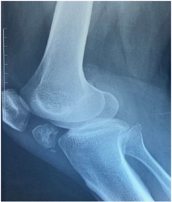
Figure 1: A lateral X-ray knee image showing an osseus mass located behind the silhouette of the patellar tendon.
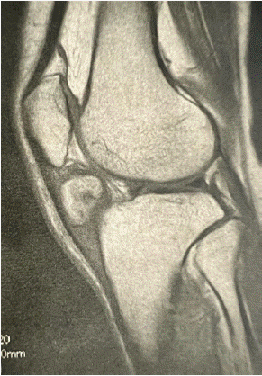
Figure 2: A T1 weighted sagittal MRI sequence showing a hyperintense mass approximatively 4 cm in wide axis Infront of the femoro-tibial joint.
With these findings a surgical approach was planned to extract the tumor, an anterior midline incision was used (Figure 3a). The mass was intracapsular but extra synovial and had completely replaced the Hoffa fat and the patellar tendon had been thinned (Figure 3b). The size of the mass was measured to be 04 cm and the color was red to yellowish from center to the surroundings (Figure 3c). On microscopic examination; the most outer surface of the mass had a thick mature cartilaginous tissue surrounded by granulated fibro-adipose areas. The center of the mass consisted mainly with well differentiated trabecular bone tissue with local hemosiderin pigmentations.
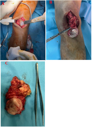
Figure 3: a: Direct transtendinous anterior medial incision, b: Dissection and excision of the mass and adherent Hoffa’s fat, c: A macroscopic appearance of the tumor.
After six weeks of postoperative follow-up, all symptoms related to Hoffa's disease disappeared, and the patient regained painless mobility of his left knee, hence a control MRI performed after one year cleared any local recurrence.
Discussion
Hoffa’s fat pad, is an intracapsular, extrasynovial structure located in the anterior knee compartment [2,3], and is richly vascularized and innervated. Inflammation and fibrosis within the IFP, due to trauma or surgery can lead to a variety of arthrofibrotic transformations including Hoffa’s disease, which is clinically represented as anterior knee pain that can often be reproduced on physical exam with maneuvers designed to produce impingement.
Our patient has described the pain as a burning located in the anterior compartment of his left knee exacerbated by the Hoffa physical test carried out with the patient in the supine position with hip and knee flexed to 90? consists in eliciting pain on palpation of the lateral and medial edges of the patellar ligament during knee extension.
At this point the differential diagnosis was difficult between Hoffa disease and other lesions of the patellar tendon, an X-ray using two diagonal incidences showed an osseous tumor taking place over the IFP.
MRI is the method of choice for analyzing Hoffa’s fat pad. The standard sequences (proton density with fat saturation and T1) are sufficient for the diagnosis of Hoffa’s disease and the most informative sections are the axial and sagittal views [4,5].
Many cases have reported with similar clinic, radiologic and pathologic fndings in the literature [6-9]. The relationship between the osteochondroma formation and Hoffa’s disease is not yet clear. Wickham et al. demonstrated the derivation of multipotent stromal cells from the IFP and they manage to illustrate that the Hoffa fat of the adult knee contains progenitor cells that have the ability to differentiate into chondrocytes, osteoblasts or adipocytes under appropriate culture conditions [10] Although this differentiation has been experienced in vitro, it still needs to clarify the etiology and pathogenesis of the osteochondroma development in Hoffa’s disease by in vivo studies.
Regarding treatment at this stage, Authors agree on surgical excision, which can be open or arthroscopic. [11,12] All the authors recommend carrying out anatomo-cytopathological analyses. In cases of Hoffa's tumor invasion, the surgical approach is recommended, thus allowing excision in one piece [13]. This excision surgery, which remains a simple procedure regardless of the approach, guarantees good clinical and functional results [11]. However, it remains to be determined whether it is a definitive solution in the context of “Hoffa's disease”.
Medical treatment can be proceeded in the Hoffa’s disease like corticoid injections or anesthetics to manage the chronic pain [14], in case of failure of this approach, a surgical excision remains necessary.
Conclusion
Hoffa's disease is a rare disease whose pathophysiology remains unclear. pain remains the main symptom of revelation. MRI is the examination of choice to map the lesion and plan the surgical procedure, which remains the treatment of choice for this pathology.
Author Statements
Clinical Message
Our case was diagnosed late and its location close to the patellar tendon made the surgical procedure delicate but naturally necessary to overcome the pain.
References
- Krebs VE, Parker RD. Arthroscopic resection of an extra- synovial ossifying chondroma of the infrapatellar fat pad: end- stage HoVa’s disease? Arthroscopy. 1994; 10: 301-4.
- Biedert RM, Sanchis-Alfonso V. Sources of anterior knee pain. Clin Sports Med. 2002; 21: 335-47.
- Bohnsack M, Wilharm A, Hurschler C, Rühmann O, Stukenborg-Colsman C, Wirth CJ. Biomechanical and kinematic influences of a total infrapatellar fat pad re- section on the knee. Am J Sports Med. 2004; 32: 1873-80.
- Jacobson JA, Lenchik L, Ruhoy MK, et al. MR imaging of the infrapatellar fat pad of hoffa. Radiographics 1997; 17: 675-91.
- Saddik D, McNally EG, Richardson M. MRI of Hoffa’s fat pad. Skelet Radiol. 2004; 33: 433-44.
- Böstman O, Karaharju E, Heikkonen L, Holmström T. Ex- traskeletal ossifying chondroma in the knee. Acta Orthop Scand. 1985; 56: 87-9.
- Bansal M, Goldman AB, DiCarlo EF, McCormack R. Soft tissue chondromas: diagnosis and diVerential diagnosis. Skelet Radiol. 1993; 22: 309-15.
- Steiner GC, Meushar N, Norman A, Present D. Intracapsul- ar and paraarticular chondromas. Clin Orthop Relat Res. 1994; 303: 231-6.
- Sakai H, Tamai K, Iwamoto A, Saotome K. Para-articular chondroma and osteochondroma of the infrapatellar fat pad: a re- port of three cases. Int Orthop. 1999; 23: 114-7.
- Wickham MQ, Erickson GR, Gimble JM, Vail TP, Guilak F. Multipotent stromal cells derived from the infrapatellar fat pad of the knee. Clin Orthop Relat Res. 2003; 412: 196-212.
- Rooney A, Wahba AJ, Smith TO, Donell ST. The surgical treatment of anterior knee pain due to infrapatellar fat pad patho- logy: A systematic review. Orthop Traumatol Surg Res. 2015; 101: 469-75.
- Takahashi T, Kimura M, Ohsawa T, Yamaguchi N, Takeshita K. A case of infrapatellar fat pad ganglion of the knee. Open Orthop J. 2017; 11: 1142-6.
- Dean BJF, Reed DW, Matthews JJ, Pandit H, McNally E, Athanasou NA, et al. The management of solitary tumours of Hoffa’s fat pad. Knee. 2011; 18: 67-70.
- Duri Z, Aichroth P, Dowd G, Ware H. The fat pad and its relationship to anterior knee pain. Knee. 1997; 4: 227-36.