
Case Report
Austin J Orthopade & Rheumatol. 2024; 11(1): 1126.
Pseudo Carpal Tunnel Syndrome with a Specific Cause: A Report of 2 Cases
Abdelhay Rabah*; Oussama Achahbar; Sbihi Mohammed; M Bousaidane; Y Benyas; J Boukhris; D Bencheba; B Chafry
Department of Orthopedic A Traumatological Surgery, Mohamed V Military Hospital, Rabat-Morocco
*Corresponding author: Abdelhay Raba Department of Orthopedic A Traumatological Surgery, Mohamed V Military Hospital, Rabat-Morocco Email: rabahabdelhay7@gmail.com
Received: November 28, 2023 Accepted: January 01, 2024 Published: January 08, 2024
Introduction
Lipomas, common benign tumors of soft tissues, are particularly rare in the hand, accounting for only 5% of upper limb locations [1]. The manifestation of a giant palmar lipoma, compressing the median nerve, is an exceptional occurrence. This rarity emphasizes the importance of Magnetic Resonance Imaging (MRI) to characterize the lipomatous nature of the lesion and clarify its relationship with palmar vasculonervous structures. Simultaneously, histology plays a crucial role in ruling out differential diagnoses, especially the most dreaded liposarcoma.
The duality between the rarity of the location in the hand and the potential for nerve complications makes palmar lipoma a unique and complex study subject. In this regard, we report two cases of lipomas with a specific digito-palmar location, presenting with dysesthesias and elective motor disturbances in the territory of the median nerve.
Clinical Observations
Case 1
A 60-year-old woman with a history of type 2 diabetes presents with a subcutaneous mass in the right palmar compartment that has been evolving and progressively increasing in size over the past 3 months (Figure 1). The patient maintains a general state of health. Associated with acroparesthesias in the first three fingers, the physical examination reveals a firm and painless mass in the thenar eminence, with a positive Tinel test.
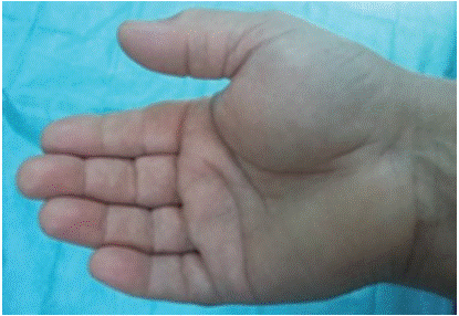
Figure 1: Clinical aspect of giant lipoma of the hand.
The standard radiograph (Figure 2) showed soft tissue opacity in the thenar compartment without bone lysis. Electromyography revealed sensory latency in the area of distribution of the median nerve.
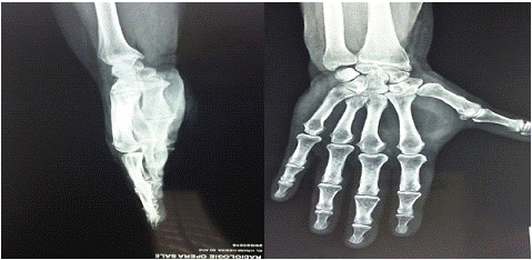
Figure 2: X-ray of the hand showed opacity of the soft parts of the thenar compartment.
The MRI (Figure 3) revealed a giant lipoma in the thenar eminence measuring 5.4cm×2.4cm×4.6 cm, extending into the carpal tunnel and compressing the flexor tendons and the median nerve.
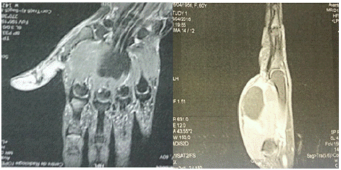
Figure 3: Giant lipoma of the hand on MRI.
The surgical treatment involved an initial biopsy followed by the resection of the lipomatous mass (Figure 4). Histopathological examination confirmed the diagnosis of a benign lipoma.
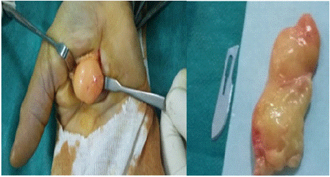
Figure 4: intraoperative aspect of giant lipoma.
Case 2
This concerns a 59-year-old patient with no significant medical history. The onset of clinical symptoms dates back ten years with the gradual development of a mass in the region of the right thenar eminence (Figure 5). One year ago, the patient began experiencing digital dysesthesias in the first two fingers, along with a decrease in pinch strength between the thumb and index finger. The examination revealed a firm, painless mass adherent to the deep plane, not pulsatile, prominent in the region of the first digital commissure, extending onto the eminence. On a symmetrical and bilateral examination, distal sensitivity in the 1st and 2nd fingers was reduced, without motor deficit, especially in the abduction, adduction, opposition, flexion, and extension of the thumb.
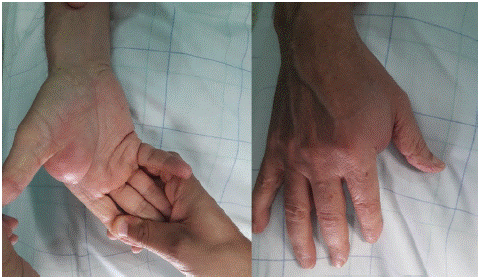
Figure 5: Clinical Aspect of the Thenar Mass in the Right Hand.
The standard X-ray showed the shadow of the soft tissue tumor, without calcification or bone involvement.
MRI revealed a fatty tumor process occupying the palmar compartment with extensions into the interosseous spaces and the thenar eminence in the submuscular region. It appeared hyperintense in T1-weighted images and hypointense in all sequences using fat signal suppression techniques. The tumor measured 112mm×82mm×27mm in size (Figure 6), and there was no contrast enhancement within the fatty tissue after intravenous injection of Gadolinium.
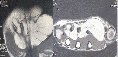
Figure 6: MRI of the hand showing a fatty tumor process occupying the palmar compartment with extensions into the interosseous spaces and the thenar eminence.
A preliminary biopsy was performed, concluding a benign conventional lipoma without cellular atypia. The patient underwent surgery under a plexus block and pneumatic tourniquet, through a longitudinal approach following the thumb opposition crease (Figure 7).
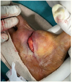
Figure 7: Incision along the thumb opposition crease.
The exploration revealed an encapsulated subaponeurotic fatty tumor, displacing the superficial palmar arch and the initial terminal branches of the lateral branch of the median nerve without invading them. It was adjacent to the distal edge of the Anterior Carpal Ligament (ACL), leading to the designation of a pseudo-carpal tunnel syndrome. The excision was complete but challenging after meticulous dissection of various elements, with the decision to section the ACL (Figure 8).
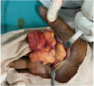
Figure 8: Macroscopic view of the giant arborescent lipoma during excision.
The macroscopic examination of the excised specimen (11cm×08cm×2.5cm) describes a benign lipoma with a homogeneous yellowish fatty appearance (Figure 9). Furthermore, microscopic analysis confirmed the diagnosis of a benign conventional lipoma without cellular atypia.
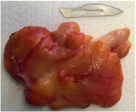
Figure 9: Homogeneous yellowish macroscopic appearance of the benign lipoma.
Both patients underwent functional rehabilitation sessions, and with a follow-up of ten months, the course was marked by complete recovery of distal sensory-motor disorders without clinically detectable local recurrence.
Discussion
A lipoma is a benign tumor composed of lobules of mature adipose cells. In the palm of the hand, it can be supra-aponeurotic and exceptionally intramuscular [2]. Giant lipomas are defined by a size exceeding 5cm, with few reports of giant lipomas in this location [3]. The first publication in 1956 documented 17 cases [4]. Depending on its location, it can cause compression of the interosseous nerve in the forearm, carpal tunnel syndrome, compression of the ulnar nerve in Guyon's canal, or digital nerve compression [5]. For our two patients, the compression involved the lateral trunk of the median nerve and its first two terminal branches. In the literature, median nerve compression is rare for small tumors, usually observed with tumors adhering to the nerve or its branches [4]. Magnetic Resonance Imaging (MRI) remains the preferred examination for studying soft tissue tumors due to its high sensitivity. This diagnostic tool helps specify the nature of the lesion, evaluate its local extension, and analyze its relationships with vasculonervous elements. The distinctive visual feature of a lipoma is a well-defined hyperintense image on T1 and T2 sequences, with a signal decrease during fat suppression sequences [6]. Additionally, in some cases, the image may show fibrous septa or calcifications. The administration of gadolinium leads to a moderate enhancement of the septa signal, while the fat signal remains unchanged [5]. Thus, due to its high sensitivity, MRI remains the gold standard for a comprehensive assessment of soft tissue tumors.
Histologically [7], Giant lipomas, considered critical size, raise concerns about their potential for malignancy. In order to differentiate benign lipomas from liposarcomas and other soft tissue neoplasms, it is imperative to perform a biopsy. Well-differentiated liposarcoma constitutes a riskier differential diagnosis for patients, with a peak incidence between 50 and 70 years of age. It appears from subcutaneous fat or cellular spaces, sometimes even from a pre-existing or recurrent lipoma. Marginal excision represents the treatment of choice. Dissection and identification of neurovascular elements must be performed with caution to avoid iatrogenic injury. Recurrences are extremely limited [8] and are generally caused by incomplete excision of the tumor.
Conclusion
The giant lipoma of the hand, due to its rarity both in terms of its size and its location, often presents signs of nerve damage. A precise diagnostic approach, using MRI and prior biopsy, is crucial to exclude the possibility of liposarcoma before resection; highlighting its favorable prognosis after successful resection.
References
- Amar MF, Benjelloun H, Ammoumri O, Marzouki A, Mernissi FZ, Boutayeb F. Anatomical snuff box lipoma causing nervouscompression: a case report. Ann Chir Plast Esthet. 2012; 57: 409-11.
- Higgs PE, Young VL, Schuster R, Weeks PM. Giant lipomas of the hand and forarm. South Med J. 1999; 86: 887-90.
- Huntley JS, McEachan J. Giant lipoma of the forearm. Hosp Med. 2004; 65: 758-9.
- Boussouga M, Bousselmame N, Lazrak KH. Lipome compressif de la loge thénar. A propos d’une observation. Chir Main. 2006; 25: 156-8.
- Flores LP, Carneiro JZ. Peripheral nerve compression secondary to adjacent lipomas. Surg Neurol. 2007; 67: 258-62; discussion 262.
- Bancroft LW, Kransdorf MJ, Peterson JJ, O’Connor MI. Benign fatty tumors:classification, clinical course, imaging appearance, and treatment. Skelet Radiol. 2006; 35: 719-33.
- Laurino L, Furlanetto A, Orvieto E, Dei Tos AP. Welldifferentiated liposarcoma (atypical lipomatous tumors). Semin Diagn Pathol. 2001; 18: 258-62.
- Balakrishnan C, Nanavati D, Balakrishnan A, Pane T. Giant lipomas of the upper extremity: case reports and a literature review. Can J Plast Surg. 2012; 20: e40-1.