
Original Article
Austin J Orthopade & Rheumatol. 2024 ; 11(1) : 1127.
Results of Locked Intramedullary Nail Application in Lower Extremity Long Bone Diaphyseal Fractures Due to High-Velocity Firearm Injuries
Namik Kemal Kilinccioglu, MD* Vedat Uruc, MD
Department of Orthopaedics and Traumatology, Faculty Medicine, Mustafa University, Tayfur Sokmen Kampusu, Turkey
*Corresponding author: Namik Kemal Kilinccioglu Mustafa Kemal University, Faculty of Medicine, Department of Orthopaedics and Traumatology, Tayfur Sökmen Kampüsü, Alahan-Antakya/Antakya/Hatay, 31060, Turkey. Tel: +90-(0)5309290713 Email: Nkkilincoglu@gmail.com
Received: February 02, 2024 Accepted: March 22, 2024 Published: March 29, 2024
Abstract
Background: The aim of this study is to determine the effects of early intramedullary nail fixation in long bone fractures such as femur and tibia that occur after firearm injuries on union and the developed complications.
Methods: In this study, a total of 82 patients who had open fractures due to firearm injuries between the years 2008 and 2020, and who were treated with intramedullary nail fixation in a single session upon their presentation to us, and followed up subsequently, were evaluated. Of the 82 major bone fractures, 48 were femoral and 34 were tibial diaphyseal fractures. These patients were examined in terms of union time, non-union, and the development of infections.
Results: Out of the evaluated 48 femoral fracture cases, infection occurred in 5 cases, with osteomyelitis developing in 2 of these patients. The average time to union was 19.2 weeks. Non-union was observed in three cases, and delayed union in 6 cases. Among the 34 tibial open fracture cases, infections of varying degrees were observed in 6 cases, with osteomyelitis developing in 1 of them. The average time to union was 22 weeks. Non-union was observed in three cases, and delayed union in 5 cases.
Conclusions: In cases of large bone open diaphyseal fractures due to firearm injuries, it is widely accepted to perform initial debridement and temporary fixation with external fixation, followed by permanent intramedullary nail application after soft tissue healing. In selected cases, we believe that with antibiotic prophylaxis and meticulous debridement in the first session, internal fixation with intramedullary nails can be applied.
Keywords: Firearm injury; Open fracture; Intramedullary nail; Internal fixation
Introduction
Various treatment modalities have been described for long bone diaphyseal fractures. However, there is no consensus on the treatment of open fractures, especially those associated with high-velocity firearm injuries. Intramedullary nail application is a preferred method due to its advantages such as shorter healing time, allowing early mobilization, and reducing the length of hospital stay.
The development of infection is the most feared complication of this treatment method. There is currently insufficient data in the literature regarding intramedullary nail application in long bone diaphyseal fractures caused by high-velocity firearm injuries. In this study, we evaluated a cohort of 82 patients with femur and tibia diaphyseal fractures following high-velocity firearm injuries during the Syrian civil war, who underwent primary intramedullary nail application. We compared our findings with the existing data in the literature.
Material and Methods
The study was initiated after obtaining approval from the Mustafa Kemal University Ethics Committee. A total of 82 patients with Gustilo Anderson type 3a open femur and tibia shaft fractures secondary to high-velocity firearm injuries during the Syrian civil war were included in the study. Subtrochanteric, intertrochanteric, supracondylar, and Gustilo Anderson type 3b fractures were not included in the study.
Upon arrival at the emergency department, patients had their open wounds cleaned with Betadine, sterile dressings were applied, and they were immobilized with a splint. Intravenous administration of a combination of first-generation cephalosporin and aminoglycoside was initiated for prophylaxis and continued for 72 hours.
Immediately upon arrival, patients underwent wound irrigation in the emergency department, followed by sterile dressing and splinting. Antibiotic prophylaxis was initiated, and radiological evaluations were performed. The diagnosis was confirmed with full-length anteroposterior and lateral X-rays, including one joint above and below the fracture. Informed written consent was obtained from all patients for intramedullary nail application. Patients were taken to surgery under spinal anesthesia. Antegrade locked intramedullary nails were used in the operations. Debridement was performed along the bullet tract, and cultures were obtained. The piriform fossa was reamed, and a guide wire was placed in the medullary canal. The guide wire was advanced into the medulla of the distal fragment after crossing the fracture line under fluoroscopic guidance. Subsequently, reaming was performed, and a locked intramedullary nail was inserted. Static locking was performed proximally and distally under fluoroscopic guidance.
In the postoperative period, patients received parenteral analgesic and anti-inflammatory treatment, with narcotic analgesics as needed. On the first day after surgery, knee bending and ankle exercises were initiated. Patients whose general condition allowed were mobilized on the first postoperative day, not fully weight-bearing, and walked with crutches. Daily dressing changes were performed, and patients were scheduled for follow-up appointments on the 11th day for suture removal and at the 3rd and 6th weeks for assessment of healing, joint movements, and weight-bearing.
Results
Between 2008 and 2019, a total of 82 patients who presented to the Hatay Mustafa Kemal University Faculty of Medicine Research Hospital Orthopedics and Traumatology Clinic due to gunshot injuries and completed their treatment were retrospectively evaluated. All 82 patients, who were injured during the Syrian civil war, were male. Out of these patients, 48 had femoral shaft fractures, and 34 had tibial shaft fractures, all of whom underwent intramedullary nail application.
The average age of the 48 patients with femoral fractures was 31.5. The average healing time was 19.2 weeks (ranging from 15 to 44 weeks). Ten patients started walking with full weight-bearing after the 3rd postoperative week, 30 patients after the 6th postoperative week, and the rest started later. In five patients, infections developed in the postoperative period (10.41%). Two of them (4.1%) were superficial infections characterized by discharge and erythema of the wound edges, starting within the first 2 weeks postoperatively. Infections resolved after debridement. Deep infection developed in the remaining three patients (6.25%). In one case, the infection improved after debridement, but in two cases (4.1%), the discharge became chronic, leading to osteomyelitis (Figure 1-4).
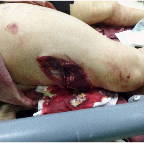
Figure 1: Femur firearm wound.
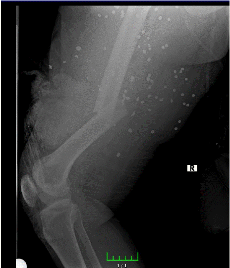
Figure 2: Preoperative x-ray.
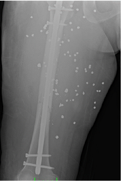
Figure 3: Early postoperative x ray.
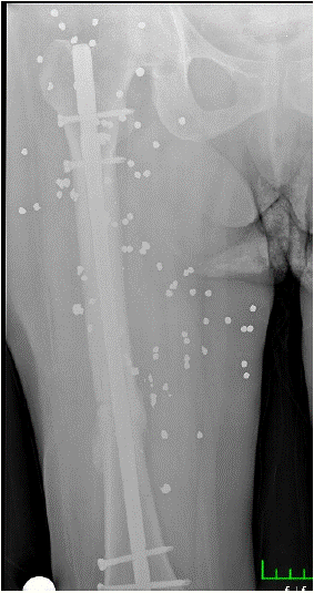
Figure 4: 24 weeks after x ray.
Three patients (6.25%) experienced non-union. One of them had osteomyelitis, and union was achieved after pseudoarthrosis surgery. Delayed union was observed in six patients (12.5%). Union was achieved by dynamization in four patients. In the other two cases, delayed union was associated with deep infection, and union was achieved after infection treatment. Shortening of the extremity occurred in three patients (6.25%) (2 patients: 2 cm, 1 patient: 1 cm) (Tabe 1).
    Complications

    Number


    nonunion

    3 patient (%6.25)


    Delayed union

    6 patient (%12.5)


    infection

    5 patient (%10.41)


    Shortening

    3 cm (%6.25)
Table 1: Complications of femur interlocking nail.
Of the thirty-four patients with tibial fractures, the average age was 34.8, and the mean follow-up duration was 24.2 months. Among the 32 patients who achieved union, the average healing time was 22 weeks (ranging from 15 to 40 weeks). Delayed union was observed in five patients (14.7%) (28-40 weeks), and union was achieved in two patients by distal dynamization. In the other three patients, delayed union was associated with deep infection, and union was achieved after the resolution of infection (Figure 5-9).
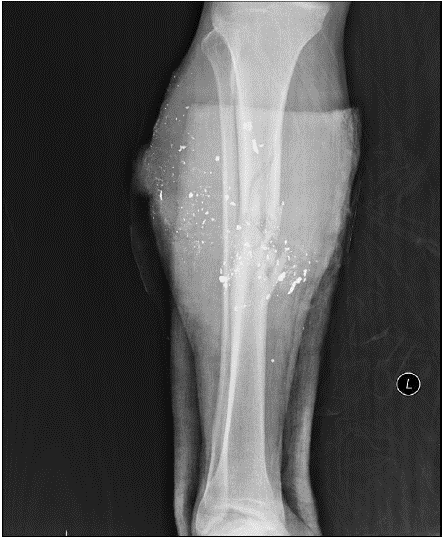
Figure 5: Preoperative tibia fracture x-ray.
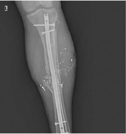
Figure 6: Early postoperative anteroposterior.
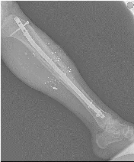
Figure 7: Early postoperative lateral x-ray.
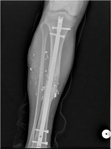
Figure 8: 24 weeks after postoperative anteroposterior x-ray.
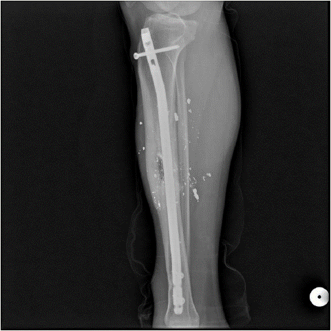
Figure 9: 24 weeks after lateral x-ray
A total of six patients (17.6%) experienced infections. Early superficial infection developed in three patients (8.8%) (within the first 2 weeks postoperatively), and these patients were treated with debridement and two weeks of intravenous antibiotics. Deep infection occurred in three patients (8.8%) 2-4 months after the initial surgery. In two patients, infection resolved after debridement and antibiotic treatment. In one patient (2.9%), osteomyelitis developed, and the implants were removed.
Eight patients started walking with full weight-bearing at the end of the 3rd postoperative week, 21 patients at the end of the 6th postoperative week, and the remaining patients were gradually mobilized later due to additional traumas.
Non-union was observed in two patients (5.88%), and union was achieved after pseudoarthrosis surgery. Shortening occurred in three patients (8.8%) (1.5 cm in two patients, 1 cm in one patient)" (Table 2).
    Complications

    Number


    Nonunion

    2 patient (%5.88)


    Delayed nonunion

    5 patient (%14.7 )


    Infection

    6 patient (%17.6)


    Shortening

    3 cm (%8.8)
Table 2: Complications of tibia interlocking nail.
Discussion
Fractures caused by firearms, especially those resulting in significant open bone fractures, pose a challenging case group for orthopedic specialists and should be considered as a health issue that requires careful attention. Both the increase in individual armament and the wars worldwide have led orthopedists to increasingly encounter firearm injuries. The primary goals in the case of large bone fractures caused by firearms, as in other open fractures, are to prevent infection, ensure functional healing of the fracture, and restore the functional alignment of the affected limb [1,2].
As with all fractures, the choice between external fixation, plate and screw osteosynthesis, and intramedullary nailing in firearm injuries depends on factors such as the type of fracture, the condition of soft tissue, the localization of the fracture, the presence of accompanying traumas, and the experience of the treating physician [3-5].
The use of intramedullary nails in complex femur fractures after firearm injuries has been considered since the 1940s, citing reasons such as non-union and malunion [6,7]. Hollman and his team applied intramedullary nails in a single stage to 19 femur fractures developed after low-velocity firearm injuries, calculating the average healing time as 4.5 months (20 weeks). Non-union occurred in one patient (5.2%), and delayed union occurred in one patient (5.2%) [8]. In a study of 68 patients with femur fractures resulting from high-velocity firearm injuries treated with locked intramedullary nails, conducted by Mian Amjad Ali and his team, the average healing time was reported as 24 weeks, and non-union was identified in four cases (5.88%) [9]. In our study, we applied intramedullary nails to 48 femur shaft fractures, mostly resulting from high-velocity firearm injuries seen in the emergency department, and calculated the average healing time as 19.2 weeks.
Six of our cases (12.5%) experienced delayed union. In two of these cases, we attributed non-union to the development of infection and achieved healing after treating the infection. In the other four femur fractures, we obtained healing by dynamizing the distal screws.
Three cases (6.25%) showed no union. Osteomyelitis was present in one of these three cases. We achieved healing in these three patients by applying pseudoarthrosis surgery.
On the other hand, Hollmann et al. mentioned the development of infection in 1 (5.2%) femur fracture case treated with intramedullary nailing [8]. In Bergman and colleagues' series of 65 femur fractures treated with intramedullary nails after firearm injuries, two cases (3%) developed serous drainage that regressed with 2-3 weeks of antibiotic therapy [5]. Tornetta reported no deep infection in femur shaft fractures treated with intramedullary nailing after firearm injuries, but cellulitis developed in one patient [10]. However, all these authors mainly examined femur fractures resulting from low-velocity firearm injuries in their studies. All the patients in our study were those who presented to our emergency department with femur diaphysis fractures resulting from high-velocity firearm injuries, most of them being wounded in the Syrian civil war.
In Mian Amjad Ali et al.'s series, infection developed in 7 cases (10.29%), with three (4.4%) being superficial and the other four (5.88%) being deep infections [9]. In our study, we evaluated 48 femur diaphysis fractures resulting from firearm injuries. Almost all patients were low self-care individuals living in refugee camps, wounded in the Syrian civil war. The etiology of all cases in our study was long-barreled firearm injuries. We detected superficial infection in 2 cases (4.1%) and deep infection in 3 cases (6.25%). After debridement and antibiotic therapy, infection regressed in four patients. However, chronic osteomyelitis developed in 2 patients (4.1%).
The tibia, due to its thinner soft tissue coverage compared to other bones, is more prone to open fractures. This condition leads to a high rate of infection and problems in wound healing.
In the literature, we did not come across a study on intramedullary nailing of tibia fractures after high-velocity firearm injuries. However, we evaluated and compared our series with four studies that assessed the use of reamed intramedullary nails in open tibia fractures [11-14]. In these studies, there were a total of 187 tibia fractures, 43% of which were type 3 open fractures. The union rate was 97%, the rate of deep infection was approximately 6.4%, the rate of chronic osteomyelitis was 0.75%, malunion was observed in 6.1% of cases, and implant failure was found to be 3%. In our study, we applied reamed intramedullary nails to a total of 34 tibia open fractures resulting from high-velocity firearm injuries. We found the average healing time to be 22 weeks. Delayed union was present in five cases (14.7%), and we achieved union by dynamizing the distal screws in two of them. In the other three cases, delayed union was associated with deep infection, and union was achieved in these cases at an average of 33.2 weeks. Non-union was identified in two cases (5.88%). We found the total infection rate to be 17.6% (6 patients). Three of these (8.8%) were superficial infections that did not progress to the fascia, and three (8.8%) were deep infections. In five patients, infection regressed after debridement and antibiotic therapy. In one patient (2.9%), osteomyelitis developed.
While external fixation followed by internal fixation appears to be the first choice in long bone fractures due to firearm injuries, intramedullary nailing in a single session can be considered as an alternative. We believe that careful debridement and antibiotic therapy, considering the overall condition of the individual and the degree of damage to soft tissues, can be applied with meticulous attention in the use of intramedullary nails in femur and tibia diaphysis fractures resulting from high-velocity firearms.
Conclusion
1. There are very few studies in the literature that investigate patients with open fractures resulting from high-velocity firearm injuries who underwent early definitive intramedullary nail placement. Our results align with the limited existing literature in terms of infection and non-union outcomes.
2 The use of external fixators appears to be initially considered as a primary treatment for large bone fractures resulting from high-velocity firearm injuries. However, several publications highlight the disadvantages of using external fixators for definitive treatment, such as the need for a second operation, frequent occurrences of pin-site infections, hindrance to early weight-bearing, and susceptibility to rotational and angular issues. Therefore, in selected cases of open fractures resulting from high-velocity firearm injuries with advanced soft tissue damage, after careful debridement and antibiotic therapy, and if the patient's overall condition permits, early definitive intramedullary nail placement can be considered as an alternative.
References
- Uzar AI, Dakak M, Oner K, Atesalp AS, Yigit T, Ozer T, et al. Comparison of soft tissue and bone injuries caused by handgun or rifle bullets: an experimental study. Acta Orthop Traumatol Turc. 2003; 37: 261-7.
- Necmioglu NS, Subasi M, Kayikçi C. [Minimally invasive plate osteosynthesis in the treatment of femur fractures due to gunshot injuries]. Acta Orthop Traumatol Turc. 2005; 39: 142-9.
- Lavine L, Grodzinsky A. Electrical stimulation of repair of bone. JBJS. 1987; 69: 626-30.
- Nicholas R, McCoy G. Immediate intramedullary nailing of femoral shaft fractures due to gunshots. Injury. 1995; 26: 257-9.
- Bergman M, Tornetta P, Kerina M, Sandhu H, Simon G, Deysine G, et al. Femur fractures caused by gunshots: treatment by immediate reamed intramedullary nailing. Journal of Trauma and Acute Care Surgery. 1993; 34: 783-5.
- BRAV EA. Further evaluation of the use of intramedullary nailing in the treatment of gunshot fractures of the extremities. JBJS. 1957; 39: 513-53.
- Carr CR, Turnipseed D. Experiences with intramedullary fixation of compound femoral fractures in war wounds. JBJS. 1953; 35: 153-71.
- Hollmann MW, Horowitz M. Femoral fractures secondary to low velocity missiles: treatment with delayed intramedullary fixation. J Orthop Trauma. 1990; 4: 64-9.
- Ali MA, Hussain SA, Khan MS. Evaluation of results of interlocking nails in femur fractures due to high velocity gunshot injuries. J Ayub Med Coll Abbottabad. 2008; 20: 16-9.
- Tornetta 3rd P, Tiburzi D. Anterograde interlocked nailing of distal femoral fractures after gunshot wounds. Journal of orthopaedic trauma. 1994; 8: 220-7.
- Court-Brown C, McQueen M, Quaba A, Christie J. Locked intramedullary nailing of open tibial fractures. The Journal of bone and joint surgery British volume. 1991; 73: 959-64.
- Kaltenecker G, Wruhs O, Quaicoe S. Lower infection rate after interlocking nailing in open fractures of femur and tibia. Journal of Trauma and Acute Care Surgery. 1990; 30: 474-9.
- Robinson C, McLauchlan G, Christie J, McQueen M, Court-Brown C. Tibial fractures with bone loss treated by primary reamed intramedullary nailing. The Journal of Bone and Joint Surgery British volume. 1995; 77: 906-13.
- Keating J, O’brien P, Blachut P, Meek R, Broekhuyse H. Locking intramedullary nailing with and without reaming for open fractures of the tibial shaft. A prospective, randomized study. JBJS. 1997; 79: 334-41.