
Case Report
Austin J Orthopade & Rheumatol. 2024 ; 11(1) : 1128.
Surgery for a Chondroma Complicated by a Pathological Fracture: Urgent or Deferred?
Badaoui R*; Ould Ghwagha O; Rachdi A; Boussaidan M; Benyass Y; Boukhriss J; Benchaba D; Chafry B
Department of Traumatologie and Orthopedic II, Military Hospital Mohammed V, Faculty of Medicine and Pharmacy of Rabat, University Mohammed V Rabat, Morocco
*Corresponding author: Badaoui R Department of Traumatologie and Orthopedic II, Military Hospital Mohammed V, Rabat, Morocco. Email: joudbadaoui17@gmail.com
Received: March 25, 2024 Accepted: April 25, 2024 Published: May 02, 2024
Abstract
Chondroma is a common primary tumor in the hand, affecting mainly the phalanges and metacarpals. The fracture is its most frequent circumstance of discovery. This represents a double drama for the patient, and double problem for the orthopedic surgeon. The treatment, consisting of filling curettage, is carried out after consolidation of the fracture. We report the experience of a case taken care of early with curettage filling protected by internal osteosynthesis by mini-plate, after fracture of the second phalanx.
The objective of this study is to report the result of this treatment allowing early rehabilitation.
Keywords: Chondroma; Pathological fracture; Early surgery; Osteosynthesis
Introduction
It is the most common benign bone tumor of the hand that develops from cartilage. It sits in the metaphyseal area and can be discovered during a hand fracture at the level of the tumor.
The tumor is most often asymptomatic and incidentally discovered during a blunt trauma to the hand (fracture, 30% according to Professor Tomeno). Avulsion of the flexor digitorum profundus may occur if localized to the 3rd phalanx. It is rare or part of a family disease to have a deforming tumor visible under the skin. Treatment may consist of simple immobilization and monitoring in case of fracture. Indeed, the cortical fracture can make the tumor disappear.
Case Presentation
It is a 35-year-old patient, with no medical history, victim a year ago of a minimal trauma at the left hand, causing her excruciating ring finger pain, which motivated her consultation. The patient underwent a front and profile X-ray exploration, which objectified a fracture, pathological in appearance, on an ovary gap, of the P2 of the ring finger (Figure 1). The MRI objectified a metaphyso-diaphyseal lesion in hyposignal T1, hypersignal T2 with cortical lysis and calcification in hyposignal (Figure 2).
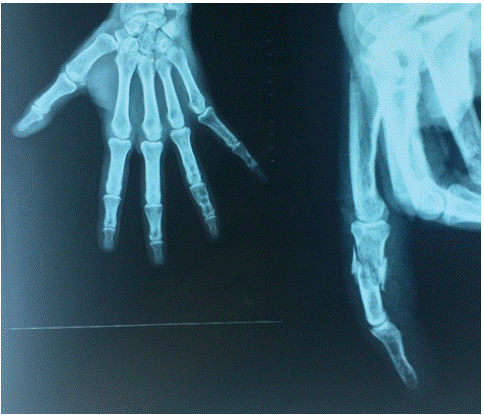
Figure 1: X-Ray of hand showing a P2 ring fracture on a lytic image.
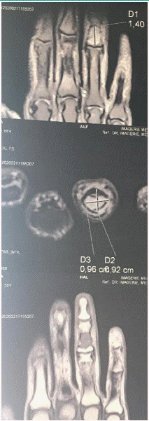
Figure 2: The MRI objectified a metaphyso-diaphyseal lesion in hyposignal T1, hypersignal T2 with cortical lysis.
The patient benefited from an external surgical intervention, where a curettage-filling by cortico-spongy grafts of the iliac bone, was carried out in the first (Figure 3 & 4), then stabilization of the fracture focus by mini plate (Figure 5).
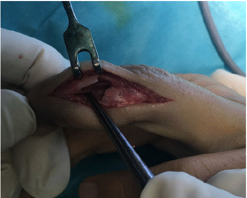
Figure 3: Curettage of the chondroma.
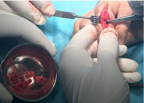
Figure 4: Filling after curettage of the chondroma by Cortico-spongy grafts.
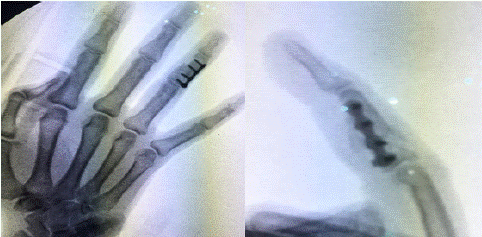
Figure 5: X-Ray of hand showing osteosynthesis of the fracture by mini- plate after chondroma treatment.
The rehabilitation was started immediately after the operation. The mobilities have recovered. The fracture was consolidated at 3 months. After an 18-month follow-up, there was no recurrence, no reduction in joint amplitudes, and no fracture.
Discussion
The chondroma corresponds to an intramedullary proliferation of mature hyaline cartilage in the metaphyso-diaphyseal regions of bones with enchondral ossification. It is a common tumor, accounting for almost 3% of all bone tumors and between 12 and 24% of benign bone tumors.
It is a tumor of the young patient with a variable age of discovery, between 10 and 40 years for Dahlin [1,2]. Chondroma is the most common tumor of the phalanges of the hands. In decreasing order of frequency, it sits in the proximal phalanx, the metacarpal bone, the intermediate phalanx and the distal phalanx, with a preference for the ulnar edge of the hand [3].
The chondroma is asymptomatic, it is the pathological fracture which is the most frequent mode of revelation since it is found in almost a third of cases, and often secondary to minimal trauma [4,5]. The management of this fracture and the underlying tumor still poses a chronological problem, most authors prefer the consolidation of the fracture then treatment of the tumor [6,7], believing that the complications are less frequent in this case [8]. But in reality, the period of disability will be greater, as well as the psychological repercussions on the patient [9,10].
Conclusion
This study shows the interest of surgical treatment with filling curettage, protected by internal osteosynthesis by mini-plate in the case of fracture on bone chondroma of the phalanges. The use of mini-plates allows early rehabilitation by stabilizing the fracture and allowing early rehabilitation preventing tendon adhesions which is responsible for stiffness.
References
- Marty FL, Marteau E, Rosset P, Faizon G, Laulan J. retrospective study of the series of 623 tumors of the hand and wrist in adults. Hand chir. 2010; 29: 183–187.
- Dalhin. General aspects and date on 8542 cases. Bone Tumors 1986; 4: 33-51.
- Takka S, Poyraz A. Enchondroma of the scaphoid bone. Arch Orthop Trauma Surg. 2002; 122: 369-370.
- Jacobson ME, Ruff ME. Solitary enchondroma of the phalanx. J Hand Surg Am. 2011; 36: 1845–7.
- Shimizu K, Kotoura Y, Nishijima N, Nakamura T. Enchondroma of the distal phalanxof the hand. J Bone Joint Surg Am. 1997; 79: 898–900.
- Marco RA, Gitelis S, Brebach GT, Healey JH. Cartilage tumors: evaluation and treatment. J Am Acad Orthop Surg. 2000; 8: 292-304.
- Ablove RH, Moy OJ, Peimer CA, Wheeler DR. Early versus delayed treatment of enchondroma. Am J Orthop. 2000; 29: 771-2.
- Yan H, He J, Chen S, Yu S, Fan C. Intrawound application of vancomycin reduces wound infection after open release of post-traumatic stiff elbows: a retrospective comparative study. J Shoulder Elbow Surg. 2014; 23: 686–92.
- Yalcinkaya M, Akman YE, Bagatur AE. Recurrent metacarpal enchondroma treated with strut allograft: 14-year follow-up. Orthopedics. 2015; 38: e647–50.
- Ablove RH, Moy OJ, Peimer CA, Wheeler DR. Early versus delayed treatment of enchondroma. Am J Orthop (Belle Mead NJ). 2000; 29: 771–772.