Case Report
Austin J Orthopade & Rheumatol. 2015; 2(3): 1020.
3 Cases of Genu Valgum in Medically Treated X-linked Hypophosphatemic Rickets
Stevens NM* and Hennrikus WL
Department of Orthopedics and Rehabilitation, Hershey Medical Center, USA
*Corresponding author: Stevens NM, Department of Orthopedics and Rehabilitation, Hershey Medical Center, 30 Hope Dr, Hershey, PA 17033, USA
Received: July 02, 2015; Accepted: September 29, 2015; Published: October 10, 2015
Abstract
X-linked hypophosphatemic rickets is a disorder of phosphate wasting in the kidney resulting in abnormal bone matrix. The classic presentation of rickets is a child with genu varum (bowed legs). The author present 3 cases of medically treated x-linked hypophosphatemic rickets presenting with atypical genu valgum (knock knees).
Keywords: Orthopaedics; Musculoskeletal; Phosphate; Calcitriol
Introduction
X-linked hypophosphatemic rickets is primarily a disorder of phosphate wasting in the kidney with secondary changes to bone. It has an estimated incidence of 1 in 20,000 individuals and is the most common form of hereditary rickets [1]. The most common mutations in these individuals are in the PHEX gene [2]. This gene is primarily expressed on the cell membrane of osteoblasts, cells which lay down bone matrix. Mutations in PHEX prevent cleavage of FGF23 (fibroblast growth factor 23) causing an increased serum level of FGF23 [3]. FGF23 then inhibits phosphate reabsorption in the kidney leading to hypophosphatemia and subsequent bone mineralization defects [3,4].
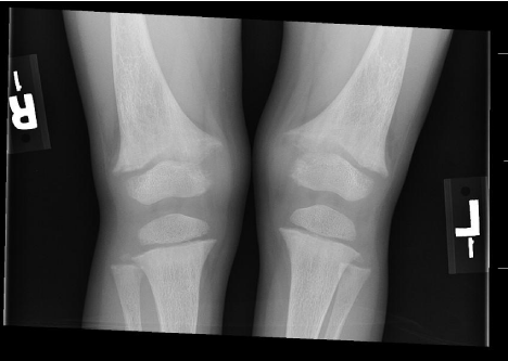
Children typically present with knee pain, bow legs and short stature. Laboratory evaluation reveals hypophosphatemia, phosphaturia, nearly normal calcium and PTH levels, and low/ normal vitamin D. Radiographs demonstrate characteristic changes in the bone, including widening of the physis and flaring of the metaphysic (Figure 1). If biopsy is performed, histology reveals distinctivehypomineralizedperiosteocytic lesions [3]. Bony abnormalities seen in untreated patients include progressive bowing, anteromedial rotational torsion of tibiae and persistent short stature [5]. Other features of the disease include dental abscesses, early tooth decay, craniosynostosis and hypertension.

    Figure 1: Radiograph of a 3 year old (Patient 2) with x-linked hypophosphatemic
rickets displaying the characteristic femoral and tibialmetaphyseal flaring and
fraying with widening of the physis.
It is particularly important in childhood for these patients to have close medical follow up with a nephrologist. Dosing of phosphate and calcitriol is altered frequently during growth. With perfect medical management, most of the clinical features of x-linked hypophosphatemia will resolve. The orthopedic surgeon is often consulted due to deformity of the lower limbs. In most cases, the deformity is genu varum; to our knowledge, this case series is the first report of genu valgum in x-linked rickets.
Case Report
Our first patient was a 16 month old female who presented to nephrology for failure to thrive. She had a strong family history of x-linked hypophosphatemic rickets. She was subsequently diagnosed and prescribed a regimen of phosphate replacement and calcitriol. In her course of follow up, the nephrologist began to notice increasing genu valgum. Orthopedics was consulted at age6 years.
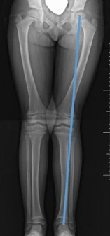
On presentation to orthopedics, genu valgus positioning of her legs was noted (Figure 2). Yearly follow up was recommended. Exam at age 7years revealed 13 degrees of genu valgum and 8 cm between her medial malleoli. By age 8 years, the intramalleolar distance had lessened to 1 cm. At age 9 years, only 3 degrees of genu valgum was noted. No orthopedic intervention was indicated.

    Figure 2: Radiograph of patient 1 at age 6 years displaying hips located and
bilateral genu valgus alignment of the femorotibial angle. This is atypical, as
rickets patients classically present with genu varus. Metaphyseal flaring can
also be seen.
Our second patient is the sister of the first patient. She was diagnosed at age 3 months and was followed by nephrology. She presented to orthopedics at age 4 years; genu valgus was noted. At age 6 years her valgus had progressed to 12 degrees, with 5 cm between her medial malleoli. At age 7 years her genu valgum had improved, with only 3 cm between her ankles. At age 8 years, her genu valgum was only 3 degrees. No orthopedic intervention was indicated.
Our third patient is unrelated to the other two. She was diagnosed at age 7.5 months and began medical treatment but was less compliant. When orthopedics was consulted at age 7 years, the exam revealed genu valgum, with 13 cm measured between her medial malleoli. She is currently followed by orthopedics on an annual basis.
Discussion
Genu varum is the most common skeletal abnormality in untreated x-linked hypophosphatemic rickets, but as demonstrated in the current case series, genu valgus may develop in medically managed x-linked hypophosphatemia. The valgus alignment may be a reflection of rickets’ propensity to augment the physiologic progression of the femoral-tibial angle during growth.
The Heuter-Volkmann principle suggests that the rate of epiphyseal growth is determined by the pressure applied at the growth plate; increased pressure inhibits growth while decreased pressure accelerates it. This can be seen as part of normal development of the femoral-tibial angle. During infancy, the legs display up to 15 degrees of physiologic bowing. Spontaneous correction of varus typically occurs by age 2.5 years. Between ages 3 and 4 years, a child will go through a period of genu valgus up to maximal angle of about 12 degrees. At about age 6, the genu valgum typically lessens to about 5 degrees, and this mild valgus alignment then persists through adulthood.
Patients with untreated rickets will have deformation of the lower extremity starting in infancy, during the period of bowing. Some hypothesize that the unmineralized bone found in rickets may be more susceptible to deformation. The result of this susceptibility is a greater degree of genu varum. If left untreated, the deformity may become permanent. In contrast, in a medically managed hypophosphatemic rickets patient, the addition of phosphate and vitamin D will strengthen the bone, minimize the classic varus deformation, and as in the current cases, reverse the varus to valgus angulations.
Patients with untreated rickets will have deformation of the lower extremity starting in infancy, during the period of bowing. Some hypothesize that the unmineralized bone found in rickets may be more susceptible to deformation. The result of this susceptibility is a greater degree of genu varum. If left untreated, the deformity may become permanent. In contrast, in a medically managed hypophosphatemic rickets patient, the addition of phosphate and vitamin D will strengthen the bone, minimize the classic varus deformation, and as in the current cases, reverse the varus to valgus angulations.
However, radiographic changes still persist in treated rickets patients’ bones [1,4].This suggests that the bone is abnormal, and may be more susceptible to physiologic stress. If true, then during the period of physiologic genu valgum, treated rickets patients may display a greater degree of genu valgus than typically seen in the normal population.
In 1993, Heath and Staheli determined that the normal range of genu valgum in white children age 2-11 years was up to 12 degrees or an intramalleolar distance up to 8 cm [2,6]. All of our patients are at the upper limit or above this range; which suggests that their bones may be more susceptible to applied force. All of the patients in the current series were successfully treated with phosphate and vitamin D3 by the nephrologist and none were indicated for orthopaedic surgery. Insufficient medical management may result in persistent deformity that may require hemi-epiphyseodesis or osteotomy.
In the current series, all three patients with medically treated x-linked hypophosphatemic rickets had a peak in genu valgum at ages 6-7 years--a few years after the typical peak age of genu valgum of 4-5 years typically seen in normal children [7-10]. The degree of disease control may correlate with the amount of genu valgum. For example, the third patient in this series was less compliant with medical treatment and developed a more significant genu valgum with 13 cm between malleoli at age 7 years. This patient is now under close follow up and treatment by the nephrologist and we will continue to follow the patient on an annual basis-- hopefully until the genu valgum corrects. Previous research indicates that early and tight medical optimization improves the alignment of lower extremities in rickets patients [1,4].
Conclusion
Treatment of x-linked hypophosphatemia with phosphate and vitamin D3manages but does not completely cure rickets. Even in well treated patients, radiographic evidence of disease may persist, suggesting that bone morphology never returns to normal. Children with rickets are susceptible to lower limb deformity deformation at any age. In the current case series, an increase in the physiologic femoral-tibial valgus angle occurred in each child during a period growth. In two out of three cases, with appropriate medical therapy, the excess valgus angular deformity of the legs resolved without surgical treatment. The third case of X-linked hypophosphatemic rickets is currently under tight medical management and will be followed carefully for any need for later surgical correction.
References
- Santos F, Fuente R, Mejia N, Mantecon L, Gil-Pe&nTilde;a H, Ordo&nTilde;ez FA. Hypophosphatemia and growth. Pediatr Nephrol. 2013; 28: 595-603.
- A gene (PEX) with homologies to endopeptidases is mutated in patients with X-linked hypophosphatemic rickets. The HYP Consortium. Nat Genet. 1995; 11: 130-136.
- Pettifor JM. What’s new in hypophosphataemic rickets? Eur J Pediatr. 2008; 167: 493-499.
- Razzaque MS, Lanske B. The emerging role of the fibroblast growth factor- 23-klotho axis in renal regulation of phosphate homeostasis. J Endocrinol. 2007; 194: 1-10.
- Carpenter TO, Imel EA, Holm IA, Jan de Beur SM, Insogna KL. A clinician’s guide to X-linked hypophosphatemia. J Bone Miner Res. 2011; 26: 1381- 1388.
- Staheli LT. The Lower Limb. Lovell and winter’s Pediatric Orthopaedics, 3rd ed. Philadelphia, PA: J.B Lippincott Co; 1990: 741-753.
- Mäkitie O, Doria A, Kooh SW, Cole WG, Daneman A, Sochett E. Early treatment improves growth and biochemical and radiographic outcome in X-linked hypophosphatemic rickets. J Clin Endocrinol Metab. 2003; 88: 3591- 3597.
- Heath CH, Staheli LT. Normal limits of knee angle in white children--genu varum and genu valgum. J Pediatr Orthop. 1993; 13: 259-262.
- Sochett E, Doria AS, Henriques F, Kooh SW, Daneman A, Mäkitie O. Growth and metabolic control during puberty in girls with X-linked hypophosphataemic rickets. Horm Res. 2004; 61: 252-256.
- McNair SL, Stickler GB. Growth in familial hypophosphatemic vitamin-Dresistant rickets. N Engl J Med. 1969; 281: 512-516.