
Review Article
Austin J Orthopade & Rheumatol. 2015; 2(3): 1021.
Intra-oral Scanners: A New Eye in Dentistry
Baheti MJ1*, Soni UN1, Gharat NV1, Mahagaonkar P2, Khokhani R2 and Dash S1
1Institute of Pravara Medical Sciences, Department of Orthodontics & Dentofacial Orthopedics, India
2Institute of Pravara Medical Sciences, Department of Prosthodontics, India
*Corresponding author: Baheti MJ, Institute of Pravara Medical Sciences, Department of Orthodontics & Dentofacial Orthopedics, oni – 413736, Maharashtra, India
Received: August 18, 2015; Accepted: November 01, 2015; Published: November 17, 2015
Abstract
Today, intra-oral mapping technology is one of the most exciting new areas in dentistry since three-dimensional scanning of the mouth is required in a large number of procedures in dentistry such as orthodontics and restorative dentistry. Since the introduction of the first dental impressioning digital scanner in the 1980’s, development engineers at a number of companies have enhanced the technologies and created in-office scanners that are increasingly user-friendly. It offers the clinician the ability to offer patients different appliances and fixed restorations of all types. The new technology has become easier to use for the clinician as well as more precise, and offers technological advances over earlier versions. These systems are capable of capturing 3D virtual images of tooth which can be used to create accurate master models on which the restorations can be made in a dental laboratory (dedicated impression scanning systems). The use of these products is rapidly increasing around the world and presents a paradigm shift in the way in which dental impressions are made. Several of the leading 3D dental digital scanning systems are presented and discussed in this article.
Keywords: Intra-oral scanner; Software; Record keeping
Introduction
The fast and continuous advances in computer sciences have resulted in increased usage of new technologies in all levels of modern society and orthodontics is not an exception to it. Paperless office is now a reality and although the transition has been slow, it has been steady [1]. Many orthodontists are joining other health professionals in using paperless patient information systems [2]. The digital Era in the orthodontics is moving at lightning speed. Digital radiographs and digital photography are becoming the norm in orthodontic records. Recent advances have now included E-study models and now the intra orally scanned study models. Orthodontists use computers for record keeping, practice management, patient education, and communication with colleagues, restorations fabrication, and many other tasks. Computers have become a necessity rather than an option.
“Impression” has different meaning in life but in dentistry, impression is negative form of teeth or other tissues of the oral cavity [3]. Impression has its importance in various aspects of dentistry like in orthodontics especially; to perform various procedures like diagnosis, model analysis, appliance fabrication, impression has to be made using different materials and techniques. Both in orthodontics and restorative area (Prosthodontics and restorative dentistry in particular), the use of plaster models are not only essential but routine practice in these clinical specialties. It has long been every dentist’s desire to be able to scan plaster models, or even patients’ teeth directly in the mouth [4]. Avoiding discomfort, speeding up work, improving communication between colleagues and prosthetic labs, and reducing the physical space needed for storing these models, are some of the alleged benefits of this technology.
The replacement of alginate and Polyvinyl Siloxane (PVS) impressions with intraoral digital scanners represents a paradigm shift in orthodontics. First introduced as an outsourced technology for storage of three-dimensional electronic study models, the digital scanner has evolved into an in-office tool with a variety of applications [5]. Using this technology, orthodontists can more accurately and efficiently fabricate clear aligners, custom braces, indirect-bonding trays, and laboratory appliances without the unpleasant experience of conventional impressions. The aim of this article is to provide an extensive review of the existing intraoral scanners for orthodontics with particular attention to the technical aspects and applications of digital impressions in dentistry, with emphasis on orthodontics.
Evolution of digital impression systems
The concept of taking impressions to make models, from which appliances could be constructed, goes back to the early 18th century [6,7]. It was not until 1856, when Dr. Charles Stent [6] perfected an impression material for use in the fabrication of the device that bears his name for the correction of oral deformities, that documentation exists of the use of an impression material other than beeswax or plaster of Paris, which had inherent problems, respectively, of distortion or difficulty of use, for creating an oral prosthesis [8].
The widely used techniques currently employed for obtaining elastomeric impressions and for creating gypsum models from those impressions have only been in use since 1937, when Sears [9] introduced agar as an impression material for crown preparations. The first elastomeric material specifically produced for the purpose of dental impression-making was Impregum™, a polyether material introduced by ESPE, GmbH in 1965. In the mere 77 years that elastic impression materials have been in use, numerous formulations have been developed, all of which have exhibited particular shortcomings in the goal of obtaining precise reproduction of the oral structures. Condensation cure silicone impression materials subsequently were developed, but these also suffered from problems with dimensional accuracy. The creation of addition silicone vinyl poly siloxane impression materials solved the issues of dimensional inaccuracy, poor taste and odor, and high modulus of elasticity, and offered excellent tear strength, superior flow ability, and lack of distortion even if models were not poured quickly.
Many dentists are reluctant to embrace the new technologies because they simply believe elastomeric impression materials and techniques have been in use for so long and work so well that they are irreplaceable. Or else, those 3D digital scanning technologies are so recent that they are not yet ready for clinical use. Actually, impression taking using elastomers, for all its inherent problems, has been used in dentistry for 77 years. Digital impression and scanning systems were introduced in dentistry in the mid-1980s and have evolved to such an extent that some authors predict that in five years most dentists in the U.S. and Europe will be using digital scanners for impression taking [10].
The advent of intraoral digital scanners coincided with the development of Computer-Aided Design and Manufacturing (CAD/CAM) technology and the 1984 introduction of Chairside Economical Restoration of Esthetic Ceramics (CEREC).The first system for the dental office was CEREC 1 in 1986. The system was developed by Prof. Dr. Werner Moermann in Switzerland and was eventually licensed to what today is Sirona Dental Systems 2. The Cerec 2 and subsequent Cerec 3 as well as the eventual the Cerec 3D system replaced the original technology in 1994, 2000 and 2003 respectively [11,12]. In 2001, Cadent introduced the Ortho CAD* system for the production of 3D digital models, virtual setups, and indirect-bonding trays. The Lava™ Chairside Oral Scanner (C.O.S.) was created at Bronte’s Technologies in Lexington, Massachusetts, and was acquired by 3M ESPE (St. Paul, MN) in October 2006. In 2006, Cadent developed the in-office iTero* digital impression system, which by 2008 was capable of full-arch intraoral scanning (Figure 1 ); in late 2009, Cadent launched the iOC* system for iTero users. The E4D Dentist system, introduced by D4D Technologies LLC (Richardson, TX) in early 2008. Align Technology purchased Cadent in 2011, allowing clinicians with iOC to begin submitting 3D digital scans in place of physical impressions for the fabrication of Invisalign appliances.
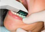
Figure 1: Intraoral digital scanner in use.
In October 2012, 3M ESPE introduced the True Definition*scanner, enabling orthodontists to submit digital scans for Incognito* custom lingual braces. Six months later, Ormco released the Lythos* digital impression system for its Insignia* and Clear guide* appliance systems. In January 2014, after much demand for further interoperability between manufacturers, the True Definition scanner qualified for Invisalign case submission, so that True Definition scans could now be used for Invisalign submission and iTero scans for the Incognito appliance system. Despite the growing popularity of intraoral digital scanners, questions remain regarding their applications and the differences among manufacturers.
Advantages of digital scanning
For the orthodontist, advantages of digital scanning include improved diagnosis and treatment planning, increased case acceptance, faster records submission to laboratories and insurance providers, fewer retakes, reduced chair time, standardization of office procedures, reduced storage requirements, faster laboratory return, improved appliance accuracy, enhanced workflow, lower inventory expense, and reduced treatment times. Benefits to the patient include an improved case presentation and a better orthodontic experience with more comfort and less anxiety, reduced chair time, and easier refabrication of lost or broken appliances, as well as potentially reduced treatment time.
Scanning technology
Digital intraoral scanners are considered Class I medical electrical devices, designed and constructed in accordance with the standards of ANSI/IEC 60601-1. Every scanner has three major components: a wireless mobile workstation to support data entry; a computer monitor to enter prescriptions, approve scans, and review digital files; and a handheld camera wand to collect the scan data in the patient’s mouth. To gather surface data points, energy from either laser light or white light is projected from the wand onto an object and reflected back to a sensor or camera within the wand. Based on algorithms, tens or hundreds of thousands of measurements are taken per inch, resulting in a 3D representation of the object’s shape. The technology used by the wand to capture surface data determines the measurement speed, resolution, and accuracy of the scanner. Four types of imaging technology are currently employed
i) Triangulation (Figure 2A).
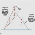
Figure 2a: Triangulation.
ii) Parallel confocal imaging (Figure 2B).
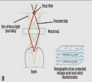
Figure 2b: Parallel confocal imaging.
iii) Accordion fringe interferometry (AFI) (Figure 2C).
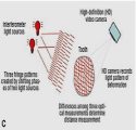
Figure 2c: Accordion fringe interferometry (AFI).
iv) Three-dimensional in-motion video (Figure 2D).
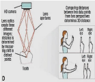
Figure 2d: Three-dimensional in-motion video.
Dedicated digital impression systems
Dedicated digital impression systems eliminate several cumbersome dental office tasks, such as selecting trays, preparing and using materials, disinfecting impressions and sending impressions to the lab. Moreover, lab time is reduced by not having to pour up plaster, place pins and replicas, cut and shape dies or articulate models.
With these systems, final restorations are produced in models created from digitally scanned data instead of plaster models made from physical impressions. Additionally, they enhance patient comfort; improve patient acceptance and understanding of the case. Digital scans can be stored on hard disks indefinitely, while conventional models, which can break or chip, must be physically stored, which requires additional office space. The Seven existing intra-oral scanning devices for orthodontics are listed below:
i) iTero
ii) Lava C.O.S.
iii) Care stream Dental’s CS 3500
iv)The True Definition Scanner
v) 3 Shape’s TRIOS
Vi) Cirona’s CEREC and Apollo System
vii) The Lythos Scanner
i) iTero: The iTero™ digital impression system (Cadent, Carlstadt, NJ) was introduced in early 2007, following 5 years of intensive research and beta testing. Based on the theory of “parallel confocal,” the iTero scanner emits a beam of light through a small hole, and any surface within a certain distance will reflect the light back toward the wand. Because the iTero software uses an open-source file type known as Landlord stl, its standard triangulation language (STL) files are compatible with the Invisalign, Harmony,*Incognito, Insignia, and Sure Smile† systems [13].
The iTero system includes a computer, monitor, mouse, integrated keyboard, foot pedal, and scanning wand organized on a well-designed mobile cart (Figure 3). Disinfection consists of replacing the disposable sleeve on the handheld scanner. The iTero wand is relatively large and bulky, weighing about 1.5lb, but has a central notched those positions the operator’s hand at the optimal balance point. Single-use [13] disposable sleeves fit over the scanning end of the wand to protect the laser light source and prevent crosscontamination. The scanner sleeve is also useful as a retractor for cheeks, tongue, and soft tissues. The parallel confocal technology allows the data capture wand to rest directly on the tooth surface, which helps steady and lighten the wand. It is important not to contact the soft tissues, however, as this could cause discomfort. The patient does not need to open the mouth wide and may even feel comfortable gently biting on the wand tip [13].
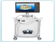
Figure 3: The iTero 3D digital impression system.
Scanning is done by quadrants, starting in the lower left buccal quadrant at the most distal molar. The wand is held at a 45° angle to the gingival margin, so that it will capture both the buccal and the occlusal landscape, and moved medially one tooth at a time, with an overlap between consecutive scans. The sequence is repeated on the lingual side, beginning with the lower left lingual quadrant. Although a full-mouth scan and bite registration takes 10-15 minutes, Align has indicated that this time will be reduced in the near future.
ii) Lava C.O.S: The Lava™ Chairside Oral Scanner (C.O.S.) was born out of the research of Professor Doug Hart and Dr. János Rohály at the Massachusetts Institute of Technology. The Lava C.O.S. was created at Bronte’s Technologies Inc (Lexington, MA) and was acquired by 3M ESPE (St. Paul, MN) in October 2006. The product was launched officially at the Chicago Midwinter Meeting in February 2008.
The method used for capturing 3D impressions involves Active Wave Front Sampling (AWS), which enables a 3D-in-Motion technique. By using AWS, however, the LAVA C.O.S. captures scanned images quickly (approximately twenty 3D data sets per second, or close to 2,400 data sets per arch) in video mode and creates a highly accurate virtual on-screen model instantaneously [14].
The Lava C.O.S. unit consists of a mobile cart (Figure 4) containing a computer, a touch screen monitor, and a scanning wand (Figure 4), which has a 13.2-mm wide tip and weighs 14 oz (about the size of a large power toothbrush). The camera at the tip of the wand contains 192 Light-Emitting Diodes (LEDs) and 22 lenses. There is no need for a keyboard or mouse, as the monitor displays a keyboard for all data input. Disinfection involves a simple wipe down of the monitor with an intermediate-level surface disinfectant designed for use on nonporous surfaces and replacement of the plastic sheath on the wand.
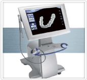
Figure 4: The Lava Chair side Oral Scanner (C.O.S.).
The Lava C.O.S. allows capturing 3D data in a video sequence and models the data in real time. After the preparation of the tooth and gingival retraction, the entire arch is dried and lightly dusted with powder to locate reference points for the scanner. During the scan, a pulsating blue light emanates from the wand head and an on-screen image of the teeth appears instantaneously. The “stripe scanning” is completed as the dentist returns to scanning the occlusal of the starting tooth. The dentist is able to rotate and magnify the view on the screen, and can also switch from the 3D image to a 2D view. The patient is then instructed to close into the Maximum Intercuspal Position (MIP), the buccal surfaces on one side of the mouth are powdered, and a scan of the occluding teeth is captured. The maxillary and mandibular scans are then digitally articulated on the screen. Then the dentist sends the data via internet to the laboratory technician, who employs customized software to digitally cut the die and mark the margin. 3M ESPE receives the digital file, generates a Stereo Lithography (SLA) model and sends it to the laboratory.
iii) Carestream dental’s CS 3500: At the American Association of Orthodontists (AAO) Annual Session in May, Atlanta-based Care stream Dental showcased its CS 3500 intraoral scanner. The CS 3500 is designed to be completely portable, requiring no trolley system to move from chair to chair (Figure 5). The wand can be plugged into any laptop via USB 2 connection. The CS 3500 is designed to give orthodontists the ability to scan a patient’s teeth and acquire a color, full arch 3D image.
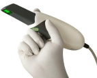
Figure 5: Care stream Dental’s CS 3500.
According to the company, the CS 3500 “offers high-angulations scanning up to 45 degrees and a depth of 16 mm, and it features a light guidance system that navigates users through the image-acquisition process.” The light guidance system allows users to look into the patient’s mouth during an image capture, rather than at the screen. It glows green when a scan has been successful and amber to indicate that rescanning is needed. A full impression takes about 5 minutes. Image accuracy measures up to 30 microns. The CS 3500 produces still image resolution of 1024 x 768 pixels and video resolution of 640 x 480 pixels.
The scanner requires no external heater to prevent fogging during image capture or powder for use. Measuring 9.6 x 1.5 x 2.4 inches and weighing 10 ounces, the wand features a slim head and an 8.2-foot cable. The wand can be fitted with different tip sizes [9].
According to the company, the CS 3500 can work as part of its CS Solutions CAD/CAM restoration portfolio or as a standalone system. It is compatible with open system CAD software, and with CS Connect-the company’s online portal-digital impressions can be sent to a lab directly.
iv) The true definition scanner: The 3M ESPE True Definition scanner, which uses the revolutionary 3D in-motion video imaging technology, is currently the only third party scanner that has been qualified for use with Invisalign’s (Figure 6). Similarly, Aligns iTero scanner is qualified for fabrication of the 3M Unitek Incognito system [5].
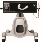
Figure 6: 3M ESPE True Definition Scanner.
The true definition setup comprises an HP‡ workstation with a 22” touch screen display, a lightweight wand, a powder dispenser, and a wireless Internet connection for uploading to the cloud. The unit weighs 73lb and, like iTero, is built on a rolling cart for easy transport. The large touch screen display eliminates the need for a horizontal surface with a keyboard and mouse, resulting in a slimmer workstation. It also serves as a virtual digital loop, enabling the operator to view, rotate, and magnify the 3D model to scrutinize the scan before approving the digital prescription. Once the scan has been sent, the clinician can log in to Invisalign’s doctor site and access the patient’s scan in a few moments.
Because a light coating of contrast powder is required prior to scanning, moisture control is critical. One technique is to place driangles into the cheek before positioning a cheek retractor; if a Nola†† retractor is used; a high-speed suction adaptor can be added to allow simultaneous use of the saliva ejector. The teeth are then lightly dusted with powder by means of the handheld dispenser connected to the work station.
The True Definition scanner seamlessly captures data in real time. The operator simply traverses the anatomy with the hand piece and observes as the video 3D mesh is accumulated. During the scan, the wand is kept about 10mm (recommended 3-17mm) from the tooth surface. The clinician may choose from a number of scanning paths, but most begin on the posterior occlusal surface of the first premolar. Scanning is divided into sextants, moving from lingual to buccal and finishing back on the occlusal. This process is repeated in the anterior sextant, starting with the first premolar to ensure an overlap of at least one tooth.
v) Ormco’s lythos scanner: Ormco Corp, Orange, Calif, launched the Lythos™ Digital Impressions System at the AAO Annual Session in May. Lythos uses AFI Technology to capture data in real time, rather than post-process stitching; meaning an entire high-resolution, dualarch scan can be completed in approximately 7 minutes.
Lythos features an open platform format-using STL files, which allow data to be easily shared with orthodontic labs and appliance manufacturers to produce a variety of custom appliances and study models. Digital impressions for every patient can be stored on Ormco’s cloud for up to 10 years through ormcodigital.com. Lythos charges no click fees or storage and management fees. Additionally, Lythos includes Ormco’s digi caste models, an online portal for digital study models that helps eliminate the cost of physical model storage.
The system is compatible with Insignia™ Advanced Smile Design™, Clear guide™ Express, and AOA lab. When Lythos scan data is used for Ormco and/or AOA products, practices earn rebates toward the payment of the impression system.
The device is portable, weighing less than 30 pounds, making transport between chairs or operatories easier. Lythos can be placed on a chairside table or on the ground. When on the ground, the screen can extend up to make the device 30 inches tall. When the screen is fully condensed, Lythos measures 15 inches tall. In addition, it features a touch screen monitor, as well as wireless Internet connectivity, which allows for patient data to be uploaded to the cloud where it can be accessed from any computer in the practice. Lythos is compatible with Apple™ products (Figure 7).
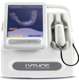
Figure 7: Lythos Scanner.
The Lythos wand is small, compact, and ergonomically designed, weighing 0.6 pounds. It features a small disposable tip. No powder coating is required for scanning.
vi) 3Shape’s TRIOS: This spring, 3Shape, Copenhagen, Denmark, unveiled the TRIOS® Color digital impression system. The intraoral scanning system features the company’s TRIOS Ultrafast Optical Sectioning Technology and Real Color Technology, creating scan images that have the appearance of real teeth and gingival. The system combines hundreds to thousands of 3D pictures to create the final 3D digital impression. TRIOS are designed to integrate with a practice’s current practice-management system and work with its established lab-order procedures. A full scan takes approximately 5 minutes [15].
TRIOS Color builds on the company’s TRIOS standard non color system. Both systems are available in TRIOS Cart or TRIOS Pod configurations. The TRIOS Pod allows for scanning with the TRIO handheld scanner and software using selected laptop PCs, giving users working in multiple locations or in a limited space more mobility and flexibility. With the TRIOS Pod configuration, users can control scanning from an iPad or mirror the 3D view on other displays in the practice, such as monitors integrated in the chair. The cart configuration features a Smart-Touch screen, allowing full control without the use of a trackball or mouse. In addition, the TRIOS Cart configuration is Wi-Fi and Bluetooth enabled, allowing for the cart to be moved throughout the office and for easy connectivity of thirdparty devices like wireless keyboards and cameras. The cart base dimensions measure 45 x 18 x 23 inches. The TRIOS Cart features an integrated training center and remote support (Figure 8).

Figure 8: 3Shape TRIOS Cart Solution.
The TRIOS system does not require the use of powder to conduct the scan. The wand has an autoclavable tip with anti-mist heater. The interchangeable tips can be flipped for upper and lower scanning.
TRIOS captures full arches and the occlusal situation as input for digital study models, treatment planning, and appliance design. The orthodontic configuration includes 3Shape’s Ortho Analyzer software, allowing practices to perform treatment simulations, virtual set-ups, and analyses on the scanned model. Scans are saved in the standard STL file format.
vii) Sirona’s CEREC and apollo systems: Sirona Dental Inc, Charlotte, NC, offers a full range of digital impression systems, including the CEREC® AC Connect with Omnicam, CEREC AC Connect with Bluecam, and Apollo DI.
Designed for the chair side, the CEREC AC with Omnicam produces full-color, 2D and 3D images and captures half-arch and full-arch impressions. Featuring Color Streaming technology, the CEREC Omnicam allows for continuous video capture of the oral cavity. Weighing 11 ounces, the hand piece has a rounded camera tube for easier rotation of the camera and a small camera tip. According to the company, the camera has an anti-shake feature and provides a uniform field of illumination. During scanning, the camera must be moved between 0 and 15 mm over the tooth surface. No powder or opaquing agent is required during the scan (Figure 9).
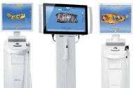
    Figure 9: Siron Dental’s CEREC Blue cam (left), Apollo DI (center), and
CEREC Omni cam (right).
Meanwhile, CEREC AC Connect with Bluecam features a blue light-emitting diode with a specific wavelength that is designed to capture better detail. Using a single image-acquisition technique to complete the scan, the CEREC Bluecam can be used on a single tooth or quadrant. A full jaw image is possible. The camera wand, which weighs approximately 9.5 ounces, must be placed directly on the teeth during use. A powder agent must be used during scanning.
Both CEREC Bluecam and Omnicam models can be used with CEREC Connect Software 4.2. Additionally, Sirona Connect is a free service-with no annual or incremental fees and unlimited usage-that can be utilized to send cases from the practice to a lab using Siron in Lab software. Once there, model information can be exported to a variety of software applications.
Rounding out the line of digital impression systems is the Apollo DI. Described as an “economical entry into the world of digital impressions,” the system includes an imaging unit, APOLLO DI software, and the APOLLO DI intraoral camera. Scans using the Apollo are black and white, but like the CEREC Omnicam, they are captured using continuous video imaging. The camera wand, which weighs approximately 3.5 ounces, is moved 2 to 20 mm over the tooth surface during scanning. Like the CEREC Bluecam, the Apollo DI requires a powder coating during scanning.
Conclusion
Intraoral digital scanners are becoming integral to the modern orthodontic office, improving both practice efficiency and the patient experience compared to conventional alginate and PVS impressions. By addressing the everyday dental office issues described above, digital impression taking, given its undeniable benefits, will transform digital intraoral scanning into a routine procedure in most dental offices in the coming years. Furthermore, digital impressions tend to reduce repeat visits and retreatment while increasing treatment effectiveness. Patients will benefit from more comfort and a much more pleasant experience in the dentist’s chair. But the only thing to worry is its cost.
With the popularization of digital systems, and the tremendous growth in two areas of dentistry that can potentially benefit from digital impression taking and digital models (orthodontics and dental implantology) one can confidently predict that in the coming years we will witness a true digital revolution in the dental office. A revolution that will benefit patients in terms of more efficient planning reduced discomfort and treatment efficiency.
References
- Lands B. The accuracy and reliability of plaster vs digital study models: A comparison of three different impression materials. University de Montréal. 2010.
- Stevens DR, Flores-Mir C, Nebbe B, Raboud DW, Heo G, Major PW. Validity, reliability, and reproducibility of plaster vs digital study models: Comparison of peer assessment rating and Bolton analysis and their constituen. Am J Orthod Dentofacial Orthop. 2006; 129: 794-803.
- Mistry GS, Borse A, Shetty OK, Tabassum R. Digital Impression System – Virtually Becoming a Reality. J Adv Med Dent Scie. 2014; 2: 56-63.
- Polido WD. Digital impressions and handling of digital models: The future of Dentistry. Dental Press J Orthod. 2010; 15: 18-22.
- Kravitz ND, Groth C, Jones EP, Graham JW, Redmond WR. Intraoral Digital Scanners. JCO. 2014; 48: 337-347.
- Guerini V. A History of Dentistry. 1909; 241-242: 305-306.
- Bremner MDK. The Story of Dentistry. New York & London, Dental Items of Interest Pub Co., Inc.; 1958.
- Ring ME. How a dentist’s name became a synonym for a life-saving device: the story of Dr. Charles Stent. J Hist Dent. 2001; 49: 77-80.
- Jacobson B. Taking the headache out of impressions. Dent Today. 2007; 26: 74-76.
- Birnbaum N, Aaronson HB, Stevens C, Cohen B. 3D digital scanners: A hightech approach to more accurate dental impressions. Inside Dentistry. 2009; 5.
- Puri S.
- Feuerstein P.
- Cadent debuts. “Next generation” iTero digital impression system. Implant Tribune. 2007; 1: 14.
- Helvey GA. The Current State of Digital Impressions. Inside Dentistry. 2009; 5.
- Doidge N. The Brain That Changes Itself. Penguin Books. 2007.