Research Article
Austin J Orthopade & Rheumatol. 2016; 3(2): 1032.
Clinical and Radiological Results of Radial Shortening Osteotomy versus Proximal Row Carpectomy in Kienböck’s Disease
Genç AS, Desteli EE and YunusImren*
1Department of Orthopedics and Traumatology, Samsun Gazi State Hospital, Turkey
2Department of Orthopedics and Traumatology, Usküdar State Hospital, Turkey
3YunusImren, MD, Department of Orthopaedics and Traumatology, Okmeydani Research and Training Hospital, Istanbul, Turkey
*Corresponding author: YunusImren, Okmeydani Training & Research Hospital, Istanbul, Turkey
Received: May 21, 2016; Accepted: July 19, 2016; Published: July 22, 2016
Abstract
In this prospective study, we aimed to evaluate the clinical and radiological results of our patients treated with Radial Shortening Osteotomy (RSO) and Proximal Row Carpectomy (PRC) together with a short review of the literature. The study included 35 patients with the diagnosis of Kienbock disease RSO was performed for 17 patients and 18 patients underwent PRC. 15 of the patients had Lichtman Stage 2, 14 patients had Stage 3A and 6 patients had Stage 3B disease. Q-DASH Score, Preoperative and postoperative Carpal Height Ratio (CHR), revised CHR, stahl index, radial inclination values were noted. Preoperative and postoperative flexion-extension range of Motion (ROM) and ulnar deviation angles were also obtained. Nakamura’s clinical evaluation system was performed to each patient. Results of clinical evaluation revealed significant progression at postoperative sixth month follow-up. Our results showed clinical improvement following surgeries of both RSO and PRC for Lichtman Stage 2, 3a and 3b disease. We consider that experience and technical familiarity of the surgeon is key factor to decide the type of the procedure to be performed.
Keywords: Kienböck’s disease; Proximal row carpectomy; Radial shortening; Lichtman’ classification
Introduction
Etiology of Kienbock disease remains unclear, this is mainly due to the rarity of the disease and lack of enough number of prospective studies. The treatment for Kienböck’s disease ranges from conservative modalities such as immobilisation to operative options such as radial shortening [1-4] ulnar lengthening [5] proximal row carpectomy [6] / silastic arthroplasty [7,8] intercarpal fusions [9] and revascularisation procedures [10,11].
Negative ulnar variance has been accepted as a predisposing factor to Kienbock disease, Hulten [12] in his study stated that in the case of negative ulnar variance, axial load on radial side of lunatum is increased which is a predisposing factor for lunatomalacia. Radial Shortening Osteotomy (RSO) is indicated in the case of negative ulnar variance as a joint leveling procedure [13-22]. Besides early stages of the disease, RSO has been found to be beneficial also in advanced stages [2,22]. Radial osteotomy has been found to increase vascularity of lunate and also made decompression [23-25]. Fragmantation and degenerative changes that occur particularly at proximal articular surface of lunate may ultimately lead collapse of the bone and the entire corpus. If carpal instability occurs in advanced disease, Proximal Row Carpectomy (PRC) can be considered [26]. PRC is a procedure used for the treatment of wrist arthritis, and has been reported to relieve pain and preserve wrist range of motion and grip strength [27-29]. Long-term outcomes of the most PRC and RSO procedures applied for treatment of Kienbock’s disease are satisfactory (Tables 1 & 2).
    Author
    Surgical method
    Number of patients
    Follow-up time
    Comments


    Liu et al. [58]
    PRC
    10
    10-29 years
    Satisfactory    results,reserved their previous work


    Lumsden et al. [44]
    PRC
    17
    15 years
    Good clinical    results


    Culp RW et al. [29]
    PRC
    20
    3.5 years
    Patients with mild    preoperative arthritic changes had better results


    Croog AS et al. [42]
    PRC
    21
    10 years
    Reliable procedure    for Lichtman Stage 3a and 3b


    De Smet L et al. [43]
    PRC
    21
    67 months
    Satisfactory    clinical results


    Ali MH et al. [51]
    PRC
    61
    19.8 years
    Unsatisfactory    results.Manyof the patients were unable to return to manual labor type    jobs.


    Lecamte F et al. [57]
    PRC
    25
    30 months
    Recommend for    Lichtman Stage 3 disease
Table 1: Long term follow-up results treated by proximal row carpectomy.
    Author
    Surgical method
    Number of patients
    Follow-up time
    Comments


    Matsui et al. [17]
    Radial shortening
    11
    10 yrs
    Satisfactory    clinical results


    Salmon et al. [18]
    Radial shortening
    15
    26 yrs
    Recommend RSO for    patients with carpal collapse accompanying severe pain


    Raven et al. [16]
    Radial shortening
    12
    22 yrs
    Recommend RSO for Lichtman Stage 3a or less disease with    a (-) ulnar variance


    Altay et al. [53]
    Radial shortening
    23
    7 yrs
    Reliable long-term    treatment for Lichtman Stage 3b disease


    Rodrigues- Pinto
    Radial shortening
    18
    10.3 yrs
    Effective for    Lichtman Stage 2,3a,3b disease.


    Axelsson et al. [19]
    Ulnar lengthening
    22
    22 yrs
    All patients    returned to work


    Trail et al. [5]
    Ulnar    lengthening and Radial shortening
    20
    11 yrs
    Satisfactory    clinical results


    Wada et al. [20]
    Radial wedge    osteotomy
    13
    14 yrs
    Good clinical    results


    Koh et al. [14]
    Radial wedge    osteotomy and Radial shortening
    25
    14.5 yrs
    Good clinical    results


    Zenzai et al. [15]
    Radial shortening    with or without ulnar lengthening
    35
    19 yrs
    Satisfactory    results,reliable method


    Viljakka et al. [13]
    Radial shortening
    16
    32 yrs
    Provides    decade-long improvement in 75% of patients


    Watanabe et al. [21]
    Radial shortening
    13
    21 yrs
    Satisfactory    clinical results
Table 2: Long term follow-up results treated by radial shortening osteotomy.
In this prospective study we aimed to evaluate the clinical and radiological results of patients treated with RSO and PRC.
Materials & Methods
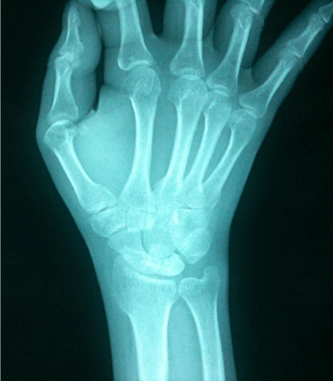
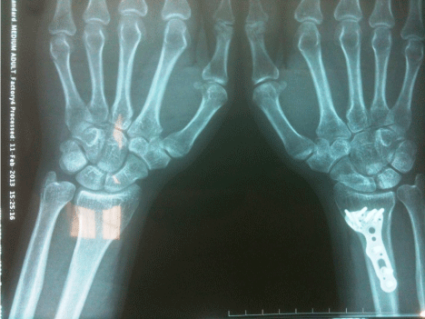
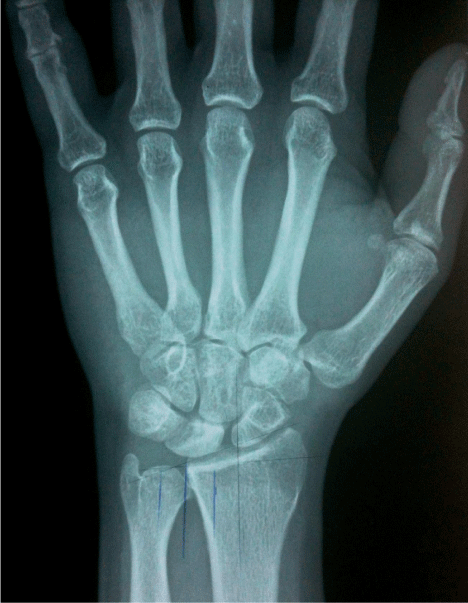
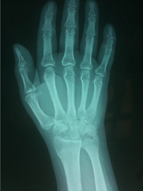
Approval was obtained from the local scientific department of our hospital and consent to study participation from all subjects for this prospective study. The study included 15 patients with the diagnosis of Kienbock disease who underwent operation between 2009 to 2012. RSO was performed for 17 patients (Group I), (Figures 1 & 2) and 18 patients underwent PRC (Group II) (Figures 3 & 4). Mean followup time was 18.4 months for Group I patients and 19.0 months for Group II patients. Mean age of Group I patients was 35 years and Group II patients was 41 years. 16 of the patients were male and 19 of them were female. 15 of 35 patients had dominant side disease and were operated from their dominant wrists. 14 of the 35 patients had a history of trauma (40%). According to Lichtman’s classification, [30] at the time of diagnosis, 15 of the patients had Stage 2, 14 patients had Stage 3A and 6 patients had Stage 3B disease. (Table 3) Palmer’s technique [31] was used to assess ulnar variance, according to this technique; 21 patients (60%) had a negative ulnar variance and 14 patients (40%) had a neutral ulnar variance.
    Stage
    Radial Shortening
    Proximal Row Carpectomy
    Overall


    1


    2
    15

    15


    3a
    2
    12
    14


    3b

    6
    6


    4


    Overall
    17
    18
    35
Table 3: Distribution of the patients according to Lichtman’s classification.
Surgical method for radial shortening
Surgery was performed through a volar approach; an incision was centered over the radial border of the FCR tendon. The tendon sheath was released and the FCR was retracted radially to protect the radial artery. The flexor pollicis longest end on was retracted to the ulnar side to protect the median nerve. The pronator quadratus was released from the radial border of the radius and retracted ulnarly. A forearm-shortening osteotomy plate was placed on the volar aspect of the radius (Figure 1). A locking screw was placed in the most distal screw hole, and a 3.5-mm nonlocking screw was placed in the reduction slot. The osteotomy was performed over the oblique lag screw hole. This area was marked on the radius with the plate in place. The plate was then removed. A transverse or oblique osteotomy to the distal radius was performed. The plate was then replaced and fixed to the radius. The most distal 3.5-mm locking screw was placed first. The radius was shortened, and the 3.5- mm nonlocking screw in the reduction slot was tightened. The remaining 3.5-mm locking screws were placed. Patients were casted until osteotomy site healed, at approximately 6 weeks.

Figure 1: Preoperative AP view before RSO.

Figure 2: Postoperative AP view after RSO (same patient).

Figure 3: Preoperative AP view before PRC.

Figure 4: Postoperative AP view after PRC (same patient).
Surgical method for proximal row carpectomy
A dorsal longitudinal incision was performed. Extensor retinaculum was divided over the fourth compartment. Extensor Digitorum Communis was retracted to the ulnar side. Extensor pollucis longus and wrist extensors were retracted to the radial side. Posterior interosseous nevre was resected, wrist capsule was opened longitudinally and lunate fossa and the articular surface of the capitate were exposed. Capsular incision was extended transversely to the either side to facilitate exposure of the scaphoid. Scaphoid was osteotomized in its mid-portion. After osteotomy of the scaphoid, its proximal pole was removed. Lunate was grasped with a towel clip and was excised. Triquetrum was removed in a similar fashion. Finally the distal pole of the scaphoid was removed. Passive flexion/extension of the hand was made in neutral and slight radial deviation to detect any impingement of the trapezium and the radial styloid. The wound was irrigated and capsule was closed and a suction drain was placed. At two weeks, the sutures were removed, and motion was started by a flexible splint.
Six months after the operation, patients were re-evaluated and modified Nakamura clinical scoring system [32] (Table 4) was used for clinical evaluation [3,4,33-38]. In Nakamura’s evaluation system, radiological evaluation makes 9 points in total. Because insufficient radiological evaluation would affect the final result, we excluded 9 points and thus total points were calculated [3]. Preoperative and postoperative angular examination was made using a goniometer. Range of motion (ROM) of wrist was noted while patient was in a sitting position and hands were put on the table. Preoperative and postoperative grip strength was examined by jamar hand dynamometer [39], dominant side was 5% decreased to equalize dominant and non-dominant sides. Grip strength was measured while shoulder was in adduction and neutral rotation, elbow at 90 degrees flexion, forearm and hand in neutral position. Test was repeated 3 times and means value was noted. Pinchmeter was used to evaluate pinch strength of 1st and 2nd fingers. Patients were in a sitting position and elbow in 90 degrees flexion. For upper extremity functional evaluation Q-DASH score [40] was used. Preoperative and postoperative Carpal Height Ratio (CHR), revised CHR, stahl index, radial inclination values were noted. Preoperative and postoperative flexion-extension Range of Motion (ROM) and ulnar deviation angles were also obtained.
    Clinical Assessment
    Points


    Pain in the wrist


    None
    10


    Mild with strenuos activity
    7


    Mild with light work
    4


    Grip strength (percentage of unaffected side)


    90%
    5


    80%
    4


    70%
    3


    60%
    2


    50%
    1


    Increase in range of flexion and extension


    >20°
    6


    10°- 19°
    5


    5°- 9°
    3


    Overall grade
    Total Points


    Excellent
    15-21


    Good
    14-Sep


    Fair
    8-Mar


    Poor
    0-2
Table 4: Content of modified Nakamura clinical scoring system.
SPSS for Windows programme 17.0 version was used for statistical analysis. Categoric comparisons were made using pearson chi square, fisher x2 and Yates x2 tests. Paired samples t-test and Wilcoxon test were used for preoperative and postoperative comparison.
Results
Results of clinical evaluation revealed significant progression at postoperative sixth month follow-up. Mean preoperative Jamarmeter value was 20.05±9.30 and postoperatively it was 29.80±14.50 for Group 1 and 17.70±4.40 and postoperatively it was 27.80±14.0 for Group 2, mean preoperative pinchmeter value was 7.05±1.70 and postoperatively it was 8.22±1.50 for Group 1 and for Group 2 preoperatively it was 4.95±1.15 and postoperative mean value was 6.30±0.90 (Table 5) Preoperative and postoperative mean flexion arc, extension arc and ulnardeviation values were found to be signicantly increased following both radial shortening osteotomy and proximal row carpectomy (p<005) (Table 6). Mean time of union for the osteotomy sites was 6.5 weeks in Group 1 patients. According to the results of radiological evaluation, carpal height ratio, revised carpal height ratio, stahl index and radial inclination did not change significantly following radial shortening osteotomy (Group 1) and Stahl index and Radial inclination values did not significantly change following proximal row carpectomy (Group 2) (p>0.05). However mean carpal height ratio and revised carpal height ratio values were found significantly different following proximal row carpectomy (p<0.05) (Table 7). Postoperative mean Q-DASH scores in Group1 and 2 patients were found to be 15.9 (4-29) and 28.4 (9-50) respectively preoperatively it was 56.3 (13-86) for Group 1 and 56.3 (25-72) for Group 2 (25-72).
    Pre-treatment Values
    Post-treatment Values
    P- Value


    Jamarmeter


    Group 1
    20.05±9.30
    29.80±14.50
    p<0.05


    Group 2
    17.70±4.40
    27.80±14.0
    p<0.05


    Pinchmeter


    Group 1
    7.05±1.70
    8.22±1.50
    p<0.05


    Group 2
    4.95±1.15
    6.30±0.90
    p<0.05
Table 5: Mean jamarmeter and pinchmeter values of both groups, pre- and
postoperatively.
    Pre-treatment (mean±SD)
    Post-treatment (mean±SD)
    P- Value


    Radial Shortening
    Range of flexion
    48.90±10.40
    65.70±6.80
    p<0.05


    Range of extension
    43.70±15.75
    59.60±6.80
    p<0.05


    Ulnar deviation
    35.30±9.40
    41.70±5.70
    p<0.05


    Proximal Row Carpectomy
    Range of flexion
    31.20±11.80
    34.80±6.40
    p<0.05


    Range of extension
    29.90±11.20
    31.80±9.80
    p<0.05


    Ulnar deviation
    29.90±11.20
    31.80±9.80
    p<0.05
Table 6: Preoperative and postoperative mean flexion arc, extension arc and ulnar deviation values of both groups.
    Pre-treatment mean±SD
    Post-treatment mean±SD
    P- Value


    Radial Shortening
    CHR
    0.50±0.05
    0.48±0.05
    0,674


    RCHR
    1.38± 0.10
    1.36± 0.12
    0,88


    SI
    0.45 ±0.10
    0.43 ±0.08
    0,58


    RI
    22.20± 3.90
    22.00 ±0.40
    0,59


    Proximal row carpectomy
    CHR
    0.48± 0.03
    0.26 ±0.05
    0,018


    RCHR
    1.30± 0.05
    1.02 ±0.05
    0,018


    SI
    0.30 ±0.04
    0.28± 0.02
    0,169


    RI
    22.40 ±3.00
    21.80± 0.05
    0,791
Table 7: Stahl index, Radial inclination, mean carpal height ratio and revised carpal height ratio values of both groups, pre- and postoperatively.
According to the results of Nakamura’s clinical evaluation system 4 patients in Group 1 had excellent result 57%) and 3 patients had good result (38%), among Group 2 patients 1 had excellent result, 3 had good results, 2 had fair results and 2 had poor results.
Discussion & Conclusion
There are several options for surgical treatment of Kienbock disease. Experience of the surgeon is key factor for decision of the type of surgery. There are conflicting results regarding the type of surgery performed for Kienbock disease. Prior studies have confirmed that consevatively treated patients had progression of the disease both clinically and radiologically [41]. Treatment method of choice at different stages of the disease still remains a debate. Some authors do not recommend radial shortening at advanced stages of lunatomalacia [39] this is due to the fact that osteotomy at this stage does not correct position of scaphoid. In their study Croog et al. [42] stated that proximal row carpectomy was a reliable surgical method for Lichtman Stage IIIA and IIIB disease. Smet et al. [43] and Lumsden et al. [44] had satisfactory outcomes in patients who they performed proximal row carpectomy, and they recommended this procedure in patients with advanced Kienbock’s disease. In their systematic review, Chim H et al. [45] recommended PRC particularly for individuals greater than 35 years of age and involved in less demanding activities with the critariae PRC preserves a functional range of wrist motion which was found to be flexion-extension arc of 73.5 degrees and radial/ ulnar deviation of 31.5 degrees in their sys tematic review and can preserve up to 68% of grip strength.
Long-term outcomes of the most PRC and RSO procedures applied for treatment of Kienbock’s disease are satisfactory (Tables 1-2).
Several studies have demonstrated pain relief and minimal functional limitation following proximal row carpectomy [46,47]. Some prior studies indicated that patients were more likely to complain of weakness in grip strength and feelings of wrist instability, however more recent studies have demonstrated good recovery of grip strength [48-50]. In their long-term follow-up study of PRC, Ali et al. [51] found that wrist motion and grip strength remained comparable to pre-operative values.
In contrary to some previous studies which did not recommend RSO for advanced Kienbock disease [34], some authors had satisfactory results [2,4,22,34,52-54]. Hulten [12] proposed that negative ulnar variance was a predisposing factor for KD however there are conflicting results in literature regarding this proposal [4,55,56]. Even though radial shortening procedure was primarily described for ulnar minus wrist joints, some authors performed radial shortening osteotomy to enable decompression of lunate rather than as a joint leveling procedure therefore they performed the procedure for ulnar zero and plus variance cases too.
In a study of 25 PRC operations, Lecomte et al. [57] recommended PRC for advanced Kienbock disease particularly for Lichtman Stage III due to risk of early degeneration of capitate and radial head. Liu et al. [58] recommended PRC in advanced disease; they had satisfactory long-term clinical results. Parallel to this result, the only radiological significance in our study observed for advanced disease was found in PRC operated patients, the difference between preoperative and postoperative values of carpal height ratio and revised carpal height ratio were found to be significantly different. Our results showed clinical improvement following surgeries of both RSO and PRC for Lichtman Stage 2, 3a and 3b disease. We consider that experience and technical familiarity of the surgeon is key factor to decide the type of the procedure to be performed.
References
- Iwasaki N, Minami A, Ishikawa J, Kato H, Minami M. Radial osteotomies for teenage patients with Kienbock disease. Clin Orthop. 2005; 439: 116-122.
- Iwasaki N, Minami A, Oizumi N, Suenaga N, Kato H, Minami M. Radial osteotomy for late-stage Kienbock’s disease. Wedge osteomy versus radial shortening. J Bone Joint Surg Br. 2002; 84: 673-677.
- Iwasaki N, Minami A, Oizumi N, Yamane S, Suenaga N, Kato H. Predictors of clinical results of radial osteotomies for kienbock’s disease. Clin Orthop. 2003; 415: 157-162.
- Nakamura R, Imadea T, Miura T. Radial shortening for Kienbock’s disease: factors affecting the operative results. J Hand Surg Br. 1990; 15: 40-45.
- Trail IA, Linscheid RL, Quenzer DE, Scherer PA. Ulnar lengthening and radial recession procedures for Kienbock’s disease. Long-term clinical and radiographic follow-up. J Hand Surg Br. 1996; 21: 169-176.
- Wall LB, DiDonna ML, Kiefhaber TR, Stern PJ. Proximal row carpectomy-study with a minimum of ten years of follow-up. J Bone Joint Surg Am. 2004; 86: 2359-2365.
- Bellemere P, Clavier MC, Loubersac T, Gaisne E, Kerjean Y, Collon S. Pyrocarbon Interposition Wrist Arthroplasty in the tyreatment of failed wrist procedures. J wrist Surg. 2012; 1: 31-38.
- Lichtman DM, Mack GR, MacDonald RI, Gunther SF, Wilson JN. Kienbock’s disease: the role of silicone replacement arthroplasty. J Bone Joint Surg Am.
- Takase K, Imakiire A. Lunate excision, capitate osteotomy and intercarpal arthrodesis for advanced Kienbock disease. Long-term follow-up. J Bone Joint Surg Am. 2001; 83: 177-183.
- Illarramendi AA, Carli P. Radius decompression for treatment of Kienbock disease. T Hand Upper Ex Surg. 2003; 7: 110-113.
- Tamai S, Yajima H, Ono H. Revascularization procedures in the treatment of Kienbock’s disease. Hand Clin. 1993; 93: 455-466.
- Hulten O. Ober anatomische Variationen des Handgelenkknochen. Acta Radiol. 1928; 9: 155-168.
- Viljakka T, Tallroth K, Vastamaki M. Long-term outcome (20 to 33 years) of radial shortening osteotomy for Kienbock’s Lunatomalacia. J Hand Surg. 2013.
- Koh S, Nakamura R, Horii E, Nakao E, Inagaki H, Yajima H. Surgical outcome of radial osteotomy for Kienbock’s disease-minimum ten years of follow-up. J Hand Surg Am. 2003; 28: 910-916.
- Zenzai K, Shibata M, Endo N. Longterm outcome of radial shortening with or without ulnar shortening for treatment of Kienbock’s disease: a 13-25 years follow-up. J Hand Surg Br. 2005; 30: 226-228.
- Raven EEJ, Haverkamp D, Marti RK. Outcome of Kienbock’s disease 22 years after distal radius shortening osteotomy. Clin Orthop. 2007; 460: 137-141.
- Matsui Y, Funakoshi T, Motomiya M, Urita A, Minami M, Iwasaki N. Radial shortening osteotomy for Kienbock disease: minimum 10-year follow-up. J Hand Surg Am. 2014; 39: 679-685.
- Salmon J, Stanley JK, Trail IA. Kienbock’s disease: conservative management versus radial shortening. J Bone Joint Surg Br. 2000; 82: 820-823.
- Axelsson R. Niveau operations in necrosis of the lunate bone. Handchirurgie. 1973; 5: 187-196.
- Wada A, Miura H, Kubota H, Iwamoto Y, Uchida Y, Kojima T. Radial closing wedge osteotomy for Kienbock’s disease: an over 10 years clinical and radiographic follow-up. J Hand Surg Br. 2002; 27: 175-179.
- Watanabe T, Takahara M, Tsuchida H, Yamahara S, Kikuchi N, Ogino T. Long-term follow-up of radial shortening osteotomy for Kienbock disease. J Bone Joint Surg Am. 2008; 90: 1705-1711.
- Weiss AP, Weiland AJ, Moore JR, Wilgis EF. Radial shortening for Kienbock’s disease. J Bone Joint Surg. 1991; 73: 384-391.
- Trumble T, Glisson RB, Seaber AV, Urbaniak JR. A biomechanical comparison of the methods for treating Kienbock’s disease. J Hand Surg Am. 1986; 11: 88-93.
- Horii E, Elias M, Bishop AT, Cooney WP, Linscheid RL, Chao EY. Effect on force transmission across the carpus in procedures used to treat Kienbock’s disease. J Hand Surg Am. 1990; 15: 393-400.
- Nakamura R, Watanabe K, Tsunoda K, Miura T. Radial osteotomy for Kienbock’s disease evaluated by magnetic resonance imaging. 24 cases followed for 1-3 years. Acta Orthop Scand. 1993; 64: 207-211.
- Lichtman DM, Lesley NE, Simmons SP. The classification and treatment of Kienbock’s disease: the state of the art and a look at the future. J Hand Surg Eur. 2010; 35: 549-554.
- Crabbe WA. Excision of the proximal row of the carpus. J Bone Joint Surg Br. 1964; 1: 708-711.
- Green DP. Proximal row carpectomy. Hand Clin. 1987; 1: 163-168.
- Culp RW, McGuigan FX, Turner MA, Lichtman DM, Osterman AL, McCarroll HR. Proximal row carpectomy: a multicenter study. J Hand Surg Am. 1993; 18: 19-25.
- Lichtman DM, Alexander AH, Mack GR, Gunther SF. Kienbock’s disease- update on silicone replacement arthroplasty. J Hand Surg. 1982; 7: 343-347.
- Palmer AK, Glisson RR, Werner FW. Ulnar variance determination. J Hand Surg Am. 1982; 7: 376-379.
- Nakamura R, Tsuge S, Watanabe K, Tsunoda K. Radial wedge osteotomy for Kienbock’s disease. J Bone Joint Surg Am. 1991; 73: 1391-1396.
- Mirabello SC, Rosenthal DI, Smith RJ. Correlation of clinical and radiographic findings in Kienbock’s disease. J Hand Surg Am. 1987; 12: 1049-1054.
- Nakamura R, Horii E, Imaeda T. Excessive radial shortening in Kienbock’s disease. J Hand Surg Br. 1990; 15: 46-48.
- Watanabe K, Nakamura R, Imaeda T. Arthroscopic evaluation of radial osteotomy for Kienbock’s disease. J Hand Surg Am.1998; 23: 899-903.
- Nakamura R, Tsuge S, Watanabe K, Tsunoda K.Radial wedge osteotomy for Kienbock’s disease. J Bone Joint Surg Am. 1991; 73: 1391-1396.
- Watanabe K, Nakamura R, Horii E, Miura T. Biomechanical analysis of radial wedge osteotomy forthe treatment of Kienbock’s disease. J Hand Surg Am. 1993; 18: 686-690.
- Nakamura R, Tanaka Y, Imaeda T, Miura T. The influence of age and sex on ulnar variance. J Hand Surg Br. 1991; 16: 84-88.
- Condit DP, Idler RS, Fischer TJ, Hastings H 2nd. Preoperative factors and outcome after lunate decompression for Kienbock’s disease. J Hand Surg Am. 1993; 18: 691-696.
- Gummesson C, Ward MM, Atroshi I. The shortened disabilities of the arm, shoulder and hand questionnaire (QuickDASH): validity and reliability based on responses within the full-length DASH. BMC Musculoskelet Disord. 2006; 7: 44.
- Keith PP, Nuttal D, Trail I. Long-term outcome of nonsurgically managed Kienbock’s disease. J Hand Surg. 2004; 29: 63-67.
- Croog AS, Stern PJ. Proximal row carpectomy in advanced Kienbock’s disease: average 10 years follow-up. J Hand Surg Am. 2008; 33: 1122-1130.
- DeSmet DL, Robijns P, Degreef I. Proximal row carpectomy in advanced Kienbock’s disease. J Hand Surg Br. 2005; 30: 585-587.
- Lumsden BC, Stone A, Engber WD. Treatment of advanced stage Kienbock’s disease with proximal row carpectomy: an average 15-year follow-up. J Hand Surg Am. 2008; 33: 493-502.
- Chim H, Moran LS. Long-term outcomes of proximal row carpectomy: a systematic review of literature. J Wrist Surg. 2012; 1: 141–148.
- Diao E, Andrews A, Beall M. Proximal row carpectomy. Hand Clin. 2005; 21: 553-559.
- Wyrick JD. Proximal row carpectomy and intercarpal arthrodesis for the management of wrist arthritis. J Am Acad Orthop Surg. 2003; 11: 277-281.
- Allende BT. Osteoarthritis of the wrist secondary to non-union of the scaphoid. Int Orthop. 1988; 12: 201-211.
- Divelbiss B, Baratz M. The role of arthroplasty and arthrodesis following trauma to the upper extremity. Hand Clin. 1999; 15: 335-345.
- Weiss AP. Osteoarthritis of the wrist. Instr Course Lect. 2004; 53: 31-40.
- Ali MH, Rizzo M, Shin AY, Moran SL. Long-term outcomes of proximal row carpectomy. Hand (NY) 2012; 7: 72-78.
- Ovesen J. Shortening of the radius in the treatment oflunatomalacia. J Bone Joint Surg [Br]. 1981; 63: 231-32.
- Altay T, Bas M, Karapinar L, Ozturk H, Us MR. Radial shortening in Kienbock’s disease. Eklem HastalikCerrahisi. 2002; 13: 15-22.
- Tatebe M, Hirata H, Iwata Y, Hattori T, Nakamura R. Limited wrist arthrodesis versus radial osteotomy for advanced Kienbock’s disease-for a fragmented lunate. Hand Surg. 2006; 11: 9-14.
- Chung KC, Spilson MS, Kim HM. Is negative ulnar variance a risk factor for Kienbock’s disease? A meta-analysis. Ann Plastic Surg. 2001; 47: 494-499.
- D’Hoore K, Smet L, Verellen K, Vral J, Fabry G. Negative ulnar variance is not a risk factor for Kienbock’s disease. J Hand Surg Am. 1994; 19: 229-231.
- Lecomte F, Wavreille G, Limousin M, Strouk G, Fontaine C, Chantelot C. Proximal row carpectomy 25 cases. Rev Chir Orthop Reparatrice Appar Mot. 2007; 93: 444-454.
- Liu M, Zhou H, Yang Z, Huang F, Pei F, Xiang Z. Clinical evaluation of proximal row carpectomy revealed by follow-up for 10-29 years. Int Orthop. 2009; 33: 1315-1321.