
Review Article
Austin J Orthopade & Rheumatol.2016; 3(3): 1039.
The Effectiveness of Radiofrequency-Based Percutaneous Microtenotomy (TOPAZ) to Treat Refractory Plantar Fasciitis
Al Bagali M¹, Al Saif M²*, Hashem F² and Sager W²
¹Department of Orthopaedics, Al Kindi Specialised Hospital, Bahrain
²Department of Orthopaedics, Salmaniya Medical Complex, Bahrain
*Corresponding author: Mohammed Al-Saif, Department of Orthopaedics, Al Kindi Specialised Hospital, Bahrain
Received: July 13, 2016; Accepted: September 16, 2016; Published: September 20, 2016
Abstract
Purpose: To assess the safety and effectiveness of radiofrequency-based percutaneous microtenotomy to treat refractory plantar fasciitis symptoms.
Study type: Prospective, non-randomized single center study.
Methods: The average age of the patients was 47 years old. All the 70 patients enrolled had refractory plantar fasciitis symptoms for more than 6 months duration, before they underwent the surgery between 2006 and 2013. The radiofrequency-based percutaneous microtenotomy was performed using TOPAZ Microdebrider device (ArthroCare, Sunnyvale, CA). Patients were followed-up for 6 months postoperatively. Pain status was recorded using Visual Analog Scale (VAS) pre-and postoperatively and Foot and Ankle Outcome Score (FAOS).
Results: Patients reported significant reduction of pain and improved function from the baseline at 6 weeks post-operatively (P = 0.05). There were no post-operative complications.
Conclusion: Radiofrequency-based microtenotomy appears to be a promising treatment option for refractory plantar fasciitis. This procedure provides a valuable alternative surgical option to those patients.
Introduction
Approximately 10 % of the population develops planter fasciitis over a lifetime [1]. It is a non-inflammatory, degenerative condition that has been linked to repetitive microtrauma and overuse [2]. Nonsurgical management has often been the more common modality of treatment, including analgesia, rest, foot orthotics and support, physiotherapy, and steroid injection [3]. Its pathology is similar to a tendinosis which is a result of failed tendon healing. Tendinosis is characterized by an absence of inflammatory cells, an abundance of disorganized collagen and fibroblastic hypertrophy, and disorganized vascular hyperplasia with avascular tendon fascicles. Vascular structures are believed to be nonfunctional [4]. Various studies have also suggested that nutritional flow through the affected tendon is compromised, resulting in a difficulty for tenocytes to synthesize the extracellular matrix which is needed for repair and remodeling [5,6]. A principal goal in treatment of tendinosis is to establish a biologic healing response [7]. It has been shown that laser and radiofrequency transmyocardial revascularization can lead to increased localized angiogenesis [8], with improvement in clinical parameters, and better histological and biochemical findings [9,10]. Fibroblastic Growth Factor (FGF), Vascular Endothelial Growth Factor (VEGF), vascularity and vascular cells were all found to be increased [11,12]. The treatment of a tendinosis by a radiofrequency -based approach might therefore be valuable. This option was studied in a pilot clinical studies and pre-clinical research which found that radiofrequency -based microtenotomy was effective in simulating an angiogenic healing response in tendon tissue. Early inflammatory response, neovascularization, elevated VEGF and a-v integrin were all shown [13,14]. Radiofrequency -based microtenotomy was thus started for the treatment of tendinosis, and has been successfully used in the management of conditions such as tennis elbow and rotator cuff tendinosis [15,16]. Improved clinical parameters in the study would include reduced pain and improved function. Improved Visual Analog Scores (VAS) and improved foot and ankle outcome scores would be indicative of a successful intervention. We thus carried out this study to determine the effectiveness of percutaneous microtenotomy using a bipolar Radiofrequency –based probe to treat chronic tendinosis of the plantar fascia. We also aim to look for any potential complications of the procedure.
Methods
Patients
This was a prospective, nonrandomized single-center clinical study. A total of 70 patients who were diagnosed with plantar fasciitis where enrolled for the study. There were 21 males and 49 females, whose average age was 47 years old (Table 1).
    Age (years)


    Mean
    47.5


    Range
    22-66


    Gender


    Female
    49(70%)


    Male
    21(30%)


    Site of Disease


    Left Foot
    50(71.4%)


    Right Foot
    20(28.6%)
Table 1:  Topaz Bipolar radiofrequency microtenotomy machine. The TOPAZ
microdebrider device (ArthroCare, Sunnyvale, CA). The figure shows the
generator which excites the electrolytes and then radicals to cause ablation
and the active tip which is usually 0.8 mm and is used to enter the areas
needed for ablation.
The study inclusion criteria included:
- Radiographic evidence of plantar fasciitis in the form of ultrasound imaging (thickened hypoechoic plantar fascia) and series of x-ray radiographs or MRI diagnosis.
- Patients had to be symptomatic for at least 6 months and had to have undergone and failed extensive conservative treatments.
- VAS (Visual Analog Scale) pain score of > 5 points on a scale of 0 to 10, during the first few minutes of walking in the morning
- Subject is willing and able to complete required follow-up
The exclusion criteria included:
- Documented lumbar radiculopathy, neuropathy or Charcot joints.
- History of diabetes mellitus, autoimmune disease, peripheral vascular disease or previous plantar fasciitis surgery, and patient with co-morbidities labeling them as high risk for general anesthesia.
- Patient is receiving worker’s compensation.
- Patient is currently involved in litigation related to the disease.
- Prior surgical treatment of the plantar fascia to be treated by this study
- Subject is not capable of understanding or responding to study questionnaires.
After undergoing a thorough pre-operative assessment, all the patients underwent an outpatient percutaneous bipolar radiofrequency microtenotomy procedure on the plantar fascia. The surgeries were all carried out by the senior author and his team. Patients were kept on full below knee cast and were instructed not to bear weight on the operated limb and subsequently were discharged on the same day. They were seen after 2 weeks to assess the foot and change the casts. The casts were removed at 6 weeks, the time of documentation of the results. They were then followed up in the clinic for 6 months. Data collection and analysis were carried out by the same surgical team.
Clinical outcomes
Patients were followed-up for 6 months after the surgery. Pain status was assessed via the Visual Analog Scale (VAS). The foot and ankle outcome score was recorded using the FAOS questionnaire [23]. Pre-operative assessment was done, and followed up at 6 weeks, 3 months and 6 months. The VAS is one-dimensional measure ofpain intensity. It was determined by using a 10-cm horizontal line that was anchored by 0 (no pain) and 10 (worst imaginable pain) at each end. Patients were asked to mark a point on the line to represent the experienced pain intensity at the time of assessment. The score was obtained by measuring from 0 (no pain) to the point the patient marked in centimeters. VAS assessment was determined in the morning with the first few steps. The FAOS was a questionnaire used at the outpatient clinics as part of assessment of the function and disability of each patient. The questions were asked by the physicians themselves and the forms were filled by the physicians as well at each visit. Points were recorded for each patient that then responded to whether there was improvement in the function
Topaz Bipolar radiofrequency microtenotomy machine
The TOPAZ Microdebrider device (ArthroCare, Sunnyvale, CA) works by aCoblation process. It is connected to a system 2000 generator. The radiofrequency energy is used to excite the electrolytes in a conductive medium, such as an electrolyte (saline) solution, to generate excited radicals within precisely focused plasma. The energized particles in the plasma thus generate sufficient energy to break up covalent molecular bonds, resulting in the ablation of soft tissues at relatively low temperatures (typically 40–70 C) [17,18]. The active tip of the TOPAZ device is about 0.8 mm (Figure 1).
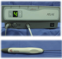
    Figure 1:  Topaz Bipolar radiofrequency microtenotomy machine. The TOPAZ
microdebrider device (ArthroCare, Sunnyvale, CA). The figure shows the
generator which excites the electrolytes and then radicals to cause ablation
and the active tip which is usually 0.8 mm and is used to enter the areas
needed for ablation.
Surgical procedure
The senior author and his team performed all the microtenotomy procedure in the study.
- Preoperatively, the area of tenderness was marked on the plantar heel. Using a template with a series of holes 5 mm apart, marks were placed throughout the area of tenderness in a grid-like pattern. Approximately 10 to 20 marks were placed within the affected area.
- Patients were placed in a supine position over the operating table and they were
- put under general anesthesia.
- A pneumatic ankle tourniquet was inflated while the surgery was carried out.
- A smooth 2 mm Kirschner (K) wire was used to puncture the skin at the marks placed around the affected area.
- The ArthroCare bipolar control unit was set at setting 4 (175 V-RMS) and the timer attached to the control units automatically sets to 500 milliseconds.
- The Topaz wand was attached to a saline drip at a rate of 1 to 2 drops per 3 seconds.
- Microtenotomy of the plantar fascia was performed by placing the Topaz wand through the percutaneous holes that was made by the smooth K-wire. The wand was advanced until resistance was felt and the radiofrequency was applied. Again, the wand was advanced further through the fascia and another radiofrequency was applied. In total, two radiofrequency applications were made in each percutaneous hole, a superficial and deep through the thickness of the fascia.
- Betadine dressing was applied on the site of surgery as well as a dry sterile dressing.
- A below knee cast was applied to assure the patients nonweight bearing.
By that the procedure has ended (Figures 2-5).
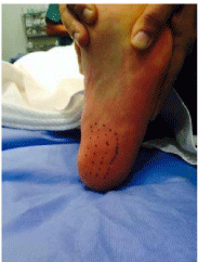
    Figure 2:  The preoperative marked sites of insertion of the active tip. They
are at the areas of origin of plantar fascia and are usually 5 mm apart. In our
practice more markings used at the medial side than lateral side, representing
the site of maximum pain.
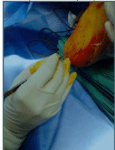
    Figure 3:  The marked areas are then incised using No 15 blade stab wounds,
opening the skin and subcutaneous tissue.
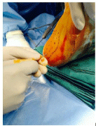
    Figure 4:  The superficial openings done by the stab wound incisions are
then dilated by inserting a 2 mm k-wire. They are then advanced until first
resistance is encountered, marking the plantar fascia.
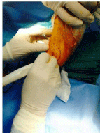
    Figure 5:  The active tip is inserted inside the opening and the pedal
connected to the generator is pressed. The current excited cause’s ablation
at the plantar fascia and the active tip then advances marking the ablation at
the fascia layer.
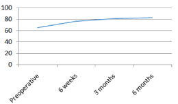
    Figure 6:  Foot and Ankle Outcome Score (FAOS) mean scores showing
improvement of the scores at the different visits. The best improvement was
noted at the 6-week mark and then a steady improvement was observed.
Statistical analysis
Non-normally distributed data were described using standard parametric statistics. Statistical assessment of scores was calculated using 95% confidence intervals, and parametric paired t-tests. SPSS version 22 for IOS was the software used. A p-value of 0.05 or less was considered statistically significant.
Results
Sixty eight out of Seventy patient experienced significant pain reduction by the 6th week post-operatively. There were no complications, including wound infection, plantar fascia rupture, and lateral column pain, alteration of foot mechanics, and nerve entrapment or neurologic deficit.
The mean VAS score was 8.1 (ranging from 6-10) pre-operatively. There was a significant improvement in the VAS score from a mean of 8.1 to 3.56 at 6 weeks and 1.34 at 6 months post-operatively (Table 2). There was no difference in the mean VAS scores and VAS score changes between males and females and Left versus right foot. There were no association between the age of the patients and the VAS scores. From the results collected, the best results were found at the initial period postoperatively at the 6-week bench mark. After then the improvement was progressive but slow as seen by the mean average scores at 6 weeks and 6 months.
    VAS    Score
    Pre-operative
    6    Weeks
    3    Months
    6    Months


    Mean (Range)
    8.1(6-10)
    3.56(2-6)
    2.16(0-5)
    1.34(0-4)


    Change in VAS from previous    time point


    Mean±SD*

    4.54±1.27
    1.4±0.73
    0.814±0.708


    95%CI

    4.24-4.85
    1.226-1.574
    0.645-0.983


    * The P-Value was <0.05.
Table 2:  Visual Analog scale measurement for pain. The table shows the mean scores of each visit. And the mean average scores and the range of scores between
brackets. The most significant improvement was seen at the 6-week visit.
The foot and ankle scores results were analyzed and showed that the preoperative mean scores for the patients in our study were 65.3 with a range of (42–78). At 6 weeks and at the beginning of weight bearing, FAOS scores increased to 77.1 (65-87). At 3 months the scores further increased but in a slow pace to 82 (69-90) and at 6 months the mean score was 83.3 (65-92). The significant increase in the scores was observed at the 6th week bench-mark and the scores then were steadily increasing in the following visits (Figure 6&7).

    Figure 7:  Showing FAOS score ranges at each visit. The figure shows
improvement of the scores throughout the different visits. Notice there was
a wider range at the 6 month visit representing observation of decreased
function and observed disability at the 6-month visit compared the 3-month
visit.
Discussion
All the patients enrolled in this study have had refractory symptoms to extensive medical treatment for plantar fasciitis, before they undergo radiofrequency-based percutaneous microtenotomy. The duration of symptoms for the majority of patients was greater than 8 months. Our primary objective was to determine overall pain relief with this minimally invasive surgical procedure for refractory plantar fasciitis. There was a significant improvement of the mean VAS score at 6 weeks postoperatively. 68 patients were satisfied with their function and pain level and have had their expectations met by 6 months follow-up. Only 2 patients in this study were unsatisfied with partial improvement of their symptoms.
One of them was found to be not compliant with the instructions. She started early weight bearing 2 weeks after the procedure. The patient was found to have a BMI of 39 and showed anxious behavior although not investigated. The other patient was later on diagnosed to have a radiculopathy from the spine and the exact impact of the procedure was difficult to determine. His results were included in the study and it was decided that no other cause other than radiculopathy and planter fasciitis contributed to the pain. The 2 cases were not further investigated.
Most patients with plantar fasciitis are still treated with multiple conservative treatments with a success rate of approximately 90% [19]. Those who fail non-surgical options may benefit from surgical options which include plantar fascia release or resection of the diseased part of the fascia. Although the outcomes of these procedures are good, they have a couple of disadvantages which include, prolong recovery rates, and increase risk of plantar fascia rupture and lateral column pain [20-22].
The percutaneous approach does not require soft tissue dissection and retraction as compared with open approach, so wound healing and recovery might be more rapid. As this procedure does not involve cutting of the plantar fascia, the risk of fascia rupture is minimized. Studies have hypothesized that rapid pain reduction could be caused by an antinociceptive effect and not revascularization and reorganization of collagen based on the observation of signs of neovascularization at 3 to 9 weeks postoperatively [8,16].. Using this new minimally invasive technique, patients were able to transition into normal shoe gear quicker within 6 weeks. No prolonged immobilization was necessary to allow incision healing (2-3 weeks non weight bearing) [13].. An advantage of this technique is the rapid return to daily activities within 3 to 6 weeks with minimal loss of time from work.
The concept of using this technique for treating tendinosis, and plantar fasciosis, was originally drawn from the research work conducted in patients treated for congestive heart failure using laser transmyocardial revascularization. The mechanism of action behind the clinical success was theorized to be associated with the localized angiogenic healing response noted to occur following the procedure. New vasculature in the laser-treated area could improve vascularization in the scar tissue [24]. The best improvements in VAS and FAOS scores were more significant at the first scoring visit 6 weeks postoperatively. After that, scores were observed to improve in a steady and slow pace (Table 2, Figure 6). Although these results would suggest benefit from the procedure, the protocol of nonweight bearing until the 6 week mark and the status of weight bearing afterwards would raise the question of the effect of weight bearing status on the efficacy of the procedure as compared with other results in other studies which had a protocol of maximum 3 weeks of non-weight bearing [3,25]. We recommend that in further studies, controlled groups should be included and more controlled weight bearing status should be established to separate the benefits achieved from non-weight bearing for period of time on the final results.
Postoperatively, patients were encouraged to continue aggressive calf stretching and to use night splinting, and this may have helped in rapid recovery as well.
The disadvantages of this study are the subjectivity of pain measurement using VAS and FAOS and the short-term followup. Long-term and prospective blinded studies using multiple and more objective pain measurement are needed in the future. Also the patients were kept non weight bearing for 6 weeks post operatively and further studies should look into the effect of non-weight bearing in affecting the overall results.
Conclusion
Radiofrequency-based microtenotomy appears to be a promising treatment option of unmanageable plantar fasciitis. The technique is simple to perform and minimally invasive. Rapid pain relief was achieved in most patients, with early return to baseline activity. As a plantar fascia sparing method, it reduces the risks associated with other procedures that transect part or the entire plantar fascia. Future studies should include the effect of post-operative non weight bearing protocol on the results of the procedure.
References
- Shea M, Fields KB. Plantar fasciitis: prescribing effective treatments. Phys Sportsmed. 2002; 30: 21-25.
- Almekinders LC. Tendinitis and other chronic tendinopathies. J Am Acad Orthop Surg. 1998; 6: 157-164.
- Sean NY, Singh I, Wai CK. Radiofrequency microtenotomy for the treatment of plantar fasciitis shows good early results. Foot Ankle Surg. 2010; 16: 174-177.
- Kraushaar BS, Nirschl RP. Tendinosis of the elbow (tennis elbow). Clinical features and findings of histological, immunohistochemical, and electron microscopy studies. J Bone Joint Surg Am. 1999; 81: 259-278.
- Ahmed IM, Lagopoulos M, McConnell P, Soames RW, Sefton GK. Blood supply of the Achilles tendon. J Orthop Res. 1998; 16: 591-596.
- Kraus-Hansen AE, Fackelman GE, Becker C, Williams RM, Pipers FS. Preliminary studies on the vascular anatomy of the equine superficial digital flexor tendon. Equine Vet J. 1992; 24: 46-51.
- Nirschl RP. Elbow tendinosis/tennis elbow. Clin Sports Med. 1992; 11: 851-870.
- Dietz U, Horstick G, Manke T, Otto M, Eick O, Kirkpatrick CJ, et al. Myocardial angiogenesis resulting in functional communications with the left cavity induced by intramyocardial high-frequency ablation: histomorphology of immediate and long-term effects in pigs. Cardiology. 2003; 99: 32-38.
- Kohmoto T, DeRosa CM, Yamamoto N, Fisher PE, Failey P, Smith CR, et al. Evidence of vascular growth associated with laser treatment of normal canine myocardium. Ann Thorac Surg. 1998; 65: 1360-1367.
- Krabatsch T, Schaper F, Leder C, Tulsner J, Thalmann U, Hetzer R. Histological findings after transmyocardial laser revascularization. Journal of cardiac surgery. 1996; 11: 326-331.
- Kwon HM, Hong BK, Jang GJ, Kim DS, Choi EY, Kim IJ, et al. Percutaneous transmyocardial revascularization induces angiogenesis: a histologic and 3-dimensional micro computed tomography study. Journal of Korean medical science. 1999; 14: 502-510.
- Yamamoto N, Gu A, DeRosa CM, Shimizu J, Zwas DR, Smith CR, et al. Radio frequency transmyocardial revascularization enhances angiogenesis and causes myocardial denervation in canine model. Lasers Surg Med. 2000; 27: 18-28.
- Tasto JP, Cummings J, Medlock V, Harwood F, Hardesty R, Amiel D. The tendon treatment center: new horizons in the treatment of tendinosis. Arthroscopy. 2003; 19: 213-223.
- Harwood R BK, Amiel M, Tasto JP, Amiel D. Structural and angiogenicresponse to bipolar radiofrequency treatment of normal rabbit Achilles tendon: A potential application to the treatment of tendinosis. Trans Orthop Res Soc Policy. 2003; 28: 819.
- Taverna E, Battistella F, Sansone V, Perfetti C, Tasto JP. Radiofrequency-based plasma microtenotomy compared with arthroscopic subacromial decompression yields equivalent outcomes for rotator cuff tendinosis. Arthroscopy. 2007; 23: 1042-1051.
- Tasto JP, Cummings J, Medlock V, Hardesty R, Amiel D. Microtenotomy using a radiofrequency probe to treat lateral epicondylitis. Arthroscopy. 2005; 21: 851-860.
- Woloszko J KM, Stalder KR. Coblation in otolaryngology. Proc SPIE. 2003; 4949: 341-352.
- Woloszko J SK, Brown IG. Plasma characteristics of repetitively-pulsed electrical discharges in saline solutions used for surgical procedures. IEEETrans Plasma Sci. 2002; 30: 1376-1383.
- Neufeld SK, Cerrato R. Plantar fasciitis: evaluation and treatment. J Am Acad Orthop Surg. 2008; 16: 338-346.
- Sammarco GJ, Helfrey RB. Surgical treatment of recalcitrant plantar fasciitis. Foot Ankle Int. 1996; 17: 520-526.
- Cheung JT, An KN, Zhang M. Consequences of partial and total plantar fascia release: a finite element study. Foot Ankle Int. 2006; 27: 125-132.
- Gerdesmeyer L, Frey C, Vester J, Maier M, Weil L, Jr., Weil L, Sr., et al. Radial extracorporeal shock wave therapy is safe and effective in the treatment of chronic recalcitrant plantar fasciitis: results of a confirmatory randomized placebo-controlled multicenter study. Am J Sports Med. 2008; 36: 2100-2109.
- Budiman-Mak E, Conrad KJ, Roach K. The Foot Function Index: a measure of foot pain and disability. J Clin Epidemiol. 1991; 44: 561-570.
- Marcelo Cardarelli, MD, MPH A Proposed Alternative Mechanism of Action for Transmyocardial Revascularization Prefaced by a Review of the Accepted Explanations. Tex Heart Inst J. 2006; 33: 424-426.
- Shah A, Best AJ, Rennie WJ. Percutaneous Ultrasound-Guided TOPAZ Radiofrequency Coblation: A Novel Coaxial Technique for the Treatment of Recalcitrant Plantar Fasciitis-Our Experience. J Ultrasound Med. 2016; 35: 1325-1331.