
Research Article
Austin J Orthopade & Rheumatol. 2016; 3(4): 1040.
Comparison of Anterior and Posterior Dislocation after Total Hip Arthroplasty through the Posterior Approach
Tian Y1*, Xu Y2 and Fu Q1
1Department of Orthopaedics, Shengjing Hospital of China Medical University, China
2Department of Anesthesiology, Shengjing Hospital of China Medical University, China
*Corresponding author: Ye Tian, Department of Orthopaedics, Shengjing Hospital of China Medical University, China
Received: August 22, 2016; Accepted: September 19, 2016; Published: September 22, 2016
Abstract
Background: Dislocation is one of the most common complications following Total Hip Arthroplasty (THA). Dislocations after THA can be divided into anterior and posterior types according to the direction of the dislocation. Reports of the difference between anterior and posterior dislocations following THA are scarce.
Aims: The aim of the study is to identify the differences in risk factors and treatment between anterior and posterior dislocation following THA through the posterior approach and explored potential risk factors and effective treatment for the two types of dislocations.
Methods: The study inclu4ded 1433 primary and revision THAs operated by the posterior approach at the Department of Orthopaedics of the Shengjing Hospital between 2005 and 2013. Clinical data of all patients was reviewed and analyzed. The study included an anterior dislocation group (n=5), a posterior dislocation group (n=23), and two matched control groups.
Results: The dislocation rate after primary THA was 1.5% (20/1304) compared with 6.2% (8/129) for revision THA. The ratio of anterior to posterior dislocation in the entire THA cohort was 1:4.6 (5:23). There was strong association between anterior dislocation and anteversion of the acetabular cup and femoral stem, size of the femoral head, and soft tissue tension. On the other hand, a higher incidence of postoperative posterior dislocation was found to relate to soft tissue tension, revision arthroplasty, and incorrect posture.
Conclusion: There are multiple factors associated with both anterior and posterior dislocation. However, the two types of dislocation have different pathologic and anatomical characteristics, as well as risk factors. It is very important to determine the true etiology of dislocation. Closed reduction and immobilization are effective treatments for most patients with either of the two dislocations before open surgery is selected.
Keywords: Anterior dislocation; Posterior dislocation; Total hip arthroplasty; Risk factor; Treatment
Introduction
Dislocation, one of the most common complications following Total Hip Arthroplasty (THA), may often result in restriction of mobility, as well as physical and mental pain [1-3]. Many risk factors are associated with dislocation, including acetabular cup and femoral stem position, femoral head size, soft tissue tension, age, gender, previous surgical history and postoperative posture [4-9].
Dislocations after THA can be divided into anterior and posterior types according to the direction of the dislocation. It has been reported that anterior dislocation is rare compared with posterior dislocation. Excessive anteversion of the acetabular cup and femoral stem is associated with a higher incidence of anterior dislocation [10,11]. On the other hand, posterior dislocation is more common after THA, whose risk factors are multiple according to previous studies [5-9]. In fact, it is very important to identify the difference between the two dislocations during the process of prevention, diagnosis and treatment of dislocations after THA. However, there are few reports of the exact difference between anterior and posterior dislocation, with respect to risk factors and treatment.
We retrospectively reviewed clinical data of 1433 patients who underwent THA between 2005 and 2013. The incidences of anterior and posterior dislocation were compared and the risk factors for dislocation were assessed. We aimed to find the difference between the two types of dislocations, as related to their associated risk factors and treatment.
Materials and Methods
Surgery technique and rehabilitation
The study was approved by the institutional research committee of the Shengjing Hospital in accordance with the Helsinki declaration and observed the specific national laws. Between 2005 and 2013, 1362 primary THAs and 133 revision THAs (86 aseptic loosening, 33 infections, 7 periprosthetic fractures, 3 recurrent dislocations) were performed by three surgeons, whose respective levels of surgical training and technique were similar, at the Department of Orthopaedics of the hospital. The posterior approach was used for all patients. After implantation of the prosthesis, the posterior capsule and short external rotators were fixed again to the greater trochanter to preserve the stability of the artificial joint.
All patients underwent our institution’s standard postoperative rehabilitation program. Briefly, an abduction pillow and a corrective shoe were used to maintain neutral 30 degrees’ abduction of the hip on the day of surgery. Patients were instructed to perform hip exercises in bed on postoperative days 1 and 2. From postoperative day 3 onward, patients were permitted to stand and walk with crutches or a walking device.
Clinical and radiographic evaluation
Patients were divided into the following groups: anterior dislocation group, posterior dislocation group, and two matched control groups without dislocation (One matched control group for anterior dislocation group, the other for posterior dislocation group). The cases in the matched groups were matched with dislocated cases for age, body weight, and preoperative diagnosis.
The method described by Ackland was used to measure inclination and anteversion of the acetabular cup by postoperative Anteroposterior (AP) pelvic radiographs. The method described by Wang was used to measure anteversion of the femoral stem by postoperative lateral femoral radiographs [12,13]. Patients’ neurologic and musculoskeletal complaints were addressed to determine soft tissue tension in relation to prosthesis stability.
Statistical analysis
Statistical analysis was performed using SPSS version 19.0 software. Statistical differences of enumeration data among groups were assessed with the Pearson chi-squared test. Numerical data are presented as the mean ± standard deviation. Statistical differences of measured data among groups were assessed with the student’s t-test. A p-value of less than 0.05 was considered statistically significant.
Results
All patients were followed up both clinically and radiologically. Fifty-six patients were lost to follow-up and four died within the first 6 postoperative months. The remaining 1433 patients were followed up for at least 6 months (mean 14 months; range 6 months to 3 years).
Rate of dislocation
Twenty-eight patients presented with dislocations of 1304 primary and 129 revision THAs performed: 20 (1.5%) occurred after primary THA and 8 (6.2%) occurred following revision THA. Of the primary THAs, 5 patients experienced anterior dislocation and 15 patients experienced posterior dislocation. All eight dislocations following revision THA were posterior dislocations (Table 1&2).
Occurring time of dislocation
We defined early dislocation as occurring less than or equal to 2 weeks after THA, middle dislocation as greater than 2 but less than or equal to 12 weeks after THA, and late dislocation as 12 weeks after THA. All anterior dislocations were early dislocations occurring between 2 and 13 (mean 5) days after THA. Twelve (52%) posterior dislocations occurred between 2 to 6 weeks after THA; nine (39%) posterior dislocations occurred between 6 to 12 weeks after THA. Two (9%) posterior dislocations occurred 12 weeks after THA.
Treatment of dislocation
All anterior dislocations were reduced successfully by closed reduction. After immobilization limiting over-extension and external rotation with a bandage in bed for greater than 4 weeks, four patients had no recurrence after a mean follow up period of 14 months. One patient experienced recurrent dislocation as she could not tolerate immobilization with a bandage. We performed revision surgery and replaced partial components for her (Figure 1). No patient had pain during the 3-year follow up period.
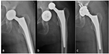
    Figure 1:  Anteroposterior radiographs of a 45-year old woman taken (a)
immediately after THA, (b) 2 days after surgery showing anterior dislocation
and (c) immediately after open reduction showing a new acetabular cup with
polyethylene liner and a longer-femoral-neck head were replaced.
For posterior dislocations, closed reduction combined with immobilization limiting flexion, adduction and internal rotation for greater than 4 weeks was successful following the first or second dislocation in 19 patients. Three dislocations could not be reduced due to soft tissue interposition, requiring open reduction to be performed (Figure 2). One patient who underwent revision THA experienced three recurrent dislocations. During revision surgery, we replaced partial components for him. The patient had no recurrent dislocation in the 3-year follow up period (Figure 3). Eighteen patients had no pain and four patients had occasional pain during the 3-year followup period. The patient with a constrained liner complained of an occasional hip click, but no pain or dysfunction.
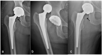
    Figure 2:  Anteroposterior radiographs of a 67-year old man taken (a)
immediately after THA, (b) 41 days after surgery showing posterior dislocation
and (c) immediately after open reduction. Arrows point to drainage.
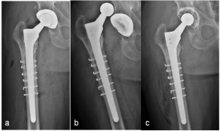
    Figure 3:  Anteroposterior radiographs of a 72-year old man taken (a)
immediately after revision THA, (b) 17 days after surgery showing posterior
dislocation and (c) immediately after open reduction showing a constrained
cemented acetabulum and a longer femoral-neck head were replaced.
Risking factors of dislocation
There was no association with cup inclination both in the anterior and posterior dislocation groups (p>0.05).
The average sum of the cup and stem anteversion angles in the anterior dislocation group was 54.4° (50°-60°) compared with 39.6° (37°-42°) in the matched control group. There was a significant difference between groups (p<0.05). However, there was no difference in the average sum of the cup and stem anteversion angles between the posterior dislocation group (20° -52°, mean 36.4°) and matched control group (30° -51°, mean 39.4°; p>0.05).
There was a significant association with femoral head size in the anterior dislocation group (p<0.05). We found no association with femoral head size between the posterior dislocation and control groups (p>0.05).
Both anterior and posterior dislocations were strongly associated with inadequate soft tissue tension (p<0.05).
There was no association between female gender and anterior dislocation (p>0.05). Also, there was no association with female gender in the posterior dislocation group (p>0.05).
For anterior dislocation, no specific cause for their dislocation was identified by the patients. On the other hand, in the posterior dislocation group, 18 patients knew which postures resulted in their dislocations. In the control group, seven patients remembered that the involved hip underwent risk postures on one or more occasions; fortunately no dislocation occurred (Table 1&2).
![]()
    Case,direction
    Gender,Age(y)
    Diagnosis
    Revision
    Inclination of cup(0)
    Anteversion of cup and stem(0)
    Size of femoral head(mm)
    Disease relating to soft tissue    tension
    Accident relating to    dislocation


    1, P
    F, 62
    SLE
    No
    52
    41
    28
    No
    Rotational movement in bed


    2, P
    M, 64
    FHN
    Yes
    48
    48
    28
    Cerebral infarction
    Getting up


    3, P
    M, 56
    RA
    No
    46
    20
    36
    No
    Deep seating


    4, P
    M, 77
    FHN
    Yes
    55
    36
    28
    Prosthesis subsidence
    No


    5, A
    F, 45
    DDH
    No
    37
    60
    28
    Poliomyelitis
    No


    6, P
    F, 78
    FHN
    No
    40
    52
    28
    No
    Getting up


    7, P
    F, 72
    FNF
    No
    43
    29
    36
    No
    Rotational movement in bed


    8, P
    M, 67
    FNF
    No
    36
    31
    36
    No
    Hyperflexion and adduction


    9, P
    M, 72
    FHN
    Yes
    53
    25
    36
    No
    Hyperflexion and adduction


    10, A
    M, 51
    FNF
    No
    50
    50
    28
    Soft tissue defect due to    previous surgery
    No


    11, P
    M, 71
    FHN
    Yes
    47
    38
    28
    No
    No


    12, P
    F, 65
    RA
    No
    45
    40
    22
    No
    Deep seating


    13, P
    M, 53
    FHN
    No
    39
    45
    28
    Poliomyelitis
    Rotational movement in bed


    14, P
    M, 74
    FHN
    No
    46
    31
    32
    Prosthesis subsidence
    No


    15, P
    F, 52
    SLE
    No
    51
    32
    28
    No
    Getting up


    16, P
    F, 68
    FNF
    No
    48
    41
    28
    Parkinsonism
    Rotational movement in bed


    17, A
    F, 51
    RA
    No
    50
    58
    28
    No
    No


    18, P
    F, 73
    FNF
    Yes
    42
    29
    32
    Prosthesis subsidence
    Hyperflexion and adduction


    19, A
    M, 52
    FHN
    No
    46
    51
    28
    Poliomyelitis
    No


    20, P
    M, 65
    FHN
    Yes
    37
    43
    32
    No
    Deep seating


    21, P
    F, 80
    FNF
    No
    39
    41
    32
    No
    No


    22, P
    M, 72
    FHN
    No
    46
    35
    36
    No
    Getting up


    23, A
    F, 56
    SLE
    No
    52
    53
    22
    Soft tissue defect due to    previous surgery
    No


    24, P
    F, 68
    FNF
    No
    35
    42
    28
    No
    No


    25, P
    F, 65
    FHN
    No
    50
    36
    28
    Poliomyelitis
    Deep seating


    26, P
    M, 75
    FHN
    Yes
    46
    45
    36
    No
    Hyperflexion and adduction


    27, P
    M, 78
    FNF
    No
    44
    31
    28
    No
    No


    28, P
    M, 71
    FHN
    Yes
    47
    27
    32
    Cerebral infarction
    Rotational movement in bed
Table 1: Clinical and radiographic data for dislocated patients.
![]()
    Parameter
    Anterior dislocation group(n=5)
    Matched control group(n=5)
    P
    Posterior dislocation group(n=23)
    Matched control group(n=23)
    P


    Gender


    0.527


    0.765


    Male
    2
    3
    13
    14


    Female
    3
    2
    10
    9


    Soft tissue tension


    0.01


    0.009


    Normal


    Defect
    1
    5
    15
    22


    4
    0
    8
    1


    Revision


    /


    0.009


    Yes
    0
    0
    8
    1


    No
    5
    5
    15
    22


    Femoral head size(mm)


    0.038


    0.555


    22, 28


    32, 36


    5
    2
    12
    10


    0
    3
    11
    13


    Inclination of cup(mean±SD)
    47.0±6.0
    45.6±4.4
    0.632
    45.0±5.5
    42.7±5.7
    0.628


    Anteversion of cup and    stem(mean±SD)
    54.4±4.4
    39.6±2.1
    0.036
    36.4±7.9
    39.4±5.8
    0.096


    Accident


    /


    0.001


    Yes
    0
    0
    18
    7


    No
    5
    5
    5
    16


    P: Posterior dislocation; A: Anterior    dislocation; SLE: Systemic Lupus Erythematosus; RA: Rheumatoid Arthritis;    FHN: Femoral head necrosis; DDH: Developmental Dysplasia of the Hip; FNF:    Femoral neck fracture
Table 2: Comparison of dislocation risk factors between anterior and posterior dislocation.
Discussion
The reason of different rates and occurring times between the two dislocations
Consistent with previous reports [9,13-16], we demonstrated that the dislocation rate after primary THA via a posterior approach is 1.8%, and is 6% following revision THA. Given the fact that there are far more posterior than anterior dislocations [10,11], surgeons in our hospital are inclined to increase the anteversion of the acetabular cup to avoid posterior dislocation, especially when using the posterior approach. In fact, this maneuver is acceptable in most of patients with normal soft tissue tension only if acetabular anteversion does not occur beyond the “safe zone” [17-19]. However, in cases of inadequate soft tissue tension, especially when a small femoral head is used, anterior dislocation may easily occur. In addition, patients are often informed that external rotation and abduction of the operated hip is safe and are advised not perform excessive internal rotation, adduction, and flexion. In fact, most cases of anterior dislocation did not excessively move their hips, and the dislocations usually occurred while turning over; some patients were even unaware of the dislocation occurring. This is why anterior dislocation after THA occurs much earlier than posterior dislocation in our hospital.
Treatment of the two dislocations
Previous studies show that most dislocations after THA, both anterior and posterior, can be treated by closed reduction and immobilization [9-11,20]. Consistent with previous research, we found that four of five anterior dislocations and 19 of 23 posterior dislocations achieved good results only through closed reduction and immobilization. As demonstrated by previous reports [20], we believe that it is very important to determine the true etiology of dislocation. In patients with a severe soft tissue tension defect and malpositioning of their prosthesis, open surgery is often necessary.
The implications of different risking factors of the two dislocations
Of the risk factors for dislocation after THA, prosthesis position is the most important, as demonstrated by multiple previous reports [21-23]. In this study, anterior dislocation is strongly associated with a larger anteversion angle of the cup and stem, as consistent with previous studies. However, the cup and stem anteversion angles did not appear to be associated with a higher incidence of posterior dislocation. The reason for this may be that surgeons in our hospital know that posterior dislocation, compared with anterior dislocation, can easily occur after THA especially when the posterior approach is utilized; they may be inclined to increase the anteversion of the cup and stem intraoperatively, which results in fewer patients with an anteversion angle less than the “safe zone” angle and anteversion both in and greater than the “safe zone” did not increase the incidence of posterior dislocation. Thus, in the posterior dislocation group, although the anteversion angles were different for different patients, we could not find a relevant association between anteversion and dislocation incidence.
It has been shown that a large femoral head is associated with a lower incidence of dislocation after THA [24,25]. In our study, a small head is strongly associated with a higher incidence of dislocation. However, a large head fails to lower the posterior dislocation rate in our study, which indicates that a risk of posterior dislocation cannot be reduced by use of a large femoral head if the involved hip undergoes excessive adduction and/or internal rotation with flexion.
We demonstrated that inadequate soft tissue tension is related to higher incidences of both anterior and posterior dislocation. This result is highly consistent with previous research [5,8,9]. If a patient with inadequate soft tissue tension has other concomitant risk factors, postoperative dislocation is inclined to occur.
We did not find a correlation between gender and dislocation, which is consistent with some reports [5,8] but not others [9,26]. Until now, a consensus on correlation between gender and dislocation has not been reached.
In the present study, adduction and internal rotation with flexion of the involved hip remains the most common risk factor contributing to posterior dislocation. Although external rotation with hip extension should be a risk factor for anterior dislocation, few patients remember the actual movement that caused their dislocation.
Conclusion
The incidence of posterior dislocation is higher than anterior dislocation following THA via the posterior approach. The reasons for both types of dislocations are multiple. However, risk factors contributing to the two dislocations are not completely the same. During the posterior approach, excessive anteversion of the acetabular cup and femoral stem combined with other destabilizing factors lead to anterior dislocation in the early postoperative period. On the other hand, posterior dislocation often results from unsuitable movement of an unstable hip, along with other risk factors in the first few postoperative weeks. It is very important to determine the true etiology during the diagnosis and treatment of dislocation. Closed reduction and immobilization are effective treatments for most patients with either of the two dislocations before open surgery are selected. Preoperative detailed patient assessment, confirmation of the lack of dislocation tendency intraoperatively, and repetitive postoperative patient education can minimize the incidence of dislocation after THA.
References
- McCollum DE, Gray WJ. Dislocation after total hip arthroplasty. Causes and prevention. Clin Orthop Relat Res. 1990; 261: 159-170.
- Brien WW, Salvati EA, Wright TM, Burstein AH. Dislocation following THA: comparison of two acetabular component designs. Orthopedics. 1993; 16: 869-872.
- Yuan L, Shih C. Dislocation after total hip arthroplasty. Arch Orthop Trauma Surg. 1999; 119: 263-266.
- Ali Khan MA, Brakenbury PH, Reynolds IS. Dislocation following total hip replacement. J Bone Joint Surg Br. 1981; 63: 214-218.
- Woolson ST, Rahimtoola ZO. Risk factors for dislocation during the first 3 months after primary total hip replacement. J Arthroplasty. 1999; 14: 662-668.
- Jolles BM, Zangger P, Leyvraz PF. Factors predisposing to dislocation after primary total hip arthroplasty: a multivariate analysis. J Arthroplasty. 2002; 17: 282-288.
- Masaoka T, Yamamoto K, Shishido T, Katori Y, Mizoue T, Shirasu H, et al. Study of hip joint dislocation after total hip arthroplasty. Int Orthop. 2006; 30: 26-30.
- Wetters NG1, Murray TG, Moric M, Sporer SM, Paprosky WG, Della Valle CJ. Risk factors for dislocation after revision total hip arthroplasty. Clin Orthop Relat Res. 2013; 471: 410-416.
- Leichtle UG, Leichtle CI, Taslaci F, Reize P, Wünschel M. Dislocation after total hip arthroplasty: risk factors and treatment options. Acta Orthop Traumatol Turc. 2013; 47: 96-103.
- Ng TP, Yau WP, Tang WM, Chiu KY. Anterior dislocation following primary total hip replacement by the posterior approach—aetiology and treatment. Hong Kong J Orthop Surg. 2003; 7: 14–18.
- Di Schino M, Baudart F, Zilber S, Poignard A, Allain J. Anterior dislocation of a total hip replacement. Radiographic and CT-scan assessment. Behavior following conservative management. Orthop Traumatol Surg Res. 2009; 95: 573-578.
- Ackland MK, Bourne WB, Uhthoff HK. Anteversion of the acetabular cup. Measurement of angle after total hip replacement. J Bone Joint Surg Br. 1986; 68: 409-413.
- Wang L, Trousdale RT, Ai S, An KN, Dai K, Morrey BF. Dislocation after total hip arthroplasty among patients with developmental dysplasia of the hip. J Arthroplasty. 2012; 27: 764-769.
- Enocson A, Hedbeck CJ, Tidermark J, Pettersson H, Ponzer S, Lapidus LJ. Dislocation of total hip replacement in patients with fractures of the femoral neck. Acta Orthop. 2009; 80: 184-189.
- Ji HM, Kim KC, Lee YK, Ha YC, Koo KH. Dislocation after total hip arthroplasty: a randomized clinical trial of a posterior approach and a modified lateral approach. J Arthroplasty. 2012; 27: 378-385.
- Brooks PJ. Dislocation following total hip replacement: causes and cures. Bone Joint J. 2013; 95: 67-69.
- Yoshimine F. The safe-zones for combined cup and neck anteversions that fulfill the essential range of motion and their optimum combination in total hip replacements. J Biomech. 2006; 39: 1315-1323.
- Amuwa C, Dorr LD. The combined anteversion technique for acetabular component anteversion. J Arthroplasty. 2008; 23: 1068-1070.
- Dorr LD, Malik A, Dastane M, Wan Z. Combined anteversion technique for total hip arthroplasty. Clin Orthop Relat Res. 2009; 467: 119-127.
- Dorr LD, Wan Z. Causes of and treatment protocol for instability of total hip replacement. Clin Orthop Relat Res. 1998; 355:144-151.
- Biedermann R, Tonin A, Krismer M, Rachbauer F, Eibl G, Stöckl B. Reducing the risk of dislocation after total hip arthroplasty: the effect of orientation of the acetabular component. J Bone Joint Surg Br. 2005; 87: 762-769.
- Higa M, Tanino H, Abo M, Kakunai S, Banks SA. Effect of acetabular component anteversion on dislocation mechanisms in total hip arthroplasty. J Biomech. 2011; 44: 1810-1813.
- Nevelos J, Johnson A, Heffernan C, Macintyre J, Markel DC, Mont MA. What factors affect posterior dislocation distance in THA? Clin Orthop Relat Res. 2013; 471: 519-526.
- Kung PL, Ries MD. Effect of femoral head size and abductors on dislocation after revision THA. Clin Orthop Relat Res. 2007; 465: 170-174.
- Howie DW, Holubowycz OT, Middleton R; Large Articulation Study Group. Large femoral heads decrease the incidence of dislocation after total hip arthroplasty: a randomized controlled trial. J Bone Joint Surg Am. 2012; 94: 1095-1102.
- Hailer NP, Weiss RJ, Stark A, Kärrholm J. The risk of revision due to dislocation after total hip arthroplasty depends on surgical approach, femoral head size, sex, and primary diagnosis. An analysis of 78,098 operations in the Swedish Hip Arthroplasty Register. Acta Orthop. 2012; 83: 442-448.