Case Report
Austin J Orthopade & Rheumatol. 2016; 3(4): 1043.
Diffuse Idiopathic Skeletal Hyperostosis (DISH) of Young Adults. Lessons to be Learnt
Mader R1,2*, Fawaz A¹, Bieber A¹ and Novofastovsky I¹
¹Rheumatic Diseases Unit, Ha’Emek Medical Center, Israel
²The Ruth and Bruce Rappaport Faculty of Medicine, Technion-Israel Institute of Technology, Israel
*Corresponding author: Mader R, Rheumatic Diseases Unit, Ha’Emek Medical Center, Rabin Rd, Israel
Received: August 29, 2016; Accepted: October 27, 2016; Published: November 01, 2016
Abstract
DISH is a condition characterized by ossification and calcification of soft tissues, mainly ligaments and entheses. The disease is poorly recognized but is often associated with metabolic and constitutional derangements, increased cardiovascular risk and at times, dreadful complications following medical procedures or minor trauma. The prevalence is variable but increases remarkably with age, and in certain elder populations may reach 35%. It has been suggested that 10 years are needed from the initiation of the process to its full radiographic manifestation. There is very little data about young individuals (ie =40 years of age) affected by the disease. We describe 4 patients affected by DISH in their 4th decade of life and in whom the process of ossification and calcifications, presumably, started to evolve in their 3rd decade of life. The clinical characteristics of the patients are discussed. Investigations of these rare cases might shed light on the pathogenetic mechanisms, and initiating factors that promote the formation of DISH.
Keywords: Diffuse idiopathic skeletal hyperostosis; Metabolic syndrome; Enthesopathy
Introduction
DISH is a condition characterized by calcifications and ossifications of soft tissues, mainly ligaments and entheses. Although the first description of DISH dates back to 1950 [1], a large body of evidence shows DISH to be of more ancient origin [2].
The etiology of DISH is unknown. However, several metabolic, genetic, and constitutional factors were reported to be associated with this condition. These include: obesity, a high waist circumference ratio, hypertension, Diabetes Mellitus (DM), hyperinsulinemia, dyslipidemia, elevated growth hormone levels, elevated insulin like growth factor-1, hyperuricemia, use of retinoids and genetic factors [3-7]. A recent study showed that patients with DISH are more often affected by metabolic syndrome and have an increased risk for cardiovascular morbidity [8].
Due to the spinal stiffness the patients affected by DISH are exposed to complications that may derive from minor trauma or medical procedures [9,10]
The condition is unequally distributed between males and females (in a ratio of ~2:1), and its prevalence rapidly increases with age [11]. The prevalence of DISH varies according to geographical location, population studied and obviously age. In an epidemiological outpatient study, the prevalence of DISH in patients over 50 years of age has been reported to be 25% for males and 15% for females [12]. A study aiming to find the prevalence of DISH in the Netherlands by screening 501 chest radiographs obtained for unrelated medical conditions corroborated these results (17% of the individuals over the age of 50 years in this study had DISH) and demonstrated that male gender and advancing age increase the probability of the development of DISH.NRR17). An autopsy study reported that in a series of 75 spines studied at autopsy 28% had DISH [11].
The reported prevalence in the fifth decade of life was extremely low ranging from 0.3 and 0.2% in males and females respectively in the Finish population to none in the female Italian population [13,14]. There is no data on patients in their 4th decade of life, probably because of its rarity. However, there have been a few description of familial cases of DISH in very young patients suggestive for a genetic basis [15,16]. Early diagnosis is important to better understand the evolution of this condition, and eventually intervene, in the future, in its course. A case series of 4 patient’s =40 years of age, diagnosed with DISH are described and their contribution to our understanding is discussed.
Case Presentation
Four patients with DISH, diagnosed at =40 years of age were identified from our data base and herein described. The cases were extracted from our data base of 200 patients fulfilling the Resnick classification criteria for DISH. The age at diagnosis has been established usually at the first or second visit in the rheumatic diseases unit. The final diagnosis has been established by a single observer (RM).

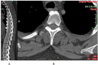
Figure 1: Chest CT showing bridging osteophytes in the sagittal plan (A) and ossification of the anterior longitudinal ligament in the transverse plan (B).

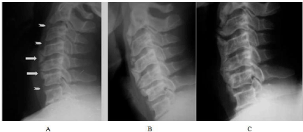
    Figure 2:  Early nuclei of ossification of the cervical spine (arrows) (A), evolving into large bridging osteophytes (B&C) (reproduced with permission from reference
15).
Case 1
A 36 years old male patient was referred for a rheumatologic evaluation for chronic low back pain. He reported difficulty in standing up, walking and although his pain was alleviated with bed rest he reported around the clock pain. He denied weight loss, fever, muco-cutaneous lesions, and family history of psoriasis, gastrointestinal or genitourinary complaints. He has been investigated by the orthopedists and has been told he had several intervertebral discs derangements. His medical history revealed that he suffered from arterial hypertension, hyperlipidemia, morbid obesity, sleep apnea, fatty liver, and hyperuricemia. He was treated with statins and several anti-hypertensive medications as well as amitriptyline and analgesics. General physical examination was unremarkable except for a BMI of 39. Musculoskeletal examination revealed a limited spinal mobility, limited hips’ internal rotation and tender heals. Chest radiographs did not show features of DISH. Revision of the previously performed CT’s of the lumbar and thoracic spine did reveal several discs’ protrusions in the lumbar spine, but also characteristic features of DISH in the thoracic spine (Figures 1A and 1B). There were no radiographic features suggestive of sacroiliitis but calcifications in the pelvic arteries were detected.
Case 2
A 38 years old female patient has been referred for evaluation of shoulders and arms pain of 2 months duration. Her previous medical history was positive for obesity (MBI 55), arterial hypertension, and hypertriglyceridemia. No history of inflammatory back pain, psoriasis, family history of psoriasis, genitourinary and/ or gastrointestinal complaints was elicited. She gave a history of maternal DM. She has been treated with aspirin, thiazide diuretic, enalapril and various NSAIDS. Her general physical examination was unremarkable except for obesity. Her musculoskeletal examination showed a limited external rotation of both hips, and tender neck and thoracic spine. Radiographs showed calcific tendinitis of the right shoulder and features of DISH involving the thoracic spine. No radiographic evidence of sacroilitis was observed. Two years later she has developed frank DM and further on underwent bariatric surgery.
Case 3
A 37 years old male patient was referred to the rheumatology outpatient clinic for evaluation of neck and low back pain of 6 months duration. The patient was otherwise healthy with normal BMI and no complaints, nor findings of other systems’ involvement. Physical examination was unremarkable except for limited range of motion in the cervical spine. Laboratory work did not show increased acute phase reactants but showed hypercholesterolemia. There were no radiographic evidence for involvement of the sacroiliac joints, but there were ossifications of the annulus fibrosus of the cervical spine, which were considered compatible with DISH even in the absence of involvement of the thoracic spine (Figures 2A, 2B and 2C). A byproduct of his investigations was calcific tendinitis of his left shoulder. Few years later, the patient has developed classical DISH involvement of the cervical spine (figure with permission). Radiographic findings compatible with thoracic spine DISH were evident only after 10 years.
Case 4
A 39 years old female patient was referred for evaluation of diffuse musculoskeletal pain of 2 years duration. Her past medical history was unremarkable except for bilateral carpal tunnel syndrome and obesity (BMI 34.8). A family history of DM was positive for both parents. No other systems’ involvement was reported or observed. Physical examination was unremarkable except the obesity. Laboratory investigations were positive for slightly elevated CRP but otherwise unremarkable. Radiographic imaging showed characteristic DISH involvement of the thoracic spine and slight enlargement of the metacarpal heads. No radiographic evidence of sacroilitis was observed. Within the following 5 years she has developed DM, hyperlipidemia and evidence of severe osteoarthritis of the knees both clinically and radiographically, and plantar enthesopathies.
Discussion
The prevalence of DISH increases with age, but is extremely variable according to the population studied, and can be as high as 26% in females and 35% in males of a hospital population [12].
Only a few studies reported the prevalence in patients before 50 years of age. However, the reported prevalence in the fifth decade of life was extremely low ranging from 0.3 and 0.2% in males and females respectively in the Finish population to none in the female Italian population [13,14]. In the case series described here of young adults, the prevalence of DISH was 2% (4/200). This figure can be considered relatively high. A single study reported a relatively high prevalence of DISH in Israel which might explain also the relatively high prevalence in young adults [17].
It was estimated that a period of at least 10 years is needed for the pathologic process to evolve completely suggesting, that for patients in their 5th decade of life, the pathologic process started in the 4th decade of life [18,19]. A study that investigated DM and HTS as risk factors for DISH, identified 12.8% of the cohort to be =50 years of age [20]. This relatively high prevalence was attributed to selection bias of patients attending a rheumatology outpatient clinic highly minded for DISH. This study demonstrated that patients with DISH, diagnosed at a relatively young age, were significantly more often affected by pain in the thoracic spine, lumbar spine tendonitis and/ or enthesopathies compared to patients with similar age and gender distribution not affected by DISH. The patients also had a significantly higher prevalence of obesity, first degree relatives with DM or HTS, and were more likely to develop DM during follow-up. These patients did not differ significantly in most aspects from patients with DISH diagnosed at an older age, except in the case of a family history of DM and HTS.
The variation in the prevalence of DISH throughout the world, suggests that genetic factors might play a part in its pathogenesis. Moreover, familial clustering of the condition and early onset (in the third decade of life) in some affected families have been observed, which are also suggestive of a genetic contribution to the disease [15,16]. Studies in dogs have revealed the overall prevalence of canine DISH to be 3.8%, whereas in the Boxer breed it is >40%, which further supports the existence of a genetic component in the risk of developing DISH [21]. So far, however, only one potential susceptibility gene (namely COL6A1, which encodes type VI collagen a chain) has been identified as a potential gene for the development of either DISH and/or ossification of the posterior longitudinal ligament [22]. Thus, although the effects of COL6A1 variants on bone metabolism have not been elucidated, it has been suggested that this protein might be involved in ectopic bone formation in DISH and OPLL.
DISH in the 4th decade of life is rare. The cases presented here, are probably sporadic and suggest that the ossification and/or calcification process of the entheses might start in the 3rd decade of life in some individuals. All the patients had at least one metabolic derangement and/or family history of DM and 3/4 were obese. In this respect they were no different than the classical elder patients with DISH.
Calcifications of other soft tissues were observed in 3/4 patients (calcification of arteries in one and shoulder calcific tendinitis in 2). Several matrix proteins were identified as protective factors in non-osseous tissues, and alterations in them were found associated with several calcium deposition diseases such as calcific tendinitis, atherosclerosis and DISH [23].These associations have not been systematically studied in DISH, but the fact that they might affect young individuals suggests an inborn defect in one of these proteins.
A recent case report of cervical myelopathy following a minor trauma in a 39 years old male patient suggests that the young patients may be affected by the same complications as the elder DISH population [24].
There are still unanswered questions. It is possible, though not established yet, that elderly patients contracted their disease earlier in life and were diagnosed late in life?. It is still debatable, whether the disease is symptomatic [25,26]. If this assumption is correct, it may well be that these patients were referred for evaluation for arthralgias due to osteoarthritis or other painful musculoskeletal complaints and diagnosed with DISH which has existed for many years. It could also be, that there might be a genetic basis, beyond the other known risk factors for DISH, such as metabolic syndrome, that put the patients at a higher risk for the development of this condition [8]. Another possibility is the limitations of plain radiographs (see case 1). In fact, it has been recently shown that CT scans of the spine have a greater yield in identifying DISH [27]. Realistically, the most common screening procedure for DISH are plain radiographs and it is not yet justified to use CT scans for that purpose. However, the number of young patients with DISH could be higher than that reported if CT scans have been employed.
There is no doubt, that at present the research of DISH is hampered by the present classification criteria that require almost “end stage” radiographic findings [28]. While awaiting new classification criteria, patients with very early DISH or patients with multiple risk factors for the development of DISH should be identified and investigated.
In conclusion, young adults may be affected with DISH and the diagnosis should not be discarded based on age alone. The metabolic and constitutional derangements of these patients are similar to elder patients and their risk to develop atherosclerotic cardiovascular diseases and DM is also similar to elder patients with DISH. At present there are no specific therapies for DISH [29]. However, interventions aimed to reduce the metabolic risk factors and lifestyle changes (i.e., weight loss, physical activity etc.) might prove useful for patients with DISH and in particular to young adults.
References
- Forestier, J. & Rotes-Querol, J. Senile ankylosing hyperostosis of the Spine. Ann Rheum Dis. 1950; 9: 321-330.
- Van der Merwe, A. E., Maat, G. J. & Watt, I. Diffuse idiopathic skeletal hyperostosis: Diagnosis in a palaeopathological context. Homo. 2012; 63: 202-215.
- Littlejohn GO. Insulin and new bone formation in diffuse idiopathic skeletal hyperostosis. ClinRheumatol. 1985; 4: 294-300.
- Denko CW, Boja B, Moskowitz RW. Growth promoting peptides in osteoarthritis and diffuse idiopathic skeletal hyperostosis-insulin, insulin-like growth factor-I, growth hormone. J Rheumatol 1994; 21: 1725-1730.
- Nesher G, Zuckner J. Rheumatologic complications of vitamin A and retinoids. Sem Arthritis Rheum. 1995; 24: 291-296.
- Kiss C, Szilagyi M, Paksy A, Poor G. Risk factors for diffuse idiopathic skeletal hyperostosis: a case control study. Rheumatology (Oxford). 2002; 41: 27-30.
- Sarzi-Puttini P, Atzeni F. New developments in our understanding of DISH (diffuse idiopathic skeletal hyperostosis). Curr Opin Rheumatol. 2004; 16: 287-292.
- Mader, R., Novofestovski, I., Adawi, M. &Lavi, I. Metabolic syndrome and cardiovascular risk in patients with diffuse idiopathic skeletal hyperostosis. Semin Arthritis Rheum. 2009; 38: 361-365.
- Mader, R. Clinical manifestations of diffuse idiopathic skeletal hyperostosis of the cervical spine. Semin Arthritis Rheum. 2002; 32: 130-135.
- Westerveld LA, Verlaan JJ, Oner FC. Spinal fractures in patients with ankylosing spinal disorders: a systematic review of the literature on treatment, neurological status and complications. Eur Spine J. 2009; 18: 145-156.
- Westerveld LA, van Ufford HM, Verlaan JJ, Oner FC. The prevalence of diffuse idiopathic skeletal hyperostosis in an outpatient population in the Netherlands. J Rheumatol. 2008; 35: 1635-1638.
- Weinfeld RM, Olson PN, Maki DD. Griffiths HJ. The prevalence of diffuse idiopathic skeletal hyperostosis (DISH) in two large americanmidwest metropolitan hospital populations. Skeletal Radiol. 1997; 26: 222-225.
- Julkunen H, Heinonen OP, Knekt P, Maatela J. The epidemiology of hyperostosis of the spine together with its symptoms and related mortality in a general population. Scand J Rheumatol. 1975; 4: 23-27.
- Pappone N, Lubrano E, Esposito-Del Puente A, D’Angelo S, Di Girolamo C, Del Puente A. Prevalence of diffuse idiopathic skeletal hyperostosis in female Italian population. Clin Exp Rheumatol. 2005; 23: 123-124.
- Gorman C, Jawad ASM, Chikanza IA. Family with diffuse idiopathic hyperostosis. Ann Rheum Dis. 2005; 64: 1794-1795.
- Burges-Armas J, Couto AR, Timms A, Santos MR, Bettencourt BF, Peixoto MJ, et al. Ectopic calcification among families in the Azores: clinical and radiological manifestations in families with diffuse idiopathic skeletal hyperostosis and chondrocalcinosis. Arthritis Rheum. 2006; 54: 1340-1349.
- Bloom RA. The prevalence of ankylosing hyperostosis in a Jerusalem population-with description of a method of grading the extent of the disease. Scand J Rheumatol. 1984; 13: 181-189.
- Mader R. Diffuse idiopathic skeletal hyperostosis: isolated involvement of cervical spine in a young patient. J Rheumatol. 2004; 31: 620-621.
- Yaniv G, Bader S, Lidar M, Herman A, Shazar N, Aharoni D, et al. The natural course of bridging osteophyte formation in diffuse idiopathic skeletal hyperostosis: retrospective analysis of consecutive CT examinations over 10 years. Rheumatology. (Oxford). 2014; 53: 1951-1957.
- Mader R, Lavi I. Diabetes mellitus and hypertension as risk factors for early Diffuse Idiopathic Skeletal Hyperostosis (DISH). Osteoarthritis Cartilage. 2009; 17: 825-888.
- Kranenburg HC, Westerveld LA, Verlaan JJ, Oner FC, Dhert WJA, Voorhout G et al. The dog as an animal model for DISH?. Eur Spine J. 2010; 19: 1325-1329.
- Tsukahara S,Miyazawa N, Akagawa H, Forejtova S, Pavelka K, Tanaka T, et al. COL6A1, the candidate gene for ossification of the posterior longitudinal ligament, is associated with diffuse idiopathic skeletal hyperostosis in Japanese. Spine. 2005; 30: 2321-2324.
- Atzeni F, Sarzi-Puttini P, Bevilacqua M. Calcium Deposition and Associated Chronic Diseases (Atherosclerosis, Diffuse Idiopathic Skeletal Hyperostosis, and Others). Rheum Dis Clin N Am. 2006; 32: 413-426.
- Galgano M, Chin L S. Central Cord Syndrome in a Young Patient with Early Diffuse Idiopathic Skeletal Hyperostosis and Ossification of the Posterior Longitudinal Ligament after Minor Trauma: A Case Report and Review. Cureus 2015; 7: 284.
- Hutton, C. DISH… a state not a disease? [editorial]. Br J Rheumatol. 1989; 28: 277-280.
- Holton KF, Denarde PJ, Yoo JU, Kado DM, Barrett Connor E, Marshall LM. Diffuse idiopathic skeletal hyperostosis and its relation to back pain among older men: The MrOS study. Semin Arthritis Rheum. 2011; 41: 131-138.
- Hirasawa A, Wakao N, Kamiya M, Takeuchi M, Kawanami K, Murotani K, et al. The prevalence of diffuse idiopathic skeletal hyperostosis in Japan the first report of measurement by CT and review of the literature. J Orthopaedic Sci. 2016; 21: 287-290.
- Mader R, Buskila D, Verlaan JJ, Atzeni F, Olivieri I, Pappone N, et al. Developing new classification criteria for diffuse idiopathic skeletal hyperostosis: back to square one Rheumatology (Oxford). 2013; 52: 326-330.
- Mader R. Current therapeutic options in the management of diffuse idiopathic skeletal hyperostosis. Expert OpinPharmacother. 2005, 6: 1313-1318.