
Case Report
Imagery. Austin J Orthopade & Rheumatol. 2019; 6(1): 1072.
The Spindle Cell Lipoma of the Thigh Simulating a Liposarcoma to the Imagery
Boussaidan M1*, Kessab A2, Badaoui R1, Boukhriss J1, Chafry B1, Benchebba D1, Bouabid S1, Oukabli M1 and Boussouga M1
¹Department of Traumatology and Orthopedics, Military Hospital Mohammed V, Faculty of medicine and pharmacy of Rabat, University Mohammed V. RABAT, Morocco
²Department of Anatomy and Pathological Cytology, Military Hospital Mohammed V Rabat, Faculty of medicine and pharmacy of Rabat, University Mohammed V. RABAT, Morocco
*Corresponding author: Boussaidane Mohammed, Department of Traumatology and Orthopedics, Military Hospital Mohammed V Rabat, Morocco
Received: March 27, 2019; Accepted: May 08, 2019; Published: May 15, 2019
Abstract
The Fusiform cell Lipoma is a rare entity of benign adipose tumors. Characterized by the presence of fusiform cells forming collagen, and can be confused with the fusocellular component Liposarcoma.
We report the case of a patient aged 46 years supported in our formation for a subcutaneous mass of the infero-internal region of the left thigh, whose appearance at ultrasound and MRI evokes at first a Liposarcoma. It is the histological and immuno-Histochemical examination that allows the confirmation of the diagnosis.
Keywords: Spindle cell lipoma; IRM; CD34
Introduction
Fusiform cell Lipoma is a rare entity of benign fat tumors [1]. Characterized by the presence of fusiform cells forming collagen [2,3], and can be confused with fusocellular component Liposarcoma [4,5], affecting mainly the young male between 45 years and 65 years, with preferential seat under posterior neck and shoulder area and 13% atypical localization [6].
Here we report the case of a patient supported in our formation for Lipoma with fusiform cells of the left thigh.
Case Presentation
It is a patient of 46 years without significant pathological history, who consults for a subcutaneous swelling of the infero-internal region of the left thigh, progressive onset and evolving for 6 months; it is a painless mass with firm consistency, movable in relation to the skin plane and adherent to the deep plane, of 8 cm of large axis. The patient benefited from an ultrasound that showed a very limited, subcutaneous ovalar formation with regular contours, of a heterogeneous tissue echostructure, respecting the adjacent muscular plane, simulating a Liposarcoma. MRI has objectified the presence of a superficial mass, measuring 50 × 43 × 21, very limited, with regular contours, in hypo signal T1et hyper signal T2; with T1 and T2 hypersignal zones. This mass is increased after injection of gadolinium, without muscular, bone or ligamentous lesions. First evoking a Liposarcoma (Figure 1,2). A surgical biopsy was performed whose anatomopathological result was in favor of a fusiform cell Lipoma. The patient has benefited a surgical resection of the tumor mass.
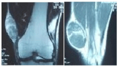
    Figure 1: MRI of the knee in frontal and sagittal section passing through the
tumor.
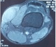
Figure 2: MRI of the knee in axial cut passing through tumor.
The anatomopathological study objectified an 8.5 × 2.5 cm tumorectomy piece surmounted by a 7.5 × 2cm skin flap (Figure 3) showing a yellowish-white appearance of elastic consistency passing flush with the deep plane and at a distance from the boundaries Side.
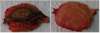
    Figure 3: Macroscopic aspect of the resection piece showing healthy
exeresis margins.
Histological examination showed tumor proliferation made of a double fusiform and Adipocyte component. The fusiform contingent is made up of cells with elongated ovoid nuclei with no atypical or mitosis, mixed with numerous Collagenous beams with an anarchic disposition. The second component is made up mainly of mature adipocytes free from cytonuclear atypies (Figure 4). An immuno- Histochemical study was carried out, showing positive staining of fusiform cells by anti-CD34 antibody (Figure 5).
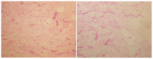
    Figure 4: Histological aspect showing a benign Fibro-adipocyte tumor
proliferation G × 20.
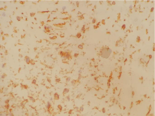
    Figure 5: Immunohistochemically study showing positive labeling of fusiform
tumor cells by anti-CD34 antibody.
Discussion
Described by ENZINGER and HAWLEY in 1975 as a separate entity. Taking the appearance of a benign subcutaneous nodule, well limited, sometimes encapsulated and rarely infiltrating the underlying muscle; most often unique [7].
It is a rare tumor that accounts for about 1.5% of all lipomas [8] which mainly affects male subjects, beyond 40 years. For Syed and al. [9] male predominance is related to the frequent detection of androgen receptors in this tumor.
The fusiform-cell Lipoma develops mainly in the cervical region, back and shoulders [10]. Other unusual locations can be seen in the face, oral cavity, pharynx, tongue, temporal region, Parotid gland, orbit, scalp, and extremities.
The clinic of these tumors is poor; the most frequent sign of appeal is the perception by the patient of a mass most often superficial, with slow and painless evolution [11.12]. The average diameter is 4 to 5 cm [3].
Imaging can evoke a Liposarcoma. It presents itself as a very limited mass of small size (less than 5 cm) with a typical adipose quota but also containing a non-specific appearance fabric. [13].
Histologically, there are three characteristic components: mature adipocytes, fusiform cells, no atypia, no mitosis, arranged in short bundles, mixed with cells and short trousbuckets of thick Collagenous fibers. In the predominant form of fusiform cells, adipocytes are rare. Rarely, vascularization is predominant and can orient to a vascular tumor. The CD34 is positive in the fusiform cells, which are otherwise S100 negative [4,5].
There are several histological variants of fusiform cell Lipoma; including Pleomorphic cell Lipoma, described by Schmookler and Enzinger in 1981. Characterized by the presence of giant cells, comprising several nuclei arranged in rosettes (floret-like giant cells) [14]. Other variants containing little or no adipocytes whose diagnosis can be very difficult are described in particular [15].
The presence of anastomotic spaces with projections of connective tissue makes it possible to distinguish another variant of the fusiform cell Lipoma: it is the pseudoangiomatous form, described in 1994 by Hawley and al [10.16].
Cytogenetic analysis revealed a loss of material from the long arms of chromosomes 13 or 16 [17].
In the case of Pleomorphic cells, the differential diagnosis remains the atypical adipose tumor and the well-differentiated Liposarcoma. It is the Immunohistochemistry and the fish analysis witch make it possible to distinguish each entity, with a overexpression of MDM2/ CDK4 in Liposarcoma. Myxoid Liposarcoma can be evoked in front of a predominance of the myxoid component, but as a rule the seat of this tumor is deep with negative CD34. The FISH analysis shows a rearrangement of the DDIT3 gene [17].
The surgical treatment consists of the complete removal of the tumor under pain of a recurrence [18].
The prognosis of these lesions is usually good, without recurrence if the initial excision is complete [1].
Conclusion
Fusiform cell Lipoma is a rare entity of benign fatty tumors with good prognosis, often unique and well-limited [2,3]. Imaging does not establish a diagnosis of certainty that can only be histological [19].
References
- Fletcher CD, Hogendoorn P, Mertens F, Bridge J. WHO classifi-cation of tumours of soft tissue and bone. 4th Edn. Lyon, France: IARC Press; 2013.
- Enzinger Fm, Weiss Sw. Benign lipomatous tumors. In: Soft tissue tumors. The C.V. Mosby Company, Third Edn, 1995, 381-430.
- Fanburg-Smith JC, Devaney KO, Miettinen M, Weiss SW. Multiple spindle cell lipomas. A report of 7 familial and 11 nonfamilial cases. Am J Surg Pathol. 1998, 22: 40-48.
- Genevay M, Coindre Jm, Guillou L. Recent entities in soft tumor pathology. Part 2. (Article in French). Ann Pathol. 2003, 23: 109-13.
- Weiss SW, Goldblum JR. Enzinger & Weiss’s Soft tissue tumors. The CV Mosby Company, Fourth Edition, 2001.
- Ko JS1, Daniels B, Emanuel PO, Elson P, Khachaturov V, McKenney JK, et al. Spindle Cell Lipomas in Women: A Report of 53 Cases. Am J Surg Pathol.2017; 41: 1267-1274.
- Enzinger FM, Harvey DA. Spindle dell lipoma. Cancer. 1975; 36: 1852-1859.
- Fletcher CD, Martin-Bates E. Spindle cell lipoma: a clinicopathological study with some original observations. Histopathology. 1987; 11: 803-817.
- Syed S, Martin AM, Haupt H, Podolski V, Brooks JJ. Frequent detection of androgen receptors in spindle cell lipomas: an explanation for this lesion’s male predominance? Arch Pathol Lab Med. 2008; 132: 81-83.
- Hawley IC, Krausz T, Evans DJ, Fletcher CDM. Spindle cell lipoma: a pseudoangiomatous variant. Histopathology. 1994; 24: 565-569.
- Latte R. Tumor of the soft tissues. Atlas of tumor pathology second series. Armed forces institute of pathology edit., Washington D.C.1981; 1: 53-150.
- Kransdorf MJ, Murphey MD. Lipomatous tumors. Imaging of soft tissue tumors. W.B saunders company edit., Philadelphia. 1997: 57-101.
- Marques MC, Garcia H. Lipomatous Tumors. In: De Schepper AM. Imaging of Soft Tissue Tumors. Springer edit., Berlin. 1997:191-207.
- Shmookler BM, Enzinger FM. Pleomorphic lipoma: a benign tumor simulating liposarcoma. Cancer. 1981; 47: 126-133.
- Billings SD, Folpe AL. MD. Diagnostically challenging spindle cell lipomas: a report of 34 ‘‘low-fat’’ and ‘‘fat-free’’ variants. Am J Dermatopathol. 2007; 29: 437-442.
- Zamecnik M, Michal M. Angiomatous spindle cell lipoma: report of three cases with immunohistochemical and ultrastructural study and reappraisal of former pseudoangiomatous variant. Pathol Int. 2007; 57: 26-31.
- Dumollard JM, Ranchère-Vince D, Burel F, Coindre JM, Tallini G, Ligon A, et al. Lipomes à cellules fusiformes et délétion 13q : apport de la cytogénétique dans l’aide au diagnostic. Ann Pathol 2001; 21: 303-310.
- Haddam Amina. Lipomes profonds des membres. Thèse de doctorat. FMPR. 2005.
- Julio AJ, Yovanny FL, Juan C, Rodríguez R, Maria E, Medina G. Lipoma con patrón infiltrativo muscular. Rev Cubana Ortop Traumatol. 2006; 20.