Case Report
Austin J Orthopade & Rheumatol. 2019; 6(1): 1073.
Triple Anterolateral Dislocation of the Talus: A Case Report
Boussaidane M*, Beniass Y, Boukhriss J, Chafry B, Benchebba D, Bouabid S and Boussouga M
Department of Traumatology and Orthopedics II, Military Hospital Mohammed V, Morocco
*Corresponding author: Boussaidane M, Department of Traumatology and Orthopedics, Military Hospital Mohammed V Rabat, Morocco
Received: June 03, 2019;Accepted: July 05, 2019; Published: July 12, 2019
Abstract
The triple dislocation of the talus is the rarest but most serious traumatic lesion of the hindfoot. We report a case of closed anterolateral enucleation of the talus, treated conservatively with a good functional result in the trauma-orthopedic II Department of the Military Hospital Mohammed V of Rabat. Currently, most authors agree on the conservative treatment of enucleations in emergency and reserves the arthrodesis for septic and osteoarthritic complications.
Keywords: Triple dislocation; Talus; Conservative treatment
Introduction
Triple dislocation of the talus or enucleation of the talus is an extremely rare and serious lesion, representing 2 to 10% of talian traumatisms [1,2]. The talus loses all its vascular connections, and its vitality is compromised [3]. The prognosis for this type of lesion is dominated by the risk of osteonecrosis [3].
Three-quarters of talus enucleation cases reported in the literature are open [4]. We report a case of closed anterolateral enucleation of the talus, treated conservatively with a good functional result in the trauma-orthopedic service II of the Military Hospital Mohammed V of Rabat.
Observation
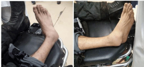
Mr. H.J, a 29-year-old man with no previous medical or surgical history, who suffered a road accident resulting in closed right foot trauma with total functional impotence and severe pain. The clinical examination showed a large distortion of the right foot in adduction, supination and inversion and a Varus attitude of the right ankle. With a talus projecting outwards and forwards (Figure 1). Without cutaneous opening or vascular-nervous complications.

Figure 1: Clinical image showing the deformity of the ankle.
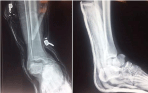
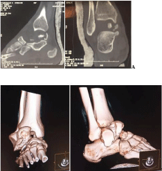
X-ray of the ankle indicated complete anterolateral enucleation of the talus (Figure 2). CT with 3D reconstruction revealed the triple dislocation of the talus associated with a parcel fracture of the taller head (Figure 3).

    Figure 2: Face and profile x-ray of the ankle showing the radiographic
appearance of the triple anterolateral dislocation.

    Figure 3: CT scans (A) and 3D reconstruction images (B) showing
anterolateral enucleation of the talus associated with a parcel fracture of the
talar head.
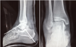
The reduction of the dislocated embankment was performed by external maneuver, by digital pressure after having put the foot in supination and in forced equine. The stability of the hindfoot was maintained by two cross wires (Figure 4). The ankle was immobilized in a plastered boot for two months followed by rehabilitation. Support was allowed in the fourth month and resumption of work in the eighth month after the accident.

    Figure 4: Fce and profile X-rays after reduction and stabilization with two
wires.
At one year of follow-up, the ankle was painless, stable with satisfactory mobility.
Discussion
Enucleation of the talus is a rare lesion that is poorly described in the literature, occurring after very serious traumas, as evidenced by the frequency of cutaneous opening. Less than 80 cases of pure enucleation have been reported in the literature, three-quarters of which are open [4].
Road accidents and falls from a high place with reception on the heels; the most common etiologies foot total dislocations of the talus [5].
The physio pathological mechanism is still debatable. According to Penal [6], anterolateral enucleation is due to a double mechanism of forced plantar flexion and inversion. Plantar flexion produces collateral ligament rupture while inversion causes rupture of talocalcaneal ligaments. However, the most precise study of the enucleation mechanisms remains that of Leitner [7]; which describes enucleation as the ultimate stage of medial taller dislocation.
The frequency of associated lesions (bone, cutaneous or less frequently vascular lesions) can be explained by the violence of the trauma [8].
The treatment of enucleation of the talus is a very controversial subject. If former authors advocated early talectomy followed by triple arthrodesis [9]; Butel and Witvoet in 1967 demonstrated the poor functional results of talectomy, and concluded the need for triple arthrodesis [8]. Currently conservative treatment remains the first choice of any therapeutic approach. Arthrodesis is reserved for secondary complications [10].
The reduction must be carried out urgently, under general anesthesia using the pulling maneuver [11,12]. Malgaigne [13] recommends exerting an impulse on the head of the astragalus to guide it towards the articular sphere. Additional post-reduction restraint is required for an average of 45 days [11,12].
Among the late complications of total enucleations, osteonecrosis is the most common. Its frequency varies from 50 to 100%, the values being much closer to the last digit [5,10,14]. Our case, escapes this type of complications.
Conclusion
The triple dislocation of the talus is the rarest but most serious traumatic lesion of the hindfoot. Currently, most authors agree on the conservative treatment of enucleations in emergency and reserves the arthrodesis for septic and osteoarthritic complications.
References
- Weston JT, Liu X, Wandtke ME, Liu J, Ebraheim NE. A systematic review of total dislocation of the talus. Orthop Surg. 2015; 7: 97-101.
- Papanikolaou A, Siakantaris P, Maris J, Antoniou N. Successful treatment of total talar dislocation with close.
- Biga N, Defives T. Fractures malléolaires de l’adulte et. luxation du cou de pied. Encycl Méd Chir. Appareil locomoteur. 1997; 10: 14-088-A-10.
- Maffulli N, Francobandiera C, Lepore L, Cifarelli V. Total dislocation of the talus. J Foot Surg. 1989; 28: 208-212.
- Copin G, Bouayed S, Kempf I. Les traumatismes graves du talus: fractures luxations, fractures communitives et énucléation. Acta Orthop Belg. 1983; 49: 698-710.
- Pennal GF. Fractures of the talus. Clin Orthop. 1963; 30: 53-63.
- Leitner B. The mechanism of total dislocation of the talus. J Bone Joint Surg. 1995; 37A: 95-98.
- Butel J, Witvoet J. Fractures et. luxations du talus. Rev Chir Orthop. 1967; 53: 494-624.
- Fabricius H, Observation LXVII. Observationum et curationum chirurgicarum centuriae. Rev Chir Orthop. 1967; 53: 494-624.
- Asselineau A, Augereau B, Bombart M, Apoil A, Feuilhade P. Les énucléations partielles ou totales du talus. Intérêt du traitement conservateur. Rev Chir Orthop. 1989; 75: 34-39.
- Patel J, Vianney Y. De la luxation sous astragalienne du pied en dedans. Revue chir orthop. 1913; 1: 1-14.
- Zimmer TJ, Johnnson KA. Subtalar dislocations. Clin orthop. 1989; 238: 190- 194.
- Malgaigne JF. Traité des fractures et des luxations. Baillére édit, Paris. 1855; 1030-1070.
- Coltart WD. Aviator’s Astragalus. J Bone Joint Surge. 1952; 34: 545-566.