Case Report
Austin J Orthopade & Rheumatol. 2019; 6(1): 1076.
Surgical Treatment by Anterior Approach of Femoral Head Dislocation Fracture (About 3 Cases and Literature Review)
Dehayni B1*, Amarir M2, Jalal Y1, Zaddoug O1, Bennis A1, Benchakroune M1, Zine A1, Tanane M1, Edderai M1 and Jaafar A1
¹Department of Traumatology and Orthopedics, Mohamed V Military Training Hospital, Faculty of Medicine and Pharmacy, Mohamed V University, Morocco
²Department of Medical Imaging, Mohamed V Military Training Hospital, Faculty of Medicine and Pharmacy, Mohamed V University, Morocco
*Corresponding author: Dehayni Badreddine, Department of Traumatology and Orthopedics, Mohamed V Military Training Hospital, Faculty of Medicine and Pharmacy, Mohamed V University, RABAT, Morocco
Received: August 05, 2019; Accepted: September 16, 2019; Published: September 23, 2019
Abstract
Femoral head dislocation fracture is a rare lesion that is often associated with hip dislocation. However, we reported a retrospective study on a series of three cases, collected in our service, which were diagnosed and treated urgently, with a good evolution in 2/3 of the cases. The literature review confirms the rarity of the lesion. Radiological diagnosis is not always easy, it should be assisted by a CT scan. Irreducibility is frequent. Choosing the right approach is difficult. Knowledge of all hip approach pathways allows the osteochondral fragment of the head to be approached for removal or fixation after reduction. Good management does not always prevent certain complications. Total hip replacement surgery from the outset always keeps its indications.
Keywords: Femoral head dislocation fracture; CT scan; Anterior approach
Introduction
Femoral head dislocation fracture is a rare lesion that is often associated with hip dislocation. And raises various problems: anatomo-radiological study of the lesion in emergency, a Difficulty of reduction and early or late complications that condition the functional prognosis of the hip.
Case Presentation
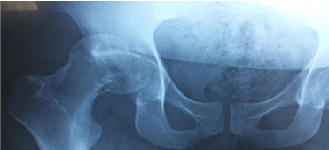
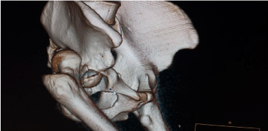
This is a retrospective study collected at the Traumatology- Orthopaedics I Department of Mohamed V Military Hospital in Rabat, over a period of 2 years, yet over a series of three young patients with no significant pathological history, it is 2 men and 1 woman, with an average age of 32 years, all of whom were victims of a road accident (high energy mechanism) with dashboard syndrome. They presented an isolated trauma of the right hip (02 cases) and left hip (01 cases). The clinical examination found a shortened lower limb, in flexion and internal rotation. Pain in active and passive mobilization. Vascular-nervous examination was without particularity, the examination of the above and underlying joints is without anomalies. Standard face pelvis, face and profile radiographs Hip supplemented by 3D reconstruction CT scan, all our patients presented a posterior superior hip dislocation with a femoral head fracture (Figures 1-3) classified as Stage II according to Pipkin’s classification. After a failed attempt to reduce hip dislocation by external maneuvers without surgery under anesthesia in two patients. These two patients received a reduction in emergency dislocation under general anesthesia by open surgery with orthopedic table with osteosynthesis by screwing the cephalic fragment by the anterior approach of the traumatized hip (Figure 3). One case had a good reduction without surgery (Figure 4), operated with a screwing 48 hours later.

    Figure 1: Radiograph of the right hip showing a dislocation fracture of the
femoral head.

    Figure 2: A hip CT scan with 3D reconstruction showing the posterior
superior dislocation fracture.

    Figure 3: Appearance of the lesion on frontal sections of the CT scan after
reduction of hip dislocation.

Figure 4: Osteosynthesis of the femoral head by screwing.
Functional rehabilitation started early with muscle strengthening and resumption of support after six weeks, a good progression in two patients with a functional PMA score (Postel-Merle d’Aubigné score) was on average 16 with 66% good results at two years of follow-up. For complications, there was one case of aseptic necrosis of the femoral head that was treated secondarily with an uncemented double-mobility total hip prosthesis, and no cases of osteoarthritis were detected.
Discussion
Dislocations associated with femoral head fracture are rare (6- 15% of posterior or anterior dislocations [1,2]. They are most often the result of a high-energy trauma in a young subject during a road accident, and are a source of joint stiffness and osteoarthritis [3]. The procedure remains poorly defined; articles on the subject are rare - about fifteen in the last 10 years - with limited series or clinical cases, since 1995, many studies have confirmed the interest of spiral CT in emergency which makes it possible to secure the diagnosis, to specify the position and size of fragments of the femoral head [4] and to propose a modernised surgical classification [5]. Several classifications in the literature, namely Pipkin’s (1957) [6], which was adopted in our study, and which describes the different types according to the size of the bone fragments, more or less voluminous, and the possible associations, either with a fracture of the acetabular wall or with a fracture of the cervix. However, it does not describe all possible types of bone fragments, including osteochondrial fragments and impact fractures. The Yoon classification in 2001 [7,8] and the AO (Swiss Association for the Study of Osteosynthesis) classification in 1987 [9-12], which better describe the different fragments but do not take into account osteochondrial fragments and associated fractures of the acetabular or cervix. Recently, Chiron proposed a classification in 2004 [3] that takes into account the size of the fragments and the associated lesions of the acetabular wall and neck. In the management of these lesions, the irreducibility of dislocation is frequent 12 times out of 24 for Duquennoy [9], 11 times out of 31 for Mehta [10] and for our series 2 times out of 3. After the reduction of dislocation, CT plays a key role in the therapeutic indication: orthopaedic treatment is indicated when the fragments of the head and possibly the acetabular wall are well reduced and there are no foreign bodies. Fragments of the head under the fovea, whether osteochondrial, quarter- or onethird the size of the head, may be removed or reduced and fixed [11]. The choice of approach should be made according to the direction of dislocation, the existence of an associated fracture of the posterior wall of the acetabulum, and the position of a fragment to be fixed. The posterior approach is most commonly used to remove foreign bodies, reduce emergency dislocation or osteosynthesize a posterior wall fracture; a medial approach on a reduced hip is described to minimally invasively approach the antero-lower part of the head, it allows removal or fixation of an antero-infero-medial cephalic fragment, but it does not allow osteosynthesis of the acetabulum [13]. Arthroscopy is a solution to remove small osteochondrial foreign bodies in the upper position [14]. In our series Hueter’s anterior approach was preferred, in order to expose the coxofemoral joint to reduce dislocation and fix the cephalic fragment. According to Chiron, the association with a femoral neck fracture or the existence of a fragment half the size of the head should, in elderly subjects, lead to the immediate offer of a total hip replacement [15]. Osteoarthritis is present in 43.7% of the series, but it is most often well tolerated [16]. Necrosis (9%) paradoxically complicates small head fractures [17,18]. In our series we have an aseptic necrosis of the femoral head that was secondarily taken over by total arthroplasty.
Conclusion
Parcel fracture of the femoral head is rare. Testimony of a violent trauma and often associated with a posterior dislocation. Fracturesluxation’s of the femoral head represent a joint trauma that involves the functional prognosis in the medium and long term. Management is often difficult because it is infrequent trauma, and experience is lacking for therapeutic decision making.
References
- Brumback RJ, Kenzora JE, Levitt LE, Burgess AR, Poka A. Fractures of the femoral head. Hip. 1987; 181-206.
- Epstein HC, Wiss DA, Cozen L. Posterior fracture dislocation of the hip with fractures of the femoral head. Clin Orthop Relat Res. 1985; 9-17.
- Tonetti J, Ruatti S, Lafontan V, Loubignac F, Chiron P, Sari-Ali H, et al. Is femoral head fracture-dislocation manage¬ment improvable ? A retrospective study in 110 cases. Orthop Traumatol Surg Res. 2011; 96: 623-631.
- Richardson P, Young JW, Porter D. CT detection of cortical fracture of the femoral head associated with posterior hip dis¬location. AJR Am J Roentgenol. 1990; 155: 93-94.
- Burdin G, Hurlet C, Slimani S, Coudane H, Vielpeau C. Traumatic hip dislocations: pure dislocation and femoral head fractures, Encycl Méd Chir (Elsevier, Paris), surgical techniques-orthopedics-traumatology. 1993; 14- 077.
- Pipkin G. Treatment of grade IV fracture-dislocation of the hip. J Bone Joint Surg Am. 1957; 39A: 1027-1042.
- Vielpeau C, Lanoe E, Hulet C, Delbarre JC, Tallier E, Locker B. Fracture dislocation of the femoral head: what should be done with the head fragment ? A series of 32 cases. J Bone Joint Surg (Br). 2001; 83B: 80-83.
- Yoon PW, Jeong HS, Yoo JJ, Koo KH, Yoon KS, Kim HJ. Femoral head fracture without dislocation by low-energy trauma in a young adult. Clin Orthop Surg. 2011; 3: 336-341.
- Duquennoy A. Traumatic hip dislocations with femoral head fracture. Rev Chir Orth. 1975; 61: 209-219.
- Mehta S, Routt Jr ML. Irreducible fracture-dislocations of the femoral head without posterior wall acetabular fractures. J Orthop Trauma. 2008; 22: 686- 692.
- Calisir C, Fishman EK, Carrino JA, Fayad LM. Fracture-dislocation of the hip: what does volumetric computed tomography add to detection, characterization, and planning treatment ? J Comput Assist Tomogr. 2010; 34: 615-620.
- Muller ME, Nazarian S. Classification of fractures of the femur and its use in the AO. Rev Chir Orthop Réparatrice Appar Mot. 1981; 67: 297-309.
- Chiron P, Laffosse JM, Paumier FL, Bonnevialle N. Un abord médial miniinvasif de la hanche : description et indication. Orthop Traumato Surg Res. 2009; 95: 218.
- Chiron P. Technique et indications de l’arthroscopie de hanche. In: Conférences d’enseignement. Cahier d’enseignement de la SOFCOT. Paris: Elsevier. 2001; 33-50.
- Chernchujit B, Sanguanjit P, Arunakul M, Jitapankul C, Waitayawinyu T. Arthroscopic loose body removal after hip fracture dislocation : experiences in 7 cases. J Med Assoc Thai. 2009; 92: S161164.
- Blankensteijn JD, Lorie CA, van der Werken C. Traumatic dis¬location of the hip with fracture of the femoral head. Neth J Surg. 1986; 38: 121-124.
- Yue JJ, Sontich JK, Miron SD, Peljovich AE, Wilber JH, Yue DN, et al. Blood flow changes to the femoral head after acetabular fracture or dislocation in the acute injury and perio¬perative periods. J Orthop Trauma. 2001; 15: 170- 176.
- Yue JJ, Wilber JH, Lipuma JP, Murthi A, Carter JR, Marcus RE, et al. Posterior hip dislocations: a cadaveric angiographic study. J Orthop Trauma. 1996; 10: 447-454.