Research Article
Austin J Orthopade & Rheumatol. 2019; 6(2): 1077.
Arthroscopic Findings in Patients with Knee Injuries in a Tertiary Care Hospital
Iqbal F*, Zia OB, Younus S, Memon N, Naveed M and Ali Shah SK
Department of Orthopaedic Surgery, Liaquat National Hospital, Pakistan
*Corresponding author: Faizan Iqbal, Department of Orthopaedic Surgery, Liaquat National Hospital, Karachi, Pakistan
Received: August 26, 2019; Accepted: October 01, 2019; Published: October 08, 2019
Abstract
Purpose: To determine the frequency of different findings on arthroscopy of knee in patients with knee injuries.
Material and Methods: This prospective observational study was conducted in orthopedic department of Liaquat National Hospital and Medical College, Karachi. The study was approved by the Ethical review committee of hospital.
Patients who encountered between 8th March 2016 to 7th September 2016 were entered. Total 156 patients of both gender, had knee injury of either side undergone arthroscopy of knee were included. Patient was Nothing Per Oral (NPO) for at least 6 hours prior to the procedure. Standard Arthroscopy portals were made for introduction of instruments and the findings were noted. Descriptive statistics were calculated. Effect modifiers were controlled by stratification. Chi-square test was applied post stratification and p-value =0.05 was considered as significant. All statistical analysis was done by using SPSS version 20.
Results: There were 111 male and 45 female patients. Mean age was 34.14±4.41 years. Mean duration of symptoms was 6.82±0.78 weeks. Left side was observed affected in 54.5% cases and right side in 45.5% cases. Articular cartilage injury was observed in 11.5% patients, meniscus injury in 31.4%, and cruciate ligament tear in 24.4% cases. Significant association of cruciate ligament tear was observed with effected sides.
Conclusion: The arthroscopy of knee joint has proved to be safe reliable with little morbidity and minor complications.
Keywords: Trauma; Knee injury; Arthroscopy; Findings of arthroscopy of knee
Introduction
Knee pain is a common complaint for which large number of patients visit to orthopaedic clinics. Knee joint is a complex hinge joint that is composed of articulation of proximal tibia, distal femur and the patella and reinforcing these bony pillars are various small and large ligaments and muscles. Trauma to any of these structures can present with knee pain [1]. X-rays is the basic imaging technique for evaluation of skeletal trauma whereas Magnetic Resonance Imaging (MRI) of the knee is a good modality in assessment of soft tissue injuries of the knee. However arthroscopy of the knee is superior in the sense that it has better sensitivity than MRI in diagnosing meniscal, synovial, ligamentous and articular cartilage pathology and can also provide therapeutic care to the patient at the same time. The knee is the joint in which arthroscopy has its greatest diagnostic and intraarticular surgical application. Arthroscopy is now considered as a gold standard for diagnosing knee joint pathologies [2].
Data regarding arthroscopic findings in patients with knee injuries in our region is limited because of the unavailability of arthroscopy register in our region. This study will determine the predictor of outcome in different findings of arthroscopy in our population and will help to relieve patient pain by assessing Visual Analogue Score (VAS) score and improve function following arthroscopy.
Therefore the purpose of this study is to determine the frequency of different findings on arthroscopy of knee in patients with knee injury.
Material and Methods
This cross-sectional study was conducted in the Department of Orthopaedics, Liaquat National Hospital, Karachi for a period of 6 months from 8th March 2016 to 7th September 2016. The study was approved by the ethics review committee of hospital (0158-2015).
During this period, 156 patients were undergone knee arthroscopy in our center. Non-probability consecutive sampling was used for the study. The sample size was calculated based on WHO formula with 95% confidence interval, margin of error of 0.045 and prevalence of 9% [3]. We define knee injury as any patient presented with history of trauma with moderate to severe pain in the knee joint between 6-8 weeks were labeled as knee injury positive. Pain was assessed using the VAS score (moderate or severe 6-10).
Inclusion Criteria:
1. All patients undergone arthroscopy of the knee joint having knee injury.
2. Age limit 20-40 years
3. Either gender
4. Either side
5. Duration of symptoms 6-8 weeks
Exclusion Criteria:
1. Patients who had undergone previous arthroscopic examination (History + examination + previous records)
2. Associated avulsion fractures confirmed on x-ray.
3. Septic knee based on history, examination, infection profile, synovial fluid analysis, bone scan, MRI.
After informed consent all the patients who fit in the inclusion criteria were undergone arthroscopic examination of knee. Demographic data and duration of disease were recorded in the proforma.
The procedure was performed by orthopaedic surgeon with 15 year experience in doing arthroscopy. Patient was Nothing Per Oral (NPO) for at least 6 hours prior to the procedure. On the day of operation patient was admitted to the hospital day surgery unit. Surgery was performed in general anesthesia. Standard Arthroscopy ports were made for introduction of instruments and the findings were noted in the proforma. To avoid examiner’s bias in making diagnosis based on arthroscopic findings, the procedure was recorded in a computer program, and cases were reviewed by another surgeon of similar experience working in the unit. Findings observed during arthroscopic examination are meniscal tear (when menisci split into two pieces). It can be medial or lateral meniscal tear, cruciate ligament tear (when ligament splits into two pieces). It can be anterior or posterior tear and articular cartilage injury (roughness of joint surface).
Data were analyzed by using SPSS version-17. Mean ± standard deviation were calculated for quantitative variables i.e. age and duration of symptoms. The frequency and percentages were calculated for qualitative variables i.e. gender, affected side (right or left), Articular cartilage injury, meniscal injury (Medial or lateral menisci tear), cruciate ligament tear (anterior or posterior). Stratification was done on basis of gender, age and duration of symptoms by applying chi square test. P value =0.05 was considered as significant.
Results
Total 156 patients of either gender with age between 20 to 40 years, had knee injury of either side and duration of symptoms 6-8 weeks, undergone arthroscopy of the knee joint were included in the study to determine the frequency of different findings on arthroscopy. Stratification was done to see the effect of modifiers on outcome. Post stratification chi square test was applied considering p≤0.05 as significant.
Overall there were 111(71.2%) male and 45(28.8%) female patients. The mean age of study subjects was 34.14±4.41 years. The age was stratified in two groups. The frequency and percentages are presented in Figure 1.

Figure 1: Percentage of patients according to age groups: (n=156).
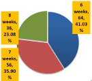
The mean duration of symptoms was 6.82±0.78 weeks. The descriptive statistics are presented in Table 1. The duration was stratified in groups. The frequency and percentages are presented in Figure 2.

Figure 2: Percentage of patients according to duration of symptoms: (N=156).
    Mean ±SD
    6.82±0.78


    95%CI    (LB – UB)
    6.70 – 6.94


    Median    (IQR)
    7.00 (1)


    Range
    2


    Minimum
    6


    Maximum
    8
Table 1: Descriptive statistics of duration of symptoms (weeks) (N=156).
The study results showed that left side was observed affected in 54.5% cases and right side was observed affected in 45.5% cases. The descriptive statistics of duration of symptoms were evaluated according to the findings and presented in Tables 2-4. The final outcome i.e. articular cartilage injury, meniscus injury, and cruciate ligament tear were evaluated in all study patients. The results showed that articular cartilage injury was observed in 11.5% patients, meniscus injury was observed in 31.4%, and cruciate ligament tear was observed in 24.4% cases.
    Yes
    No


    (n=18)
    (n=138)


    Mean    ±SD
    7.06±0.80
    6.79±0.77


    95%CI    (LB – UB)
    6.66 – 7.45
    6.66 – 6.92


    Median    (IQR)
    7
    7


    -2
    -1


    Range
    2
    2


    Minimum
    6
    6


    Maximum
    8
    8
Table 2: Descriptive statistics of duration of symptoms (weeks). According to
articular cartilage injury (N=156).
    Medial
    Lateral
    No


    (n=23)
    (n=26)
    (n=107)


    Mean ±SD
    6.91±0.84
    6.50±0.70
    6.88±0.77


    95%CI (LB – UB)
    6.55 – 7.28
    6.21 – 6.79
    6.73 – 7.03


    Median (IQR)
    7
    6
    7


    -2
    -1
    -1


    Range
    2
    2
    2


    Minimum
    6
    6
    6


    Maximum
    8
    8
    8
Table 3: Descriptive statistics of duration of symptoms (weeks). According to
meniscus injury (N=156).
    Anterior
    Posterior (n=17)
    No


    (n=21)
    (n=118)


    Mean ±SD
    6.76±0.76
    6.82±0.80
    6.83±0.78


    95%CI (LB – UB)
    6.41 – 7.11
    6.41 – 7.24
    6.69 – 6.97


    Median (IQR)
    7
    7
    7


    -1
    -2
    -1


    Range
    2
    2
    2


    Minimum
    6
    6
    6


    Maximum
    8
    8
    8
Table 4: Descriptive Statistics of Duration of Symptoms (Weeks). According to
cruciate ligament tear (N=156).
The stratification according to gender, age, duration symptoms, and affected side was done. Post stratification, associations of findings were observed with these modifiers using chi square test considered p value ≤0.05 as significant. The results showed that significant association of cruciate ligament tear was observed with affected sides (p=0.033) as shown in Table 5. Significant associations of other findings were not observed with gender, age, duration of symptoms, and affected sides.
    CRUCIATE LIGAMENT TEAR
    P-Value


    ANTERIOR
    POSTERIOR
    NO (n=118)
    TOTAL


    (n=21)
    (n=17)


    LEFT (n=85)
    6
    9
    70
    85
    0.033*


    RIGHT (n=71)
    15
    8
    48
    71


    TOTAL
    21
    17
    118
    156


    Chi square    test was applied.
      P-Value    =0.05 Considered as Significant
      *Significant At 0.05    Level
Table 5: Frequency and association of cruciate ligament tear according to
affected sites (n=156).
Discussion
Painful knee joint is the most common orthopedic problem nowadays. Many procedures have been suggested for its diagnosis by different researchers. Till the recent past, clinical evaluation, Magnetic Resonance Imaging (MRI), Examinations under Anesthesia (EUA) and arthroscopy have all been used for the diagnosis of knee joint problems [4-7]. After the series of papers published in the last three decades, the significance of MRI and arthroscopy has been accepted due to their high percentage of diagnostic accuracy. Arthroscopy is now well established as a method of diagnosing meniscal lesions [8-10]. Its advantages have been pointed out in several reports. Arthroscopy is now one of the primary means of diagnosis and treatment of knee lesions. Arthroscopy provides safe, quick and precise method of diagnosis [4,11,12]. Many researchers have tried to prove that MRI has an edge over arthroscopy for the diagnosis of knee joint problems. This may be true for developed countries where the cost of surgery and hospital stay is much higher [5,13].
Hence, a patient, after going through an MRI of the knee joint and showing no need for arthroscopy surgery, saves the relatively higher cost of arthroscopy examination. However, in Pakistan cost of MRI is almost the same as of diagnostic arthroscopy. In a government setup, where the facilities to perform arthroscopy are available, arthroscopy is much cheaper compared to the private sector hospitals. Considering the above-mentioned facts it is more feasible to bypass MRI and opt for arthroscopy examination in case the clinical diagnosis fails to identify underlying pathology in a symptomatic knee joint. During diagnostic arthroscopy, if there is any need of surgical intervention the surgical procedures can be done as a continuation of diagnostic arthroscopy in the same setting without any additional arrangements [4,8].
In a study, the role of diagnostic arthroscopy was reviewed in patients ‘with symptomatic knee joint. This study was conducted to highlight the significance of arthroscopy in diagnosis of knee joint problem. The result of their study show only 65.6% accuracy of clinical diagnosis so the need for performing arthroscopy before surgery is essential [14].
The comparison was done between clinical diagnosis and arthroscopy diagnosis in 64 patients with a correct clinical diagnosis of 65.6%. Mean age of patients was 34 years that is comparable with the study conducted by Carmichael, et al. [15] in 1979, in which they showed the mean age of 34 years. In a similar study male to female ratio was approximately 1:4. Spires et al showed 77% clinical diagnosis accuracy rate that is higher than our results (65.6%) [16]. Suman et al reported a 55% accuracy rate of the clinical diagnosis in a similar study in1984, [5,15] but their study comprised of the patients between ages 14 to 19 years. In a similar study by Anis et al in 1996 found 72% accuracy rate of the clinical diagnosis in comparison with arthroscopic diagnosis in 25 patients [16].
According to Magee, et al. [17]comparison between arthroscopy and MRI presented sensitivity for meniscal injuries of the knee of 89% and demonstrated that signal abnormalities seen on MRI gave information about morphological alterations of injuries. In their study, the sensitivity and specificity values for MRI and arthroscopy were respectively 70.4% and 50% for meniscal injuries. Brooks et al, [18] demonstrated that MRI did not have the capacity to decrease the number of negative arthroscopy procedures, given that the physical examination had concordance of 79% with the arthroscopic findings and MRI showed con-cordance of 77% with arthroscopy.
Din S has reported in his study that out of 64 patients who underwent knee arthroscopy after injury, 28% had meniscal injury, 21% had cruciate injury while 9% had osteoarthrosis (articular cartilage injury 3).
In a study conducted by Maffuli N on 378 patients with complete anterior cruciate ligament tear, 157 showed at least one lesion of the articular cartilage. The medial femoral condyle showed the highest frequency of articular cartilage lesions, especially in the weightbearing portion [19].
The main limitations of the present study include a single-center experience, low female representation and nonrandomized study design. One of the limitations of this study is that it was conducted with small sample size and in urban environment therefore, the results might not be generalizable to larger populations.
Conclusion
It is concluded from the study that Arthroscopy of knee joint has proved to be safe reliable with little morbidity and minor complications. Knee joint with mechanical symptoms with locking should always be considered for arthroscopy.
References
- Phillips BB. Arthroscopy of the lower extremity. In: canale ST, Beaty JH. Campbell’s Operative Orthopaedics. 11th ed., Philadelphia, Mosby: 2812.
- Rossbach BP, Pietschmann MF, Gulecyuz MF, Niethammer TR, Ficklscherer A, Wild S. Indications requiring preoperative magnetic resonance imaging before knee arthroscopy. Arch Med Sci. 2014; 10: 1147-1152.
- Din S, Ahmad I. Arthroscopy: A relieable Diagnostic tool in lesions of Knee Joint. J Postgraduate MedIns. 2011.
- Jackson RW. The role of Arthroscopy in the management of disorders of the knee: An analysis of 200 consecutive examinations. J Bone Joint Surg Br. 1972; 13: 310-322.
- Arnoczky SP. Physiologic principles of ligament injuries and healing. Ligament and Extensor Mechanism Injuries of the Knee: Diagnosis and Treatment. 1991; 67-81.
- Sherman OR, Fox JM, Synder SJ, Pizzo WD, Friedman MJ, Ferkel RD. Arthroscopy: No problem surgery. J Bone Joint Surg Am. 1980; 68: 256-265.
- Suman RK, Stother G, Killingworth. Diagnostic-Arthroscopy of the knee in children. J Bone Joint Surg Br. 1984; 66: 535-543.
- Roy K, Adam H, Steven E, Deborah Mc K. Arthroscopic debridement for osteoarthritis of the knee. J Bone Joint Surg Am. 2006; 88: 936-943.
- Ilamberg P. Gillquist J, Lysholm. A comparison between Arthroscopic meniscectomy and modified open meniscectomy. J Bone Joint Surg. 1984; 66: 189-192.
- Miller MD. What’s new in sports medicine. J Bone Joint Surg Am. 2004; 86: 653-661.
- Glinz W. Diagnostic and operative arthroscopy of the knee joint. Hogrefe & Huber. 1980.
- Vangsness CT, Ghaderi BA, Hohl MA, Moore TM. Arthroscopy of meniscal injuries with tibial plateau fractures. Bone & Joint Journal. 1994; 76: 488-490.
- Carmichael W, MacLeod AM, Travels J. MRI can prevent unnecessary arthroscopies. J Bone Joint Surg Br. 1994: 49: 624-628.
- Shahab-ud-din IA. Arthroscoiy. A reliable diagnostic tool in lesions of knee joint. 2006; 4: 352-325.
- Selesnick FH, Noble HB, Bachman DC, Steinberg FL. Internal derangement of the knee: diagnosis by arthrography, arthroscopy, and arthrotomy. Clin Orthop. 1985; 198: 26-30.
- Anis Y. Diagnosis of the medial meniscal injury. Arthroscopy VS clinical assessment. J Pak Orth. 1996; 2.
- Magee T, Shapiro M, Williams D. MR accuracy and arthroscopic incidence of meniscal radial tears. Skeletal Radiol. 2002; 31: 686-689.
- Brooks S, Morgan M. Accuracy of clinical diagnosis in the knee arthroscopy. Ann R Coll Surg Engl. 2012; 84: 265-268.
- Maffulli N, Binfield PM, King JB. Articular cartilage lesions in the symptomatic anterior cruciate ligament-deficient knee. Arthroscopy: J Arthrosco Related Surg. 2003; 19: 685-690.