
Case Report
Austin J Orthopade & Rheumatol. 2019; 6(2): 1079.
Fibroma of Flexor Tendons Sheath of the Forearm as a Rare Cause of Carpal Tunnel Syndrome: Case Report and Review of Literature
Abaza SE*
Gulf Diagnostic Center Hospital Abu Dhabi, UAE
*Corresponding author: Safaa-eldin Abaza, Gulf Diagnostic Center Hospital Abu Dhabi, UAE
Received: September 27, 2019; Accepted: October 25, 2019; Published: November 01, 2019
Abstract
Fibroma of Tendon Sheath (FTS) is a rare, painless solid, benign tumor is commonly found in upper extremity specially the hands and fingers. FTS of the forearm is further rarer and unusual variety which poses a preoperative challenge. We herein report a case of a 36 years old lady presented with painless swelling at the left forearm associated with symptoms and signs of carpal tunnel syndrome with no predisposing factor of its occurrence .histopathological examination turn to be a Solitary Fibroma of Flexor Tendons Sheath of the forearm (FTS) We reviewed the available English literature and recommend that FTS should be included in the differential diagnosis as one of the causes of carpal tunnel syndrome in young ages ,though rare it may pose anatomical and neurological problems if not treated properly and in time.
Background
Fibroma of the Tendon Sheath (FTS) is a rare benign soft tissue tumor that arises from the flexor tendon sheath of the hand and fingers even rarer in the forearm. Despite its having a distinct microscopic appearance, it is difficult to suggest the diagnosis of fibroma of the tendon sheath prospectively because the lesion shares imaging features with those of other tumors-most notably, giant cell tumor of the tendon sheath.
It usually presents as a painless solitary, slow-growing, firm, small nodule that shows strong attachment to the tendon or flexor tendon sheath over the volar aspect of the fingers, palm, wrist and forearm.
Clinical Presentation and radiological evidence by ultrasound and MRI are non-diagnostic prospectively and share a lot of differential diagnosis specially the most common lesion of giant cell tumor of the tendon sheath and more challenging when associated with symptoms and signs of carpal tunnel syndrome in young age female (male to female ratio of 3:1).
First described by Geschickter and Copeland in 1949, later described as a separate clinic pathological entity by Chung and Enzinger. In their series, 98% of the lesions were in the extremities, 82% in the upper extremities.
The predominant lesion in the upper extremities were the fingers usually (49%); the hand, (21%); and the wrist (12%). Lesions were typically located on the flexor surfaces, occurring most common in men (75%) 20-50 years old with male to female ratio of 3:1., and to be located in the right hand.
Surgery for local excision should be performed carefully, as the recurrence rate is 24%.
Case Presentation
A 36 years old married Egyptian lady having 3 children nonsmoker or alcoholic presented in Jan 2019 with her main complaint are tolerable numbness in the radial three fingers left hand specially at night and a painless soft tissue swelling at the volar aspect lower third of left forearm (Figure 1) insidious onset slowly progressive course over 8 month duration with no history of trauma either direct or indirect, with occasional feeling of pain on incidental pressing on the tumor by hard object ,her main concern is the slowly growing swelling as the numbness is tolerable otherwise she is not complaining of any chronic illness (patient statement).
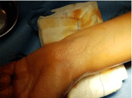
Figure 1: Soft tissue tumor volar aspect lower 1/3 left forearm.
On examination ,Phalen,reverse Phalen test and Tinel sign are all positive for right carpal tunnel syndrome with vague ill-defined soft tissue swelling occupying volar and to the ulnar side aspect of lower 1/3 of left forearm with restricted mobility to the deeper structure but not attached to the normal overlying skin otherwise no peripheral neuro or vascular deficit [1-10].
Investigation
• Systemic examination, blood chemistry and serology tests are all within normal limits.
• Nerve conduction study for median nerve with all figures within normal limits .
• Ultrasound examination in the clinic revealed large hypo and anechoic mass surrounded the flexor tendons both the superficialis and deeper profundus flexor tendons and going deep to the median nerve but not attached to it.
Differential Diagnosis
• Tenosynovial giant cell tumor
• Inclusion body fibromatosis
• Nodular fasciitis
• Desmoplastic fibroblastoma
• Palmar/plantar fibromatosis
Treatment
Under G.A tourniquet ,no isthmach and via zigzag volar skin incision (Figure 2,3) including flexor surface of the lower 1/3 left forearm and extending distally parallel to the thenar crease in the palm, the median nerve and its superficial palmar branch were isolated over vessel loop (Figure 4,5) ,the tumor was found as a nodular dirty brownish and reddish coloration soft tissue encircle the flexor tendons both the superficialis and the deeper profundus flexor tendons extending distally to the carpal tunnel along the flexor tendons occupying the space between the median nerve and the ulnar neurovascular bundle (Figure 6,7),first the carpal tunnel was opened totally (Figure 8) and the tumor was carefully dissected and excised from flexor tendons first the superficialis (Figure 9) then deep to the profundus flexor tendons (Figure 10) down to the pronator quadrates (Figure 11) and extending from the median nerve laterally to the ulnar neurovascular bundle medially (Figure 12), there was also tenosynovitis and synovial thickening which was removed (synovectomy of both superficialis and profundus flexor tendons (Figure 13A,13B), hemostasis and closure in layers (Figure 13C), the excised tumor and synovia was sent to histopathology examination.
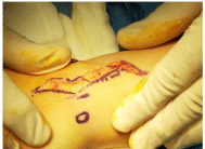
Figure 2: Volar Zigzag skin incision.
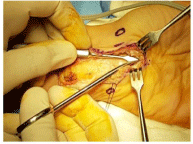
Figure 3: Carpal tunnel opening and release.
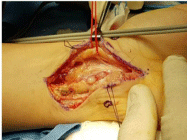
Figure 4: Isolation of the median nerve and its palmar branch.
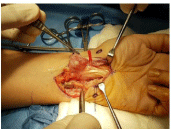
Figure 5: The tumor in the flexor superficialis tendons.
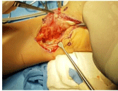
Figure 6: The tumor extend to the deeper flexor profundus tendons.
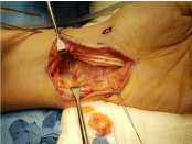
Figure 7: Excision of the tumor en mass.
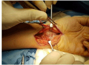
    Figure 8: Excision of the tumor in both the superficialis and profundus flexor
tendons down to pronator quadratus muscle.
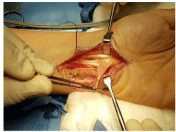
Figure 9: Flexor tendons synovectomy.
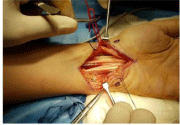
Figure 10: Complete clearance of the tumor and synovectoy.
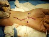
Figure 11: Closure in layers.
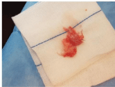
Figure 12: Well circumscribed, small fibrous multinodular mass.
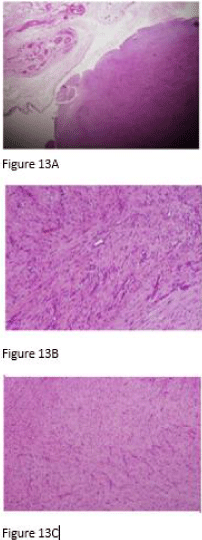
    Figure 13: Showing Well circumscribed nodules of dense fibrous tissue with
occasional spindle or stellate mesenchymal cells in S or C shaped patterns:
A: Cells have scant cytoplasm and elongate nuclei with evenly distributed
fine chromatin.
B: Often dilated or slit-like channels/clefts resembling tenosynovial spaces.
C: Varies from cellular to paucicellular.
Outcome & Follow-up
Post-operative passed uneventful with no postoperative complaint, stitched were removed after 2 weeks wound closed with primary intension and patient felt relief of the night numbness.
The result of the histopathology examination confirm the diagnosis of tendon sheath fibroma with well circumscribed nodules of dense fibrous tissue with occasional spindle or stellate mesenchymal cells in S or C shaped pattern, cells have scanty cytoplasm , elongated nuclei with fine chromatin with presence of dilated or slit like channels and clefts resemble tenosynovial spaces, no atypical mitosis, necrosis or hyperchromatosis, the nearby synovial tissues shows signs of inflammation with tenosynovitis.
Features are in keeping with fibroma of tendon sheath with nearby tenosynovitis and no granuloma and no malignancy can be seen.
Discussions and Brief Review of Similar Cases
Fibroma of the tendon sheath is a rare benign tumor of the fingers ,palm and wrist even more rarer in forearm mostly presented in men, aged 30-50 years old(male to female ratio 3 to 1).
Our case is young female with her main complaint is symptoms of tolerable carpal tunnel syndrome in her non dominant left hand associated with noticeable soft tissue swelling volar aspect lower 1/3 left forearm with localized tender to incidental pressure by a hard objects.
Clinically the tumors presents as solitary, slow-growing subcutaneous nodules. About one third of the cases present with localized tenderness and pain due to compression of the underlying nerves.
Even it is not our case but worth mentioning that Fibroma of tendon sheath has also been reported to cause a “trigger wrist” an impingement of the tumor adhering to the flexor tendons in the carpal canal, resulting in snapping fingers or carpal tunnel syndrome. It has several causes including tumors, tenosynovial thickening and abnormal muscle bellies. The tumors known to cause “trigger wrist” are FTS and tenosynovial giant cell tumor, both tend to adhere to the flexor tendons.
Trigger wrist usually complain about the following symptoms: snapping and clicking or triggering around carpal tunnel with or without mild to moderate median neuropathy. There are a total of five cases in literatures of trigger wrist: three cases of anomalous muscle belly of flexor digitorum superficialis and two cases of fibroma around flexor tendon sheath within carpal tunnel.
FTS is most confused with giant cell tumor (GCT) of the tendon sheath. Both lesions may occur in similar location although as compared to GCT, FTS is more common in younger in males and tend to occur in upper extremity.
Microscopically, FTS is hypocellular, with slit like vascular channels within a dense collagen matrix, whereas GCT of the tendon sheath is more cellular, cells have histocyte -like nuclei, prominent giant cells, foam cells and hemosiderin but no slit-like vascular spaces and no extensive hyalinized stroma (Table 1).
    Tenosynovial Giant Cell Tumor
    Fibroma of Tendon Sheath


    Oval histiocyte-like nuclei
    Elongate nuclei


    Giant cells nearly always    present
    Giant cells rare


    Foamy histiocytes common
    Histiocytes rare


    Hemosiderin common
    Hemosiderin rare


    No slit-like vascular spaces
    Slit-like vascular spaces    common


    Smooth muscle actin negative
    Smooth muscle actin strong    positive


    Desmin may be positive
    Desmin negative


    Printed    from Surgical Pathology Criteria: https://surgpathcriteria.stanford.edu/ 2005.
      Stanford University    School of Medicine.
Table 1: Surgical pathology criteria.
Learning Points and Take Home Messages
1. Prospective diagnosis of FTS like any tumor essentially by histopathological examination as it has to be differentiated from other common soft tissue tumor of forearm and wrist mostly tenosynovial giant cell tumor.
2. FTS has to be in the deferential diagnosis of the carpal tunnel syndrome in young age associated with apparent or otherwise hidden soft tissue tumor of forearm and wrist as any space occupying lesion e.g. a mass of flexor tenosynovitis, anomalous of flexor muscle belly, or benign tumors inside carpal tunnel, often cause median neuropathy. Surgeons probably will perform an early release of carpal tunnel only. Although the symptoms may be solved via this procedure, but the main problem still remains.
3. Complete wide excision with its capsule is the treatment of choice with preserving the nearby important structure especially neurovascular bundles and considering recurrence rate in literatures about 24%.
4. Although it is a rare benign tumor but it may pose anatomical and neurological complications if not diagnosed and treated properly in time.
References
- Garrido A, Lam WL, Stanley PR. Fibroma of a tendon sheath at the wrist: a rare cause of compression of the median nerve. Scand J Plast Reconstr Surg Hand Surg. 2004; 38: 314-316.
- Jaggi Rao, Achilleas Thoma and Sam Salama, Fibroma of Tendon Sheath as a Cause of Carpal Tunnel Synrome, Canadian Journal of Plastic Surgery. 1997.
- Satti MB. Tendon sheath tumours: a pathological study of the relationship between giant cell tumour and fibroma of tendon sheath. Histopathology. 1992; 20: 213-220.
- Crossref.
- Arora K. Fibroma of tendon sheath. Pathology Outlines.com website. 2019.
- Millon SJ, Bush DC, Garbes AD. Fibroma of tendon sheath in the hand. St louis: 2001; 1361-1388.
- Kosuge, D, Narin, D. Flexor tendon fibroma as a cause of wrist triggering and carpal tunnel syndrome.
- Srivastava P, Srivastava S. Fibroma of Flexor Tendon Sheath of Finger: A Rare Tumor of Hand-Revisited. J Neoplasm. 2018; 3: 1.
- Heckert R, Bear J, Summers T, Frew M, Gwinn D, McKay P. Fibroma of the tendon sheath-a rare hand tumor. Pol Przegl Chir. 2012; 84: 651-656.
- Fibroma of the tendon sheath is a lesion composed of tightly packed spindle cells surrounded by collagen fibers.