Case Report
Austin J Orthopade & Rheumatol. 2019; 6(2): 1080.
The Polytraumatised Shoulder: A Case of Triple Disruption of the Shoulder Superior Suspensory Complex
Rehman H*, Jun Wei LIM and Kumar K
Department of Orthopedics and Trauma, Aberdeen Royal Infirmary, UK
*Corresponding author: Haroon Rehman, Department of Orthopaedic and Trauma, Aberdeen Royal Infirmary, Aberdeen, UK
Received: October 29, 2019; Accepted: November 26, 2019; Published: December 03, 2019
Abstract
Multiple injuries to the shoulder’s superior suspensory complex can result in significant morbidity and disability. Concomitant fractures of the acromion and the coracoid process of scapula in association with acromioclavicular dislocation is a rare injury, usually resulting from direct trauma to the shoulder. Triple disruptions of the shoulder’s superior suspensory complex can have debilitating consequences for patients if treated inadequately. Surgeons can be distracted by the more common and more obvious injuries such as acromioclavicular joint dislocation and missed significant fractures of the shoulder’s superior suspensory complex. We reported an interesting case of triple disruption to the shoulder superior suspensory complex. We described the surgical procedure and postoperative care in this care report. We hope our case will draw attention to the significance of a polytraumatised shoulder, which may only have subtle features on plain film x-rays.
Keywords: Coracoid fracture; Acromion fracture; Acromioclavicular joint dislocation; Triple disruption; Shoulder superior suspensory complex
Introduction
Multiple injuries to the Shoulder’s Superior Suspensory Complex (SSSC) can result in significant morbidity and disability. The SSSC includes the glenoid fossa, the coracoid process, the coracoclavicular ligament, the distal end of the clavicle, the acromioclavicular joint, the coracoacromial ligament, and the acromion. The integrity of this complex is essential to normal shoulder biomechanics and operative intervention is indicated in cases where multiple disruptions to the SSSC have occurred [1]. Combined injuries of the shoulder can be missed at initial presentation if simple and careful evaluation of the patient and radiographs is not performed.
Coracoid fractures account for approximately 2-13% of scapular fracture and approximately 1% of all fractures [2,3]. Approximately 8-9% of all scapular fractures involve the acromion [4]. Concomitant fractures of the acromion and the coracoid process of scapula in association with acromioclavicular dislocation is a rare injury, usually resulting from high force trauma to the shoulder.
An extensive literature review reveals little discussion regarding triple disruption of the SSSC. We report an interesting case of triple disruption to the SSSC involving a fracture of the coracoid process with concomitant acromion fracture and acromioclavicular joint dislocation. This case report also provides further discussion on SSSC injury mechanism, clinical evaluation, operative treatment and subsequent functional outcomes.
Case Presentation
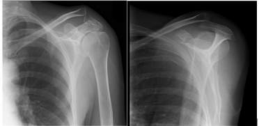
Our patient was an independent, high functioning, 56-year-old female who presented after falling down a flight of stairs onto a tiled floor. She had no significant past medical history. She sustained a left sided shoulder injury initially misdiagnosed as isolated acromoclavicular dislocation on plain radiographs (Figures 1a & 1b), in addition to multiple rib fractures and a small apical pneumothorax.

    Figure 1: 1a: Left shoulder X ray (AP view); 1b: Left shoulder X ray (lateral
view).
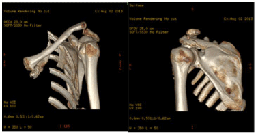
At her 2-week outpatient follow up, the extent of her shoulder injury became apparent. She had sustained acromioclavicular joint separation with superior migration of the clavicle; a displaced acromion fracture and a Type 1 (Ogawa Classification) coracoid fracture [3]. Further imaging with computed tomography was obtained to confirm the fracture pattern and characteristics (Figures 2a & 2b). She was admitted to a tertiary orthopaedic trauma unit for open reduction and internal fixation.

    Figure 2: 2a: CT 3D reconstruction of left shoulder (anterior); 2b: CT 3D
reconstruction of left shoulder (posterior).
Surgical Technique
Position and preparation: The patient was positioned in the beach chair position and underwent her operation under general anaesthetic. The left upper limb was prepared and draped as per standard protocol.
Procedure: An anterolateral approach to the shoulder with deltoid split was used. The corocoid process was identified but we had difficulty accessing its base. A clavicle osteotomy had to be performed and the coracocloavicular ligament was partially divided. The deltoid muscle was then reflected inferiorly for better visualisation of the fracture site. The corocoid fracture was exposed, reduced and lagged with 2 x 4.0mm partially threaded screws. The proximal extension of the wound was used for approach to the acromion. The acromion was fixed with a 1/3 tubular plate. The clavicle osteotomy was reduced and fixed with fibre wire. A fibre wire was also used to reattach the coracoclavicular ligaments. Standard layered closure was performed.
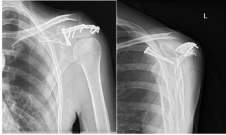
Post-operative care: Postoperatively, the left upper limb was immobilised in a sling with an abduction wedge for 3 weeks. Active abduction was restricted for 6 weeks from surgery. Clinical review and check radiographs were performed at 1,3 and 6 weeks postoperative; and subsequently at 3, 6, 12 and 18 months postoperative (Figures 3a & 3b). At 3 weeks postoperative, the patient was allowed to commence passive range of movement exercises with the physiotherapist. At 6 weeks postoperative, the sling was removed and unrestricted physiotherapy was commenced.

    Figure 3: 3a: Left shoulder postoperative X ray (AP view); 3b: Left shoulder
postoperative X ray (lateral view).
Outcomes: At 3 months postoperative, the patient had 40 degrees of active forward flexion, 40 degrees of active abduction and 10 degrees of active external rotation. On passive range of movement examination, she had 80 degrees of forward flexion and 80 degrees of abduction.
At 6 months postoperative, the patient had 100 degrees of active forward flexion, 60 degrees of active abduction, 40 degrees of active external rotation and was able to touch the L3 vertebra for internal rotation. On examination of the contralateral (unaffected) limb, she was capable of 90 degrees of external rotation. A plateau in her progress with physiotherapy was noted. Therefore, arthroscopic capsular release and removal of metalwork was subsequently performed at 8 months after her primary operation.
3 weeks following her capsular release operation (9 months post primary procedure), she had 120 degrees of active abduction and 40 degrees of active external rotation. At 18 months post injury, she had 170 degrees of active forward elevation, 140 degrees of active shoulder abduction, and 60 degrees of active external rotation. She had an oxford shoulder score of 38/60 and had returned to her routine activities (Appendix 1).
Discussion
Triple disruptions of the SSSC can have debilitating consequences for patients if treated inadequately. Surgeons can be distracted by the more common and more obvious injuries such as acromioclavicular joint dislocation and missed significant fractures of the SSSC. We seek to draw attention to the significance of a polytraumatised shoulder, which may only have subtle features on plain film x-rays.
In isolation, disruptions to the SSSC are often innocuous and suitable for conservative management. Unlike bony pelvis, the bony and soft tissue ring of the SSSC can be broken at a single point as the force is dissipated via the acromioclavicular and coracoclavicular ligament. However, a high energy force transmission can still cause failure of the complex at multiple points. Untreated of such injuries can lead to functional deficit, malunion, non-union, pain, impingement, fatigue, weakness, neurovascular injury and/or early osteoarthritis [5,6].
Although double disruptions of the SSSC have been previously reported [5,6], there is a paucity of literature on triple disruption. Goss describes three separate cases of double disruption to the SSSC including: Type V clavicle fracture; combined glenoid and clavicle fractures; acromion and coracoid process fractures [6]. Goss promotes surgical intervention to achieve good outcomes due to the inherent instability of this kind of injuries [6,7]. We have identified only one other case report on triple disruption of the SSSC [8].
Four mechanisms of injury resulting in acromion fractures have been described in the literature: direct force; transmitted force via humeral head through traumatic superior displacement, or superior migration as a result of rotator cuff arthropathy; avulsion fracture secondary to forceful deltoid muscle contraction; and stress fracture [4]. Acromion fracture is quite often undisplaced or minimally displaced and thus amenable to conservative treatment with sling immobilisation. Non-operative treatment is also indicated in displaced fractures in cases where the subacromial space is not diminished [9]. Isolated fractures of the acromion can be readily accessible using a posterior approach alone. An incision is made over the posterior border of the acromion, down through the fascia separating the deltoid and trapezius. This approach allows the deltoid to be reflected inferiorly and permits adequate visualisation of the acromion. Although our patient’s fracture was relatively distal, the reduction was adequately maintained with 1/3 tubular plate and screws alone. The distal end of the acromion is thinner and some authors have advocate tension band wiring as method of fixation [10]. In cases where the base of the acromion is fractured, surgeons should consider fixation with reconstruction plate. Other treatment options include suture, staples and kirschner wires.
Coracoid fracture can occur in isolation or as in our case, part of an injury complex with acromion involvement. In adult, the fracture occurs most commonly at the base of the coracoid [3,11]. Several mechanisms of injury of coracoid fracture have been described in the literature. Isolated coracoid fracture can occur through direct trauma to anterolateral of the shoulder. Avulsion fracture from the coracoid base can occur due to sudden contraction of the conjoined tendon in resisted flexion of the arm [12,13]. Avulsion injury of the coracoid can also be caused by acromioclavicular dislocation whereby there is a caudad displacement of the clavicle results in avulsion through pull of intact coracoclavicular ligaments. Epiphyseal separation of the coracoid with acromioclavicular ligament sprain is commonly described in adolescents [14]. The largest series of coracoid fractures in adults describes associated acromioclavicular disruption, acromial fractures, clavicle, scapular and glenoid fractures [13]; such injuries should be actively excluded on presentation. It is not unusual for coracoid fracture to present with concomitant acromioclavicular separation [13,15]. Ogawa et al found that 37 of 67 coracoid fractures were associated with ipsilateral acromioclavicular joint dislocations [3]. There is no consensus on the treatment of coracoid fractures in the literature. Operative treatment in the form of screw fixation has been described and is indicated in those cases where displacement has occurred [5,11,16]. The coracoid process can be approached through direct incision through the skin overlying it, which form an extension of the deltopectoral approach in cases where the glenoid fossa requires attention too. The base of the coracoid must be identified to allow anatomic reduction. This requires dissection down to and along the cephalad slope of the coracoid process. The fracture can be adequately stabilized with a single lag screw. Occasionally additional stability is conferred using 1/3 tubular plate fixed at the cephalad slope of the fracture. However, it was not indicated in our case based on intraoperative findings.
In our case, the acromion fracture and acromioclavicular separation was probably caused by direct trauma, resulting in superior displacement of the ipsilateral clavicle with subsequent avulsion of the coracoid through the pull of intact coracoclavicular ligaments. Standard plain film radiography consisting of 3 shoulder views may not be enough to diagnose this injury. Careful attention should be given to the acromion and coracoid process; the aid of specific views such as stryker notch view may be required. Surgeons should have a low threshold to request further imaging such as radiographs with a 450 to 600 cephalad tilt, computed tomography or magnetic resonance imaging as further investigation.
Conclusion
We present a case of a successfully treated triple injury to the shoulder superior suspensory complex. We hope our case will encourage vigilance when examining seemingly isolated SSSC fractures, especially in cases where high force trauma is involved.
References
- Oh C, Jeon I, Kyung H, Park B, Kim P, Ihn J. The treatment of double disruption of the superior shoulder suspensory complex. Int Orthop. 2002; 26: 145-149.
- Ada JR, Miller ME. Scapular fractures: analysis of 113 cases. Clin Orthop. 1991; 269: 174-180.
- Ogawa K, Yoshida A, Takahashi M, Ui M. Fractures of the coracoid process. Journal of Bone & Joint Surgery. 2011; 79: 17-19.
- Rockwood CA, Green DP, Bucholz RW, Heckman JD. Rockwood and Green’s fractures in adults. Lippincott Williams & Wilkins. 2006.
- Goss TP. The scapula: coracoid, acromial, and avulsion fractures. Am J Orthop. 1996; 25: 106-115.
- Goss TP. Double disruptions of the superior shoulder suspensory complex. J Orthop Trauma. 1996; 7: 99-106.
- Lim K, Wang C, Chin K, Chen C, Tsai C, Bullard MJ. Case report: concomitant fracture of the coracoid and acromion after direct shoulder trauma. J Orthop Trauma. 1996; 10: 437-439.
- Kim SH, Chung SW, Kim SH, Shin SH, Lee YH. Triple disruption of the superior shoulder suspensory complex. Int J Shoulder Surg. 2012; 6: 67.
- Kuhn JE, Blasier RB, Carpenter JE. Fractures of the acromion process: a proposed classification system. J Orthop Trauma. 1994; 8: 6-13.
- Anavian J, Wijdicks CA, Schroder LK, Vang S, Cole PA. Surgery for scapula process fractures: good outcome in 26 patients. Acta orthopaedica. 2009; 80: 344-350.
- Eyres KS, Brooks A, Stanley D. Fractures of the coracoid process. J Bone Joint Surg Br. 1995; 77: 425-428.
- Rounds RC. Isolated fracture of the coracoid process. The Journal of Bone & Joint Surgery. 1949; 31: 662-663.
- Asbury S, Tennent T. Avulsion fracture of the coracoid process: a case report. Injury. 2005; 36: 567-568.
- Montgomery SP, Loyd RD. Avulsion fracture of the coracoid epiphysis with acromioclavicular separation. Report of two cases in adolescents and review of the literature. J Bone Joint Surg Am. 1979; 59: 963-965.
- Hak DJ, Johnson EE. Case Report and Review of the Literature: Avulsion Fracture of the Coracoid Associated with Acromioclavicular Dislocation. J Orthop Trauma. 1993; 7: 381-383.
- Dawson J, Fitzpatrick R, Carr A. Questionnaire on the perceptions of patients about shoulder surgery. J Bone Joint Surg Br. 1996; 78: 593-600.