
Research Article
Austin J Orthopade & Rheumatol. 2020; 7(1): 1083.
Outcome of Clavicular Hook Plate in Management of Unstable Lateral Clavicular Fractures
Alghamdi AA*
Department of Surgery, Faculty of Medicine, Baha University, Saudi Arabia
*Corresponding author: Ahmed Abdullah Alghamdi, Surgical Department, Faculty of Medicine, Baha University, Saudi Arabia
Received: February 07, 2020; Accepted: February 18, 2020; Published: February 25, 2020
Abstract
Background: Lateral clavicular fracture is a topic in orthopedic fracture care that has been heavily debated over the last decades. The aim of the current study was to assess the results of hook plate in management of lateral clavicular fractures.
Patients and Methods: Twenty one lateral clavicular fractures, in 21 patients were prospectively included in the current study. There were 15 males and 6 females, with a mean age of 32.8 years. A clavicular hook plate was used in reduction and fixation of the fractures in all patients. The plate was electively removed after 6 months postoperatively.
Keywords: Lateral clavicular fractures; Hook plate; Clavicular plate
Introduction
Fractures of the clavicle are common fractures involving 10% of adult fractures, and about one third of fractures involving the shoulder girdle in adult, with the majority involving the midshaft and the lateral end is involved in about 28% [1]. The latter was classified according to the relationship of the fracture line to the Coracoclavicular (CC) ligaments and the extension into the Acromioclavicular (AC) joint. Type I fractures occur lateral to the CC ligaments, are usually stable. In type II the fracture line occurs medial to the CC ligaments resulting in displacement of the medial fragment. Type III are intra-articular fracture involving the AC joint, the majority are not displaced [2].
Conservative treatment remains a reasonable option for type I and III lateral clavicular fractures with favorable outcome. Unstable type II fractures carries a high risk of symptomatic non-union in about half of the cases [3,4]. Indications for surgery included unstable fractures, open fractures, flail shoulder, and associated neurovascular injuries [2].
Many fixation techniques have been described for treatment of displaced lateral clavicular fractures, including transacromial Kirschner wires, tension band wires [5,6] coracoclavicular screws or sutures, and plate fixation. None of these techniques is regarded as the gold standard. Clavicular hook plate fixation has been used recently providing rigid fixation and good bony union rates. However conflicting data regarding complications have been reported. The aim of the current study was to evaluate outcome of fixation of unstable lateral clavicular fractures using clavicular hook plate.
Patients and Methods
Between May 2013, and May 2016, 21 lateral clavicular fractures in 21 patients were treated by clavicular hook plate at Alhada Military Hospital. Inclusion criteria included displaced lateral clavicular fractures. Displacement was defined either clinically by deformity (skin tenting or impending skin penetration) or radiologically by more than 15 mm displacement in anteroposterior view. Exclusion criteria were open fractures and associated shoulder girdle fractures.
Fifteen were males and 6 were females with a mean age of 32.8 years (range, 22 to 48). Ten patients injured their dominant shoulder. All fractures were acute with the average time to surgery was 5 days (range, 2 to 14). The mechanism of injury was a fall on the shoulder 13 patients, and motor-vehicle accidents in 8. The study was approved by institutional ethical board of Benha University and all patients have signed an informative consent.
Patients were evaluated clinically and radiologically using at least Antero Posterior (AP), lateral, and axial views. Patients were classified according to Neer classification [2]. Ct scans were done for patients with suspected Ac joint involvement or suspected glenoid fractures. There were 16 patients with Neer type II fractures, and 5 with type III.
Surgical technique: Patients were positioned in the beach chair position with the affected arm draped free, on an orthopedic radiolucent table, with access for intraoperative radiography.
Longitudinal skin incision along the anterior border of the lateral half of the clavicle was used (Figure 1). The deltoid insertion with the periosteum was incised along the skin incision exposing the fracture. The posterior border of the AC joint was identified and the hook of the plate was passed through 5 mm snip in the trapezium passing under the acromion. The plate was then used to indirectly reduce the medial clavicle into position. Reduction was checked by fluoroscopy and the screws were sequentially inserted from medial to lateral.
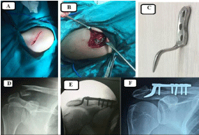
    Figure 1: A: Longitudinal skin incision; B: Insertion of plate hook posterior
to ac joint with fixation of medial screws; C: The clavicular hook plate; D:
preoperative radiograph; E: Intraoperative radiograph; F: Postoperative
radiograph.
Post-operative care: The shoulder was immobilized in arm sling, and passive and active assisted movements were started as tolerated. Active movements were allowed after 2 weeks. Radiological assessments were done immediately after surgery, at 6 weeks, and every 2 weeks till union then every 3 months till first year, then annually.
Clinical assessment was done using Visual Analog Scale (VAS) for pain (composed of 10 points where zero is no pain at all and 10 is maximum unbearable pain), shoulder range of movement, impingement tests, and Constant functional shoulder score. The plate was routinely removed after 6 monthes from all patients except one who refuse to undergo subsequent surgeries. Results at 6 months (before removal of the plate), and at last follow-up were analyzed.
Results
The average duration of follow-up was 31 months (range, 24 to 41). All fractures were united at an average of 8.7 weeks (range, 6 to 12). The average active shoulder movements in 6 months’ postoperative follow-up were as follows: abduction 135 (range, 100 to160), forward flexion 150 (range, 100 to170), external rotation 30 (range, 30 to35) and internal rotation 50 (range, 40 to 75). The average active shoulder movements in the latest follow-up were as follows abduction 1158 (120 to170), forward flexion 160 (range, 130 to170), external rotation 35 (range, 30 to 45) and internal rotation 50 (range, 30 to 65). There was a statistically significant improvement in range of motion after plate removal (p‹0.001).
The mean age adjusted constant score was 88 (range 82 to 94) in the 6 months follow up, which improves significantly to 94 (range, 84 to 100) in the last follow-up (p‹0.01). The average VAS for pain was 2.3 (range, 0 to 4) at 6 months and 2.2 (range, 0 to 4) at final followup with no statistical significant differences. There was no significant differences between type II and type III fractures as regard union time or outcome scores.
Complications
One patient had superficial skin infections, which was resolved by antibiotics and dressing. sixteen patients had impingement signs and symptoms at the 6 months’ follow-up. Symptoms improved in 14 patients 4 to 6 weeks after removal of the plate, while in two patients symptoms persist and both had arthroscopic repair of infraspinatus tendon tear evident by MRI. None of our cases developed radiologic evidences of osteolysis in the acromion or arthritic changes in AC joint.
Discussion
Management of the lateral clavicle fractures had been a matter of debate for many decades. There is agreement about fixation of displaced Neer types II and III fractures owing to high non-union rates in conservatively managed fractures. However, there is no consensus about the gold standard fixation techniques. That explain the presence of many fixation methods from K wires to plates.
The gold standard method in management of unstable fractures, remain anatomical reduction of the fracture and stable fixation to allow early mobilization. In the current series, we present the results of 21 unstable lateral clavicular fractures treated by clavicular hook plate. Union was achieved in all fractures at an average of 8.7 weeks (range, 6 to 12) with favorable shoulder function as manifested by an average age adjusted constant score of 94 (range, 84 to 100) at final follow-up. The results obtained in the current series were similar to those obtained in similar case series [7-9] using the clavicular hook plate (Table1).
![]()
    Study
    No of Patients
    Union time
    Union rate
    Constant score at final follow    up
    Complications


    Meda et al. (2006) [8]
    31
    12 weeks (6-18)
    100%
    92 (84-100)
    2 cases superficial infection,    6 impingement, 5 osteolysis


    Tiern et al. (2012) [9]
    28
    _
    96%
    97
    9 impingeemnt, 7 osteolysis,    one nonunion


    Good et al. (2012) [7]
    36
    12 week (8-16)
    95%
    83.8 (44-100)
    2 fracture clavicle at medial    edge of plate, one nonunion


    Current study (2017)
    21
    8.7 weeks (6-12)
    100%
    94 (84-100)
    1 superficial infection, 16    impingement
Table 1: Shpwig results of different studies using clavicular hook plate.
Neer and Watson-Jones used AC K-wires and reported good results as regard fracture union. However, later reports of complications including hardware failure, breakage, and migration, and prolonged immobilization resulted in poor functional results. Rockwood and Lyon concluded that K-wires is a poor option in management of lateral clavicular fractures [10]. Adding tension band to the acromioclavicular wires either metal or sutures, didn’t reduce the risk of wire migration and breakage. Hsu et al. compared results of 35 patients fixed by hook plate and 30 patientsfixed by transacromial tension band wiring. They reported comparable union time and functional outcome at 6 months in both groups but wires migration and failure in 5 patients of the tension band group. Transacromial fixation is seldome used nowadays because it violates the AC joint with possible subsequent arthritis and prevent the normal AC joint movement resulting in early implant failure.
Coracoclavicular screws traditionally used for AC joint dislocations- was used to indirectly reduce and fix unstable distal clavicular fractures. Very few studies have showed good results and few complications [11,12]. However, the technique didn’t gain popularity owing to the potential risks of neurovascular injury or coracoid fractures, and the prolonged immobilization for fear of screw failure. Alternatively, a less rigid coracoclavicular fixation using many types of sutures and bands have gained popularity with comparable good results and less complications. Most of these band fixations could be done arthroscopically [13,14]. However coracoid fracture and failure of fixation were reported [14]
The clavicular hook plate was designed to allow indirect fixation of the small or comminuted lateral clavicular fragment and at the same time allowing a stable construct that allow union but didn’t omit acromioclavicular movements. Stegeman et al, published a meta-analysis of different fixation techniques in treatment of unstable lateral clavicular fractures [15]. There was no significant outcome difference between the clavicular hook plate and the other fixation methods. But the hook plate fixation was associated with a 24-fold increased risk of complications compared to coracoclavicular suture fixation and an 11-fold increased risk of major complications compared to acromioclavicular fixation.
As regard complications, 16 patients developed impingement signs that resolved after removal of the plate. Routine clavicular hook plate removal is not universally regarded as essential, and there are reports of good outcomes with retained plate [8,16]. Our results have shown that range of shoulder movement, impingement signs and functional outcomes scores significantly improved after removal of the plate. No hardware failure was recorded in the current series. In the largest published series of 222 patients, Zhu, et al. reported 7 (1.50%) clavicular stress fracture, 5 (1.07%) hook cut -out and 3 (0.64%) hook breaks [17].
Conclusion
Fixation of unstable lateral clavicular fractures using clavicular hook plate yielded good results and complications, however routine plate removal is mandatory to avoid rotator cuff problems.
Level of evidence: Type IV therapeutic case series.
References
- Nordqvist A, Petersson C. The incidence of fractures of the clavicle. Clin Orthop Relat Res 1994; 300: 127-132.
- Neer CI. Fractures of the distal third of the clavicle. Clin Orthop Relat Res. 1968; 58: 43-50.
- Chalidis B, Sachinis N, Samoladas E, Dimitriou C, Christodoulou A. Acute management of clavicle fractures. A long term functional outcome study. Acta Orthop Belg. 2008; 74: 303-307.
- Robinson C, Cairns D. Primary nonoperative treatment of displaced lateral fractures of the clavicle. J Bone Joint Surg Am. 2004; 86: 778-782.
- Badhe S, Lawrence T, Clark D. Tension band suturing for the treatment of displaced type 2 lateral end clavicle fractures. Arch Orthop Trauma Surg. 2007; 127: 25-28.
- Hsu T, Hsu S, Chen H, Wang S. Comparison of hook plate and tension band wire in the treatment of distal clavicle fractures. Orthopedics. 2010; 33: 879.
- Good D, Lui D, Leonard M, Morris S, McElwain J. Clavicle hook plate fixation for displaced lateral-third clavicle fractures (Neer type II): a functional outcome study. J Shoulder Elbow Surg. 2012; 21: 1045-1048.
- Meda P, Machani B, Sinopidis C, Brownson P, SPFrostick. Clavicular hook plate for lateral end fractures: A prospective study. Injury. 2006; 37: 277-283.
- Tiren D, Bemmel A, Swank D, Linden Fvd. Hook plate fixation of acute displaced lateral clavicle fractures: mid-term results and a brief literature overview. J Orthop Surg Res. 2012; 7: 2-1.
- Lyons F, Rockwood C.Migration of pins used on the shoulder. J Bone Joint Surg Am. 1990; 72: 1262-1267.
- Macheras G, Kateros K, Savvidou O. Coracoclavicular screw fixation for unstable distal clavicle fractures. Orthopaedics. 2005; 28: 693-696.
- Fazal M, Saksena J, Haddad F. Temporary coracoclavicular screw fixation for displaced distal clavicle fractures. J Orthop Surg. 2007; 15: 9-11.
- Nourissat G, Kakuda C, Dumontier C, Sautet A, Doursounian L. Arthroscopic stabilization of Neer type 2 fracture of the distal part of the clavicle. Arthroscopy. 2007; 23: 671-674.
- Cho C-H, Bae K-C, Kim DH. Coracoclavicular Stabilization Using a Suture Button Device for Neer Type IIB Lateral Clavicle Fractures. Arthroscopy: The Journal of Arthroscopic & Related Surgery. 2017; 33: 59-60.
- Stegeman S, Nacak H, Koen HH, nger B. Surgical treatment of Neer type-2 of the distal clavicle, a meta-analysis. Acta Orthop. 2013; 84: 184-190.
- Faraj A, Ketzer B. The use of a hook-plate in the management of acromioclavicular injuries. Acta Orthop Belg. 2001; 67: 448-451.
- Zhu Y, Cui H, Jiang P, Wang J. Complications of treatment of acromioclavicular joint dislocation and unstable distal clavicular fracture with clavicular hook plate. Zhongguo Gu Shang. 2013; 26: 927-931.