
Research Article
Austin J Orthopade & Rheumatol. 2020; 7(2): 1087.
Clinical Profile of Plasmoytomas: About 11 Cases
Rachdi I*, Aydi Z, Hannach E, Zoubeidi H, Hamrouni S, Dhaou BB, Daoud F and Boussema F
Internal Medicine Department, Habib Thameur Hospital, Faculty of Medicine of Tunis, Tunis el Manar University, Tunis, Tunisia
*Corresponding author: Imène Rachdi, Internal Medicine Department, Habib Thameur Hospital, 1008, Ali-Ben-Ayed Street, Montfleury, Tunis, Tunisia
Received: June 08, 2020; Accepted: July 07, 2020; Published: July 14, 2020
Abstract
Aims of the Study: Determine the clinical, topographic, therapeutic and evolutionary peculiarities of of plasmocytomas in a department of internal medicine.
Methods: Retrospective study over 5 years from January 2014 to January 2018 collecting 11 cases of plasmocytomas of different localizations hospitalized in a department of internal medicine. The diagnosis was confirmed histologically in all cases.
Results: We retained eleven observations of plasmocytomas. The average age was 64.09 years. The sex ratio was 1.2. The plasmocytoma revealed MM in all cases. The seat of the plasmocytoma was osseous in 6 cases (iliac n = 2, costal n = 1, sternocostal n = 1, vertebral n = 1 and medullary n = 1). Extra-osseous locations were found in 5 cases (n orbital = 2, n = 1 retroperitoneal, n = 1 and axillary n = 1). Renal failure was noted in five cases. MM was classified stage III according to the Salmon and Durie classification in five cases. The treatment was based on chemotherapy associated or not with decompressive radiotherapy that was prescribed in 7 patients. The evolution was favorable in 5 cases. In two patients, there was a progression of the mass despite radiotherapy. Two cases of death were reported due to disseminated intravascular coagulation. One patient was lost to sight and another patient was referred to oncology.
Conclusion: The diagnosis and management of patients with solitary plasmocytoma require the same procedure as for patients with multiple myeloma. Multidisciplinary management is necessary to improve the functional and vital prognosis.
Keywords: Plasmocytomas; Multiple myeloma; Radiotherapy
Introduction
Plasmocytomas are monoclonal, solitary or accompanied by Multiple Myeloma (MM), medullary or extramedullary myeloma [1,2]. They are characterized by a non-specific clinical picture. They may remain asymptomatic for a long time. They may be prognostic, functional or life-threatening due to complications.
The objective of our study is to determine the clinical, topographical, therapeutic and evolutionary characteristics of plasmacytomas in patients hospitalized in a department of internal medicine.
Patients and Methods
This is a retrospective study collecting 11 observations of patients hospitalized in a department of Internal Medicine for plasmacytomas of different locations, over a period of five years from January 2014 to January 2018. The diagnosis of plasmacytoma was histologically confirmed in all cases.
Results
Eleven observations were retained. The mean age was 64 years with extremes ranging from 49 to 79 years. The gender M/F ratio was 1.2. The plasmacytoma revealed an MM in all cases. The mean time to diagnosis was 9 months (2-24 months). The most common telltale sign was pain (n=10). Other warning signs were altered general condition (n=3), spinal cord compression (n=1) and exophthalmos (n=1). The plasmocytoma site was bony in six cases. It was an iliac (n=2) (Figure 1), costal (n=1), vertebral (n=1) (Figure 2), sternocostal (n=1) (Figure 3) and medullary (n=1) site. Extraosseous locations were found in five cases (Table 1). Renal failure was noted in five cases. MM was classified as stage III according to the Salmon and Durie classification in five cases. Treatment was based on chemotherapy with or without decompressive radiotherapy indicated in seven patients. The evolution was favorable with radiological stability in five cases. In two patients, there was a progression of the mass despite radiotherapy. Two cases of death were reported in a Disseminated Intravascular Coagulation Chart. One patient was lost to follow-up and another patient was referred to oncology. The mean follow-up time was 56 months (12- 120 months). The follow-up was biological (blood count, protein immuno-electrophoresis, sedimentation rate) and radiological. All patients progressed to MM after a mean time of 9 months. Bone MRI: Iliac Plasmocytoma with vertebral collapse (Figure 1) ; Lumbar CT scan: Plasmocytoma at vertebral level L 3 (Figure 2); Thoracic MRI: Sterno-costal expansive process (Figure 3); Orbito-cerebral scan: Tissue mass of posterolateral wall of the orbit with intra-orbital extension (Figure 4); Abdominal scan: Retroperitoneal tissue process with L 3 vertebral lysis (Figure 5).
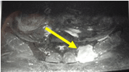
Figure 1: Bone MRI: Iliac Plasmocytoma with vertebral collapse.
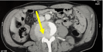
Figure 2: Lumbar CT scan: Plasmocytoma at vertebral level L 3.
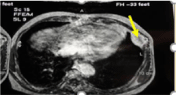
Figure 3: Thoracic MRI: Sterno-costal expansive process.
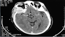
    Figure 4: Orbito-cerebral scan: Tissue mass of posterolateral wall of the orbit
with intra-orbital extension.
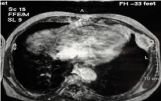
    Figure 5: Abdominal scan: Retroperitoneal tissue process with L 3 vertebral
lysis.
![]()
    Extraosseous locations
    Number (n)


    Orbital (Photo 4) location
    1


    Retro-peritoneal (Photo 5)    location
    1


    Renal location
    1


    Axillary location
    1
Table 1: Extra-osseous plasmocytomas.
Discussion
Plasma cell tumours are a group of neoplasias characterized by monoclonal proliferation of B lymphocytes that produce immunoglobulin in a homogeneous manner [1,2]. They account for 1-2% of all human neoplasia and may be medullary or extramedullary in origin. A classic distinction is between the bony solitary plasmacytoma variant of MM, which presents as a single bony lesion, the extramedullary plasmacytoma with soft-tissue-seated lesions, and MM. Thus, plasmacytomas can be solitary or within the framework of MM [2,3].
Bone or intramedullary Plasmacytoma (BP) is characterized by a proliferation of malignant plasma cells derived from a single clone of B lymphocytes, located in a circumscribed area of the body. BP is solitary when there is no diffuse medullary invasion [4]. BP accounts for 4% of the plasma cell proliferative syndromes dominated by multiple myeloma [3]. Solitary BP are in the age range between 40 and 50 years. The male predominance is clear, with a sex ratio of 3 to 4. This is consistent with the results of our series [3]. Spinal location is the most frequent. The PSO can also reach the ribs, sternum, iliac bone and long bones [5]. Iliac involvement such as that observed in two of our patients was rarely described. Histopathological study of the BP is the key to diagnosis [3]. Magnetic Resonance Imaging (MRI) is very efficient for the evaluation of spinal cord invasion and soft tissue extension. It shows T1 iso-signal and T2 hypersignal bone lesions that are raised after injection of contrast material [5].
Plasmocytoma is a radiosensitive tumor. Treatment is based on complete surgical removal combined with local postoperative radiotherapy. The dose administered varies from 35 to 50 Gy in 15 to 25 sessions over 3 to 5 weeks. This allows local control in 86 to 100% of cases. The most common progression to MM is within 3-5 years after the initial diagnosis [6].
Extra-Medullary Plasmacytomas (EMP) are very rare [7]. They have the same histological and immunological features as MM but differ in the absence of spinal cord invasion, their occurrence in soft tissue, and the localized, single or, more rarely, multiple nature of the proliferation. They are rare and account for 3-5% of all plasmacytomas [8]. As in our series, they are most prevalent in middle-aged men aged 55 years and older. They are mainly found in the upper airways (nasal fossa, sinus, oropharynx, salivary glands and larynx) and the digestive tract in about 80 to 90% of cases. Renal and retroperitoneal localizations such as those observed in our patients have been rarely described [9,10]. In a multi-center American series, orbital localization was noted in 30 cases including 18 cases of MM [11]. The characteristics of PO and POM were reported in Table 2. Therapeutically, because POMs often originate in the head and neck, radical surgery with curative aim for this localization is often mutilating and not indicated because of the radiosensitivity of the tumor. Radiotherapy is the treatment of choice for EMP of the head and neck. For other localizations, surgery is often performed as a first-line treatment with postoperative radiotherapy when the section slices are invaded [12,13]. Chemotherapy may be offered for orbital plasmacytoma alone or in combination with radiotherapy.
![]()
    OP
    EMP


    Mean Age
    55
    55


    Sex-Ration Male/Female
    1-2
    1-3


    Predominant Site
    Axial Squeletton


    Especially Vertebraes
    Head and Neck


    % With Protein M
    60
    <25


    % Of Development of Mm
    >75
    <30


    % Survival at 10 Years
    40-50
    70
Table 2: Clinical characteristics of OP and EMP [8].
Conclusion
The diagnosis and management of patients with plasmacytoma requires the same approach as for patients with multiple myeloma. Radiation therapy is an effective treatment for bone plasmacytoma, allowing local control in most cases. The prognosis is affected by the association with myeloma. The value of chemotherapy as adjuvant therapy in plasmacytomas of any location is variously evaluated. It has been proposed for the management of histologically aggressive forms.
References
- Mendenhall W M, Mendenhall C M, Mendenhall NP. Solitary Plasmacytoma of Bone and Soft Tissues. Am J Otolaryngol. 2003; 24: 395-399.
- Ben Salah H, Hdiji S, Makni S, Ghorbel AM, Boudawara T, Elloumi M. Plasmocytomes extra-osseux. Extra-medullary plasmocytomas. Cancer/ Radiothérapie. 2012; 16: 282-287.
- Bousnina S, Zendah I, Marniche K. Plasmocytome solitaire à localisation costale: une tumeur rare à ne pas méconnaître. Rev Pneumol Clin. 2006; 62: 243-246.
- Ketataa W, Triki F, Msaada S. Une localisation rare de plasmocytome solitaire. Revue de Pneumologie clinique. 2009; 65: 165-168.
- Masmoudi K, Elleuch E, Akrout R, Mnejja MA, Feki A, Ezzeddine M, et al. Le plasmocytome solitaire osseux: à propos de 3 cas et revue de la littérature. Le Pan Afr Med J. 2016; 25: 219.
- Brouchet L, Ballouhey Q, Brouchet A. Tumeurs primitives de la paroi thoracique. EMC (Elsevier Masson SAS, Paris), Pneumologie. 2010; 6-002- G-69.
- Kim S, Kim T, Sohn J, Yoon H, Shin D, Kim I, et al. Primary pulmonary plasmacytoma presenting as multiple lung nodules. Korean J Intern Med. 2012; 27:111-3
- Sautar R. Guidelines on the diagnosis and management of solitary plasmocytoma of bone and solitary extramedullary plasmocytoma. BJH 2004; 124: 717-726.
- Fan F, Deauna-Limayo D, Brantley Thrasher J, Damjanov I. Anaplastic plasmacytoma of the kidney. Histopathology. 2005; 47:432-3.
- Tejido Sánchez A, Hernández Martínez E, Ortíz MC, et al. Renal plasmacytoma. Report of a new case. Arch Esp Urol. 2001; 54: 718-22.
- Thuro BA, Sagiv O, Shinder R, Debnam JM, Ozgur O, Ng JD, et al. Clinical Presentation and Anatomical Location of Orbital Plasmacytomas. Ophthal Plast Reconstr Surg. 2017.
- Soutar R, Lucraft H, Jackson G, Reece A, Bird J, Low E, et al. Guidelines on the diagnosis and management of solitary plasmacytoma of bone and solitary extramedullary plasmacytoma. Clin Oncol. 2004; 16: 405-413.
- Caers J, Paiva B, Zamagni E, Leleu X, Bladé J, Kristinsson SY, et al. Diagnosis, treatment, and response assessment in solitary plasmacytoma: updated recommendations from a European Expert Panel. J Hematol Oncol. 2018; 11: 10.