
Case Report
Austin J Orthopade & Rheumatol. 2022; 9(1): 1109.
Lipomas of the Hand: Rare Location, About 4 Cases and Review of the Literature
Mokhchani Y1,2*, Rabbah A1,2, Fahl M1,2, Chafry B1,2 and Boussouga M1,2
¹Department of Orthopedic Surgery and Traumatology II, Mohammed V Military Teaching Hospital, Rabat, 10000, Morocco
²Faculty of Medicine and Pharmacy, Mohammed V University, Rabat, 10000, Morocco
*Corresponding author: Youness Mokhchani, Department of Orthopedic Surgery and Traumatology II, Mohammed V Military Teaching Hospital, Rabat, 10000, Morocco; Faculty of Medicine and Pharmacy, Mohammed V University, Rabat, 10000, Morocco
Received: February 21, 2022; Accepted: March 12, 2022; Published: March 19, 2022
Abstract
Lipoma is a benign mesenchymal tumor that develops in abundant areas of fatty tissue. Its location in the hand is rare. The first lipoma of the hand was described by Stein in 1959. The targeted localizations in the hands concern the thenar and hypothenar eminences, more rarely digital localizations. The clinical symptomatology varies according to the location, with progressively increasing size leading to a hold on the mobility of the fingers and a risk of neurological complications. Ultrasound or better Magnetic Resonance Imaging (MRI) is necessary for diagnosis. The anatomopathological study is essential to confirm the histological nature and eliminate a malignant tumor. Surgical excision remains the treatment of choice for lipomas. Postoperative evolution is generally favorable without local recurrence and with restoration of neurological signs.
Keywords: Benign tumor; Lipoma; Hand; Fatty tumor
Introduction
Benign tumor pathology of the hand is common. It is dominated by synovial cysts. Lipomas constitute only 1 to 3.8% of cases. The hand represents 5% of localizations of upper limb lipomas. We present four cases of lipomas of the hand and we try to develop the different clinical and paraclinical particularities and the therapeutic modalities of this pathological entity. We report the case of four lipomas of different locations in four patients (two women and two men), with no particular pathological history: two lipomas of the thenar compartment, a palmar location at the level of the middle segment, and a last case at the postero-external level of the wrist.
Case Presentation
Patient A: 54-year-old woman who has presented for 2 years with a painless soft mass in the thenar lodge of the right hand, gradually increasing in size, without neurological signs but with difficulties in gripping.
Patient B: 66-year-old man, who presents with a huge mass at the level of the first commissure of the right hand, evolving for 3 years, not painful, but hampering the mobility of the thumb.
Patient C: 58-year-old woman, presenting with a mass in the middle segment of the left hand, and experiencing discomfort when mobilizing the 2nd, 3rd and 4th fingers, with reduced sensitivity in the territory of the median nerve.
Patient D: 55-year-old patient presenting with a large mass, progressively evolving over the past 3 years, occupying the posteroexternal edge of the left wrist, and reporting paresthesia affecting the dorsal surface of the 1st interdigital space.
Standard X-rays were performed for all patients, having objectified thickening of the soft tissues of the segments concerned, in the form of shadows of tissue images without bone lesions. All our patients also benefited from ultrasounds which specified the fatty nature of the masses; these ultrasounds were supplemented by Magnetic Resonance Imaging (MRI) for better structural and topographic analysis.
For the first three patients, surgical excision of the masses was performed by incisions following the flexion creases of the hand, while respecting the palmar vasculo-nervous pedicles and the underlying flexor tendons. While for the 4th patient, the approach was direct posterolateral on the mass, with careful dissection so as not to damage the terminal threads of the sensory branch of the radial nerve as well as the superficial radial vein. Histological studies of the excision pieces, which measured between 2.5 by 3cm for the smallest, and 3 by 6cm for the largest, concluded that there were lipomas without signs of malignancy. The clinical evolution was favorable for the 4 patients without local recurrence and with disappearance of the neurological signs (Figure 1-6).
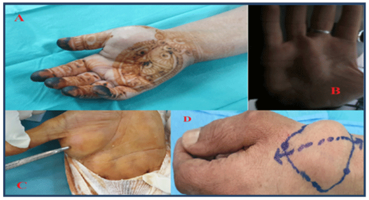
Figure 1: Clinical images: hands of the four patients.
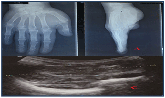
    Figure 2: Standard X-ray of the hand of patient “A” showing thickening of the

soft tissues. Ultrasound of the soft tissues of the hand of patient “C” showing a

mass isoechoic of the thenar eminence seat of hyperechoic spans measuring

6cm by 2.5.
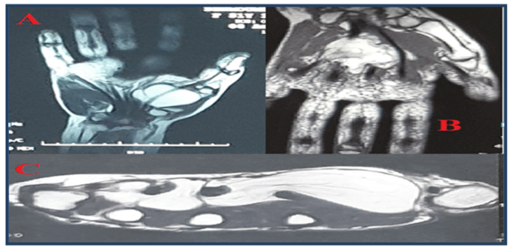
    Figure 3: MRI aspects of patients “A, B, C” showing homogeneous and welldefined

deep lipomas sheathing the flexor tendons.
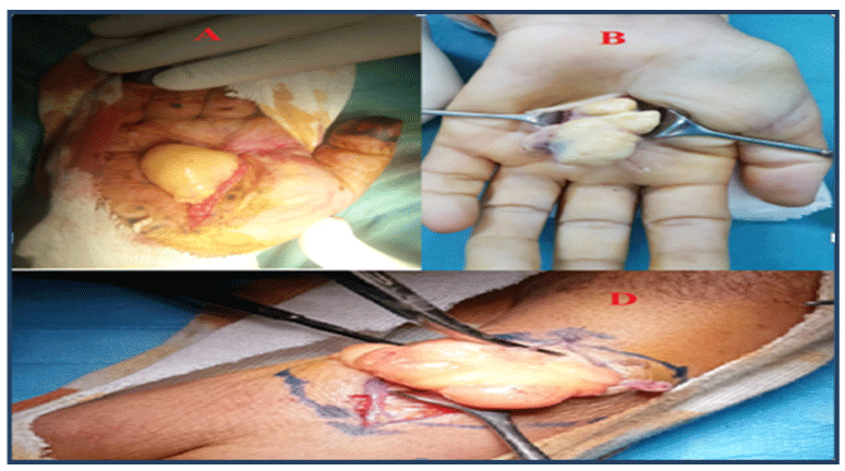
Figure 4: Intraoperative images of patients “A, B, D”.

Figure 5: Macroscopic aspects of lipomas after excision, patients “A, B, D”.
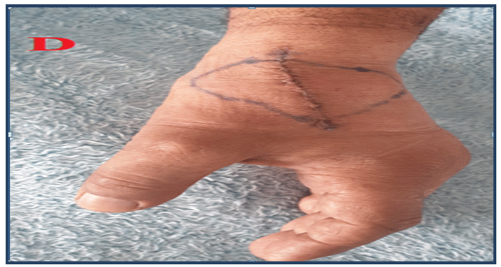
Figure 6: Postoperative image on D2: wrist of patient “D”.
Discussion
A lipoma is a benign tumor consisting of mature fat [1]. Lipomas make up approximately 16% of soft tissue mesenchymal tumors [2]. They are nevertheless rarely encountered in the hands and more rarely in the fingers. Lipomas are the most common benign form of soft tissue tumor in the body [3]. They are called “giants” when the excision piece exceeds 5 cm in diameter [4]. They appear especially around the fifth and sixth decades. These tumors may be superficial, arising from subcutaneous tissue or, less frequently, of subaponeurotic origin, arising from deep within Guyon’s lodge, carpal tunnel, or deep palmar space. Finally, in some cases, they can come from juxtaarticular regions or near the periosteum (para-osteal lipoma), they can reach the bone and cause cortical hyperostosis [5]. Clinically, superficial lipomas are often asymptomatic, usually manifesting as a tumor of fluctuating, lobulated and mobile soft consistency. Posch [6] described the clinical test of applying ice to the tumour, which in the case of a lipoma causes the mass to solidify. The usual evolution is a slow growth, which can stabilize spontaneously. Depending on its location, the lipoma can lead to carpal tunnel syndrome [7-10], compression of the ulnar nerve in Guyon’s canal [11], or digital nerves [12], or even finger jump [13]. These are the subaponeurotic lipomas that compress the branches of the median nerve and the ulnar nerve. Vascular compression with distal ischemia has not been reported in the literature. Signs of nerve compression are not correlated with tumor size. In the series consulted, compression of the median nerve was sometimes observed for small tumours. The nerve compression, in these cases, was explained by the intraoperative discovery of a tumor adhering to the nerve or of a tumor crossed by the nerve branches [8] (as is the case for our 4th patient).
Radiological investigations make the diagnosis of lipoma in 71% of cases. Ultrasound and especially magnetic resonance imaging (MRI) are useful in the evaluation of these lesions. MRI is the reference examination for soft tissue tumours, due to its high sensitivity. It specifies the nature of the lesion, its local extension and its relationship with the neurovascular elements. The characteristic image of the lipoma is a well-limited hypersignal image on the T1 and T2 sequences, with reduced signal on the fat suppression sequences. On a series of 134 MRI of tumors and pseudotumors of the wrist and hand, the preoperative diagnosis by MRI of benign lipoma was confirmed by histology in 94% of cases for Capelastegui et al. [14]. MRI by analyzing the fat content of tissues can provide arguments differentiating lipoma from liposarcoma. The latter is the differential diagnosis with the greatest risk for the patient. It is the most common soft tissue sarcoma in adults; its frequency varies from 1.1 to 2.5/1000,000 with a peak between 50 and 70 years [15]. It is the most common soft tissue sarcoma in adults. The differential diagnosis arises with other soft tissue tumors such as ganglion cysts, giant cell tumors, myxomas, angiolipomas, intraneural lipofibroma [16].
All the authors agree that in the presence of a deep fatty tumor of more than 5 cm, it is necessary to perform a biopsy or a cytopuncture to rule out a liposarcoma [14]. Fine-needle aspiration cytology can establish the differential diagnosis between lipoma and liposarcoma in 95% of cases for Kooby et al. [15], advantageously replacing the biopsy which carries a risk of local dissemination of the tumour. Histological examination confirms the benignity of the lesion and eliminates the sarcomatous component. It is true that benign tumors are more frequent than malignant tumors in the hand, but the malignancy of tumor lesions must always be suspected and pathological evidence is essential to adapt therapeutic management. The majority of cases reported in the literature have demonstrated that surgical excision of lipomas has good results for functional recovery. The classic digitopalmar approach according to Bruner is recommended, given the advantages it offers to healing without retractions. Marginal resection is the treatment of choice for benign lipomas, since it causes less than 5% tumor recurrence. Identification and dissection of vasculonervous elements must be careful to avoid iatrogenic damage. The excision must be as complete as possible in order to minimize the risk of local recurrences. That said, these remain exceptional [4,5]. The consent of the patient and the prior explanation of the risks is necessary before any therapeutic management. There is no rehabilitation protocol necessary, but just active movements, especially in the event of joint or tendon non-invasion. The paresthesias felt initially can be suppressed after removal of the tumour.
Conclusion
Hand lipomas represent rare benign tumor pathology. MRI represents the most interesting exploration, with a diagnostic aim and a certain therapeutic influence. Only histological examinations can confirm the histological nature of the lesion, which means that the malignant component must always be suspected. The proximity of the relationship with the vasculo-nervous structures must lead to the greatest caution during surgical dissection. Patient information is necessary for adequate care.
References
- Calandruccio JH, Jobe MT. In: Canale ST, editor Campbell operative. Orthopaedics 9th edition, St Louis: Mosby-Year Book, Inc. 1998; 4: 3704- 3705.
- Ersozlu S, Ozgur AF, Tandogan RH. Lipoma of the index finger. Dermatol Surg. 2007; 33: 382-384.
- Lisenda L, van Deventer S, Pikor T, et al. A propos d’un cas: lipome géant de la main. SA Orthop J. 2013; 12: 46-48.
- Fnini S, Hassoune J, Largab A. Lipome géant de la main. Rev Chir Main. 2010; 29: 44-47.
- Chronopoulos E, Nikolaos P, Karanikas C, Kalliakmanis A, Plessas S, Neofytou I, et al. Patient presenting with lipoma of index finger. Cases journal. 2010; 3: 20.
- Posch JL. Tumors of the hand. J Bone Joint Surg. 1956; 38A: 517-540.
- De Smet L, Bande S, Fabry G. Giant lipoma of the deep palmar space, mimicking persistent carpal tunnel syndrome. Acta Orthop Belg. 1994; 60: 334-335.
- Cribb GL, Cool WP, Ford DJ, Mangham DC. Giant lipomateus of the hand and forearm. Br J Hand Surg. 2005; 30B: 509-512.
- Babins DM, Lubahn JD. Palmar lipomas associated with compression of the medial nerve. J Bone Joint Surg. 1994; 76A: 1360-1362.
- Hironori S, Mitsuhiro T, Hirofumi T, Tsutomu H, Hiroshi T. Carpal tunnel syndrome and trigger wrist caused by a lipoma arising from flexor tenosynovium: A case report. J Hand Surg. 2002; 27A: 1056-1058.
- Zahrawi F. Acute compression ulnar neuropathy at Guyon’s canal resulting from lipoma. J Hand Surg. 1984; 9: 238-239.
- Boussouga M, Bousselmame N, Lazrak KH. Lipome compressif de la loge thénar. À propos d’une observation. Chir Main. 2006; 25: 156-158.
- Brand MG, Gelberman RH. Lipoma of the flexor digitorum superficialis causing triggering at the carpal canal and median nerve compression. J Hand Surg. 1988; 13: 342-324.
- Laurino L, Furlanetto A, Orvieto E, Del Tos AP. Well-differentiated liposarcoma (atypical lipomatous tumors). Semin Diagn Pathol. 2001; 18: 258-262.
- Kooby DA, Antomescu CR, Brennan MF, Singer S. Atypical lipomatous tumor/ well-differentiated liposarcoma of the extremity and trunk wall: Importance of histological subtype with treatment recommendations. Ann Surg Oncol. 2004; 11: 78-84.