
Case Presentation
Austin J Orthopade & Rheumatol. 2022; 9(2): 1111.
Tibial Tubercule Avulsion Fracture in Child Soccer Player: A Conservative Approach
Rosado D*, Gálvez Aranda D, Mondragón R and Medina Porqueres I
Department of Orthopedics, School of Sports Medicine, Spain
*Corresponding author: Daniel Rosado, Department of Orthopedics, School of Sports Medicine, Spain
Received: June 30, 2022; Accepted: July 25, 2022; Published: August 01, 2022
Abstract
Tibial Tubercle Avulsion Fracture (TTAF) is an uncommon disease. They are usually found during childhood-adolescent age and described as non-traumatic fractures during running with direct physis affection. In order to avoid growth complications, which would lead to harmful consequences for the patient, an accurate diagnosis following a solid treatment has to be carried out.
We describe a case report of a 12-year-old football player boy with a tibial tuberosity avulsion following a conservative treatment. Pharmacological treatment and a rehabilitation program was prescribed with a satisfactory evolution. Even though one of the physician consulted suggested open reduction and internal fixation, the medical staff for his team decided a more conservative approach which came out as an adequate management for this case. After a two years follow up the evolution was successful and the player was able to perform every football specific activity pain free and without any kind of limitation.
Keywords: Tibial tuberosity fracture; Conservative management; Adolescent age
Introduction
Soccer is practiced worldwide as sport at all ages. Children active during sensible growth spurt may develop different disorders from exposing the joints to repeated mechanical stress. Knee injuries in young soccer players appear mainly secondary to and inadequate load of work; and if we consider the architectural fragility of the epiphysis at this age it is easy to have in mind the outcome of epiphysitis. In some cases epiphysitis can be underestimated and miss diagnosed leading to a more serious event: tibial tubercule avulsion fracture.
The second largest epiphysis of the human body is the proximal epiphysis of the tibia. During the first few days after birth most newborns develop the epiphyseal Secondary Ossification Centre (SOC) and by the third month after birth it is present in all infants. Its spherical shaped located in a central position. At around 10 years old it will expand with growth to a more elliptical shape, and then finally create the concavity of the tibial plateau.
From ages 7 to 12 another centre of ossification develops: the anterior tibial tuberosity´s.
We have then two different ossification centres:
The epiphyseal SOC:
1. Perpendicular to the longitudinal axis of the tibia.
2. Shapes the proximal epiphysis contributing to is growth allowing the modelling for the joint to articulate
3. And the ossification centre of the anterior tibial tuberosity:
4. Tangential to the longitudinal axis of the tibia.
5. Provides a reinforcement in order for the patellar tendon to insert.
By ages between 14 and 17 years both ossification centres fuse together to merge with the tibial metaphysis.
TTAF are unusual and represent less than 3% of all proximal tibial fractures, being most prevalent in adolescent [1], and Osgood- Schlatter lesion may induce TTAF injuries, however, further studies are still needed to fully determine this association [2].
It also has to be considered as a significant element, the biomechanics of the patellar tendon insertion.
In the event of a TTAF, the potential risk of ischemia following fracture is very low as different arteries that penetrate the physis from different angles and directions [3] guarantee the vascular supply.
Case Presentation
A 12-year-old male patient with no previous pathological history reported a sudden pain and weakness in his left knee while just running without an identified trauma during a regular soccer practice.
Immediate pain, deformity of the left knee with swelling making him unable to walk showed up leading to functional limitation.
On examination, the knee presented a defensive passive 20 degrees flexion, anterior deformity with increase in anterior tibial region volume, swelling and tenderness. Severe pain made it impossible to explore active or passive mobility arches of the knee with little tolerance to palpation surrounding the anterior tibial tuberosity area.
He showed no signs or symptoms of compartment syndrome; so it was ruled out. Tibialis posterior and dorsalis pedis pulsations were felt and there was no neurovascular deficit. Immobilization bandage was performed and radiograph of the knee was taken. In imaging, avulsion of the left tibial tuberosity was detected, (Figure 1), with no extension up to metaphysis 4 months after first Rx (Figure 2) new Rx was taken. It showed a reduction of the free fragment and it had emigrated upward. It was prescribed then an MRI in order to have more accurate data about location and effect over the surrounding soft tissue (Figure 3).
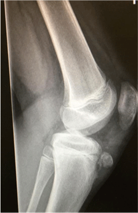
Figure 1: In imaging, avulsion of the left tibial tuberosity was detected.
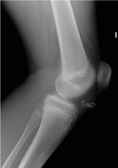
    Figure 2: New Rx was taken. It showed a reduction of the free fragment and

it had emigrated upward.
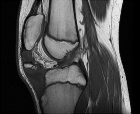
    Figure 3: It was prescribed then an MRI in order to have more accurate data

about location and effect over the surrounding soft tissue.
Discussion
Tibial tubercle avulsion fracture in children are usually found in actions such as kicking or jumping when the quadriceps muscle is contracting repeatedly against a force or in situations involving knee load flexion with a quadriceps contraction [1-3] (landing actions).
1. TTAF consists of a traumatic physis separation in the deep plane from the ossification nucleus of the tibial tuberosity. It shows a relation with the growth process of the secondary ossification centre of the tibial tuberosity. There are four stages when classifying its growth [4,5].
2. The cartilaginous stage: children from 0 to 11 years, before the appearance of tibia tubercle’s secondary ossification centre
3. The apophyseal stage: children from 11 to 14
4. The epiphyseal stage: children from 14 to 18 years, begins with the formation of anterior tongue of bone that the secondary ossification centre of proximal tibia joins to the tubercle
5. The finally the bony stage: children older than 18 years. Resulting in bony fusion [6].
Having in mind that Osgood Slatter Disease has been reported to occur more frequently in the apophyseal stage of development, it is considered this period of time especially sensible to traction forces [7].
Physical closure starts from the centre of proximal tibial physis, and proceeds to the distal end of the physis, and avulsion fractures occur in the area that is too weak to resist the tension produced by muscle loading [1].
It is very important a close follow-up of pediatric patients with this lesion, weather they are treated or not surgically, as there is a high risk of severe complications. The stress in quadriceps has been presented to be 289 to 307 kN/m² during jumping, 292 kN/m² during landing, and 241 kN/m² during running [8].
Its incidence has been reported to between 0.4 y 2.7% of all epiphyseal injuries and less than 1% of all growth plate injuries [9]. It is more common in the adolescent age groups. Adolescent soccer players are exposed to repeated biomechanical stress developing many musculoskeletal disorders [10]. At this age the muscle, ligament, and tendons are stronger than bone [11]. They are also more frequently described in males, probably due to their to their more prevalent participation in sports activities and the fact that physeal closure occurs later in males than in females [2].
Other risk factors have been related to TTAF such as obesity. Obese patients were more likely to sustain fractures involving the physis [12]. It has also been reported that obesity is associated with more frequent fracture in children [13] and weakness of physis relative to the tension applied during intense muscle loading.
TTAF traumas regarding its biomechanics could lead to patellar tendon rupture, meniscal tears, cruciate ligament damage and collateral injuries [14] the type and extension of the injury will be determined by the mechanism and secondary ossification stage.
There are multiple classification systems with multiple modifiers. Watson-Jones [15] classification determines 3 types:
1. Type 1: avulsion of the apophysis without injury to the tibial epiphysis
2. Type 2: epiphysis is lifted cephalad and incompletely fractured
3. Type 3: displacement of the proximal base of the epiphysis with the fracture line extending into the joint Ogden [16] proposes a modification of Watson-Jones classification:
4. Type I: fracture of the secondary ossification centre near the patella tendon insertion
5. Type II: fracture between the primary and secondary ossification centres
6. Type III: fracture that traverses the primary and secondary ossification centres (most common type)
7. Type IV: fracture through the entire physis
8. Type V: avulsion of the periosteal sleeve Modifiers
1. A – non displaced and
2. B - displaced
The goal of the treatment is the anatomical reduction of the fragment with restoration of tibial articular surface leading to best recovery of the extension mechanism of the injured knee. Conservative treatment with immobilization has been reported in type I injuries with small free fragments [17].
Conclusion
TTAF are a rare pathology rare but they have to be considered severe in childhood due to the high possibility of serious complications. Its treatment might be influenced by misdiagnosis or lack of knowledge leading to poor prognosis. However, the therapeutic diagnostic approach for this injury it is usually quite straightforward. Upon early physical examination, image studies should be carried out. Anteroposterior and lateral knee X-rays study will address a correct diagnosis and posterior classification. This will guide the appropriate treatment for the patient taking in consideration age and sports modality.
In case of uncertainty or suspicion of other injuries, complementary studies such as MRI or CT should be requested.
References
- Edmonds EW, Mubarak SJ. Proximal tibial physical fractures in children. Eighth ed. Philadelphia, PA: Wolters Kluwer Health. 2015: 1057–1074.
- Jakoi A, Freidl M, Old A, Javandel M, Tom J, Realyvasquez J. Tibial tubercle avulsion fractures in adolescent basketball players. Orthopedics. 2012; 35: 692–696.
- Nelson GE, Kelly PJ, Peterson LFA, Janes JM. Blood supply of the human tibia. J Bone Joint Surg Am. 1960; 42: 625–636.
- McKoy BE, Stanitski CL. Acute tibial tubercle avulsion fractures. Orthop Clin North Am. 2003; 34: 397–403.
- Ehrenborg G, Engfeldt B. The insertion of the ligamentum patellae on the tibial tuberosity. Some views in connection with the Osgood-Schlatter lesion. Acta Chir Scand. 1961; 121: 491–499.
- Ducher G, Cook J, Lammers G. The ultrasound appearance of the patellar tendon attachment to the tibia in young athletes is conditional on gender and pubertal stage. J Sci Med Sport. 2010; 13: 20-23.
- Blankstein A, Cohen I, Heim M, Diamant L, Salai M, Chechick A. Ultrasonography as a diagnostic modality in Osgood-Schlatter disease. Arch Orthop Trauma Surg. 2001; 121: 536-553.
- Thorpe SK, Li Y, Crompton RH. Stresses in human leg muscles in running and jumping determined by force plate analysis and from published magnetic resonance images. J Exp Biol. 1998; 201: 63–70.
- Abalo A, Akakpo-numado KG, Dossim A. Avulsion fractures of the tibial tubercle. J Orthop Sur. 2008; 16: 308-311.
- Rössler R, Junge A, Chomiak J, Dvorak J, Faude O. Soccer injuries in players aged 7 to 12 years. Am J Sports Med. 2016; 44: 3309-3179.
- Alvulquerque RP, Campos AS, de Araújo SGC, Gameiro VS. Fracture of tibial tuberosity in an adult. BMJ Case Rep. 2013.
- Dimitri P, Bishop N, Walsh JS. Obesity is a risk factor for fracture in children but is protective against fracture in adults: a paradox. Bone. 2012; 50: 457– 466.
- Gilbert SR, Maclennan PA, Backstrom I. Altered lower extremity fracture characteristics in obese pediatric trauma patients. J Orthop Trauma. 2015; 29: 12–17.
- Jalgaonkar AA, Dachepalli S, Al-Wattar Z. Atypical tibial tuberosity fracture in an adolescent. Sports Medicine. 2011; 34: 215-218.
- Makram Z, Heidi A, Taoufi kA. Acute tibial tubercle avulsion fractures in the sporting adolescent. Ach Orthop Trauma Surg. 2008; 128: 1437-1442.
- Ogden JA, Southwick WO. Osgood-Schlatter´s disease and tibial tuberosity development. Clin Orthop. 1976; 116: 180-189.
- Tachdjian O. Ortopedia Pediátrica. Fracturas que abarcan la fisis tibia proximal y la apófisis del tubérculo tibial y fracturas avulsión de la apófisis del tubérculo tibial. Editorial Interamericana-McGraw-Hill. 1994; 5: 3539-3540.