Case Report
Austin J Otolaryngol. 2014;1(4): 2.
A Temporal Bone Fracture Involving the Superior Semicircular Canal
Hong Chan Kim, Hyung Chae Yang, Yong Beom Cho and Chul Ho Jang*
Department of Otolaryngology-Head and Neck Surgery, Chonnam National University Hospital, South Korea
*Corresponding author: Chul Ho Jang, Department of Otolaryngology-Head and Neck Surgery, Chonnam National University Medical School and Chonnam National University Hospital, 42 Jebong-ro, Dong-gu, Gwangju, 501-757, South Korea
Received: October 10, 2014; Accepted: November 14, 2014; Published: November 17, 2014
Abstract
Temporal bone fractures typically take the path of least resistance, which is along structurally weakened points such as the various foramina perforating the skull base. The superior semicircular canal has a relatively thicker bony structure than surroundings. As a result, the fracture proceeds otic capsules typically fractures the surrounding structures. Here, we present the first case of a temporal bone fracture involving the superior semicircular canal and demonstrate its high-resolution computed tomography image.
Keywords: Head injuries; Closed; Temporal bone; Semicircular canals; Temporal bone fracture
Case Presentation
A 45 year-old man who fell off from a height of 3m was referred to the ENT department for otolaryngologic evaluation. He was admitted to the intensive care unit because of a closed head injury with multiple scalp lacerations, right hearing impairment and headache. The skull X-ray showed a right temporal bone fracture. The initial brain computed tomography (CT) scan revealed an acute epidural hematoma (EDH) along the right parieto-temporo-occipital convexity with pneumocephalus, and right temporal bone fracture.
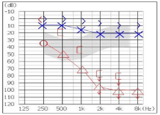
The patient was alert and oriented; however, he had a severe headache at that time. His responses to the doctor’s orders were somewhat restricted. No facial paralysis was noted. He complained of non-whirling type dizziness and right sided hearing impairment. Computed dynamic postulography showed a vestibular pattern; however, nystagmus was absent. Pure tone audiometry revealed a mixed type severe hearing impairment (Figure 1A).
Figure 1A : Pure tone audiometry which was checked on post-trauma day 1 showed right-sided mixed type severe hearing impairment.
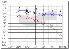
Figure 1B : The 7-month post-treatment follow-up pure tone audiometry showed an improved PTA threshold but there was still mild hearing impairment. The air bone gap was closed.
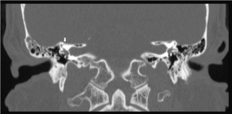
For further evaluation of the temporal bone fracture, a high-resolution temporal bone computed tomography (TBCT) was checked. The TBCT revealed fractures of the right longitudinal temporal bone fracture. The fracture ran through the mastoid (longitudinal fracture), and the squamous and tympanic portions along the axial plane. In addition, it passed through the superior semicircular canal (Figure 2) to the tegmen. The ear ossicles and bony facial canal remained intact.
Figure 3 : Coronal CT scan showed the right-sided temporal bone fracture involving the superior semicircular canal (white arrow). Note that the tegmen tympanum and other parts of otic capsules were remained intact.
He was treated with close clinical observation and serial imaging. A follow-up CT which was checked at 7 months after the diagnosis showed spontaneous resolution of the epidural hematoma. The follow-up PTA threshold improved to 45 dB with a closed air-bone gap (Figure 1B). His dizziness subsided.
Discussion
Injuries to the temporal bone occur in 2 to 10% of cases involving blunt head trauma [1], and only 2.5% to 5.8% of fractures disrupted the otic capsule [2,3]. To produce a fracture of the temporal bone, a tremendous amount of force is required [4]. The fracture typically takes the path of least resistance, which is along structurally weakened points such as the various foramina perforating the skull base [5]. The superior semicircular canal (SSCC) is a small structure which is surrounded by a relatively short and dense bony canal, and near the SSCC, there are structurally weak points such as the vestibule, round window, oval window, internal auditory canal, facial hiatus, etc. The SSCC involvement by a temporal bone fracture is very rare, in addition, little is known about the clinical feature of the semicircular canal fractures.
Temporal bones require a great force for them to fracture. Recent dynamic loading studies reported that fractures need 6000 to 8000 N of lateral impact [6]. Fractures typically take the path of least resistance, which is along structurally weakened points such as the various foramina perforating the skull base [5]. Consequently, a fracture line takes the path of tegmen tympanum, oval or round window instead of SSCC which is enclosed by thick bone. According to the several large studies regarding lesions of traumatic perilymphatic fistula, the oval and round window were mainly involved. There were few reports of lateral semicircular canal involvement [7,8]; however, there were no report of SSCC fracture. In addition, semicircular canals were involved only in the cases with the transverse fracture, while there was no involvement with the longitudinal fracture [9].
The leading cause of temporal bone fractures is motor vehicle accidents [2] and the incidence of temporal bone fractures is 2% to 10% of consecutive head injury patients [1]. Temporal bone fractures are usually combined with other bodily injuries, and consequently, the initial evaluation and management is focused on urgent lift-threatening issues such as controlling an intracranial hemorrhage, as with this patient [5].
Temporal bone fractures have traditionally been divided into transverse and longitudinal fractures. However, a recent study questioned the clinical predictive ability [10] of the traditional classification, and pointed out ambiguities in it [11]. A new structural scheme classifies fractures according to whether they disrupt or spare the otic capsule and which offers the advantage of radiographic utility and stratification of clinical severity [5,12]. Otic capsule violating fractures tend to show more severe clinical features such as intracranial hemorrhage, cerebrospinal fluid leakage, facial weakness and profound hearing loss [3,13].
The reported patient experienced mixed type severe hearing impairment, tinnitus, disequilibrium and headache. However, facial weakness and obvious cerebrospinal fluid leakage which were frequently observed in other reports of temporal bone fractures were not noted. Disequilibrium and tinnitus of the patient were improved over the time. The signs and symptom of perilymphatic fistula were absent.
He finally had mild to moderate hearing loss in mid frequencies (1,000-2000 Hz), however profound hearing loss in high frequency (8000 Hz). We could assume two possible reasons for low tone hearing preservation. First, the endolymphatic space of the utricle and semicircular canals is isolated from the endolymphatic space of the cochlea and saccule by a valve of Bast. Second, animal experiment revealed that labyrinthine concussion causes significant pathological changes in the cochlea, especially to the organ of Corti in the upper basal turn, the region serving 4000 hz frequency [14]. These may be the reason for the relative hearing preservation in the case; however exact mechanism is still unknown. Further studies are needed to identify the hearing preservation mechanism in the patients with isolated semicircular fractures.
References
- Ghorayeb BY, Rafie JJ. [Fracture of the temporal bone. Evaluation of 123 cases]. J Radiol. 1989; 70: 703-710.
- Brodie HA, Thompson TC. Management of complications from 820 temporal bone fractures. Am J Otol. 1997; 18: 188-197.
- Dahiya R, Keller JD, Litofsky NS, Bankey PE, Bonassar LJ, Megerian CA. Temporal bone fractures: otic capsule sparing versus otic capsule violating clinical and radiographic considerations. J Trauma. 1999; 47: 1079-1083.
- Travis LW, Stalnaker RL, Melvin JW. Impact trauma of the human temporal bone. J Trauma. 1977; 17: 761-766.
- Diaz RC, Kamal SM, Brodie HA. Middle Ear and Temporal Bone Trauma. In: Johnson JT, Rosen CA, editors. Bailey's Head & neck surgery-otolaryngology. 2. 5th ed. Philadelphia, PA: Lippincott Williams & Wilkins; 2013; 2410-2431.
- Yoganandan N, Pintar FA, Sances A Jr, Walsh PR, Ewing CL, Thomas DJ. Biomechanics of skull fracture. J Neurotrauma. 1995; 12: 659-668.
- Yaniv E, Traub P. Traumatic perilymphatic fistulae of the lateral semicircular canal. J Laryngol Otol. 1988; 102: 521-523.
- Weider DJ, Johnson GD. Perilymphatic fistula: a New Hampshire experience. Am J Otol. 1988; 9: 184-196.
- Wysocki J. Cadaveric dissections based on observations of injuries to the temporal bone structures following head trauma. Skull Base. 2005; 15: 99-106.
- Yanagihara N, Murakami S, Nishihara S. Temporal bone fractures inducing facial nerve paralysis: a new classification and its clinical significance. Ear Nose Throat J. 1997; 76: 79-80, 83-6.
- Ghorayeb BY, Yeakley JW. Temporal bone fractures: longitudinal or oblique? The case for oblique temporal bone fractures. Laryngoscope. 1992; 102: 129-134.
- Little SC, Kesser BW. Radiographic classification of temporal bone fractures: clinical predictability using a new system. Arch Otolaryngol Head Neck Surg. 2006; 132: 1300-1304.
- Magliulo G, Ciniglio Appiani M, Iannella G, Artico M. Petrous bone fractures violating otic capsule. Otol Neurotol. 2012; 33: 1558-1561.
- Emerson LP. Hearing Loss in Minor Head Injury. In: Naz S, editor. Hearing Loss: InTech. 2012.


