
Case Report
Austin J Otolaryngol. 2024; 10(1) : 1132.
Frontal Ectopic Accessory Thyroid: An Exceptional Case Report
Moad El Mekkaoui¹*; Zakaria El Hafi¹; El Mehdi Hakkou²; Hafsa Elouazzani³; Razika Bencheikh¹; Anas Benbouzid¹; Abdelilah Oujilal¹; Nadia Cherradi³; Leila Essakalli¹
¹ENT- Head and Neck surgery Departement, Hospital of Specialties, Mohammed V University, Rabat, Morocco
²Neurosurgery Department, Hospital of Specialties, Mohammed V University, Rabat, Morocco
³Laboratory of Anatomical Pathology, Hospital of Specialties, Mohammed V University, Rabat, Morocco
*Corresponding author: Moad El Mekkaoui ENT- Head and Neck surgery Departement, Hospital of Specialties, Mohammed V University, Rabat, Morocco Email: moad.elmekkaoui@gmail.com
Received: December 27, 2023 Accepted: January 30, 2024 Published: February 06, 2024
Abstract
Thyroid ectopia is a rare malformation related to a failure of the thyroid gland migration during embryonic development, resulting in the presence of thyroid tissue at locations other than the normal locations in the anterior neck region. The combination of thyroid ectopy and a thyroid in normal cervical position is uncommon. The case study is of great interest, in this case to clarify the diagnostic difficulties and therapeutic modalities of an exceptional localization of accessory ectopic thyroid, located on the forehead.
Keywords: Accessory ectopic thyroid; Forehead; Otorhinolaryngology; Head neck surgery; Case report
Introduction
The thyroid gland is the first endocrine gland developed during fetal embryology from the endoderm, which begins between the third and fourth week of gestation [1]. Thyroid ectopia is a rare malformation related to a failure of the thyroid gland to migrate during embryonic development, resulting in the presence of thyroid tissue at locations other than the normal locations in the anterior neck region, between the second and fourth tracheal cartilages. The combination of thyroid ectopy and a thyroid in normal cervical position is exceptional [2]. 90% of the cases of ectopic thyroid are located at the level of the tongue, other localizations at the level of the head and neck have been found in the literature [3-5]. We will report in this case, in line with the SCARE 2020 criteria [6], an exceptional location of ectopic accessory thyroid located in the forehead of incidental discovery during surgery, associated with a thyroid nodule classified EU-TIRADS 5.
Case Report
This is a 53-year-old patient, of North African origin, housewife, hypertensive on treatment, never operated, with no specific family history, who initially presented with a left frontal swelling evolving for 2 years, progressively increasing in volume without associated neurological signs. The patient presented in consultation. The clinical examination revealed a painless left frontal mass, 2 cm long, well limited, firm, mobile in relation to the superficial and deep planes, without thrill to palpation. The neurological examination was strictly normal. Cervical examination did not reveal any cervical adenopathy or cervical swelling.
A cerebral CT scan without contrast injection was performed, which showed an isodense frontal intraosseous formation at the level of the diploe lysing the anterior bony cortex and respecting the posterior bony cortex (Figures 1,2). Preoperative tissue diagnosis was not performed.
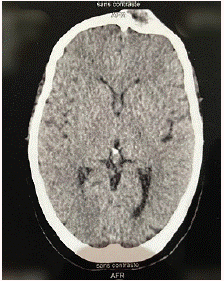
Figure 1: Cerebral CT scan without contrast injection in axial section showing frontal isodense formation.
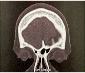
Figure 2: Cerebral CT scan without contrast injection in coronal section showing frontal isodense formation.
A preoperative workup was performed, including a blood count, ionogram, hemostasis workup, electrocardiogram and chest X-ray. Surgical removal of the mass was performed under general anesthesia by the neurosurgeon. The procedure involved making a bitragal skin incision, removing the tumor and a thin layer of surrounding bone without reconstruction, followed by fat filling to avoid aesthetic damage. Anatomopathological examination was in favor of an ectopic thyroid tissue without histological signs of malignancy (Figure 3,4).
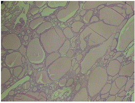
Figure 3: Microphotograph showing a well-differentiated regular thyroid tissue formed by vesicles of variable shape and size with homogeneous colloid content. HE AU GX10.
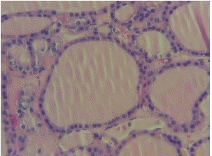
Figure 4: The vesicles are lined by a layer of regular thyreocytes without atypia. Interstitial tissue is sparse. HE AT GX40.
A cervical ultrasound was then requested, finding a normal volume thyroid gland (11.6 ml) with bilateral spongiform nodules classified as EU-TIRADS 2 and a right medio-lobar nodule longer than wide and strongly hypoechoic classified as EU-TIRADS 5. A thyroid biology test was performed, the patient was euthyroid with strictly normal TSHus, T4 and T3. However, thyroglobulin was elevated (135ng/ml). An iodine 124 scan was requested but no fixation was found that could suggest other ectopic thyroid tissues.
A total thyroidectomy under general anaesthesia was performed by the ENT surgeons, with extemporaneous examination of the right lobe, which revealed hyperplastic nodules of benign appearance. The final anatomopathological examination of the surgical specimen confirmed benign normo-vesicular multi-nodular hyperplasia (Figure 5).
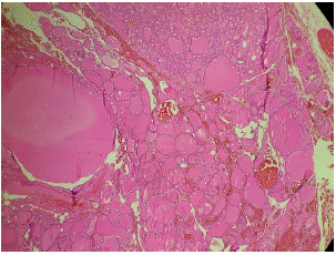
Figure 5: Microphotograph showing hyperplastic thyroid parenchyma consisting of nodules surrounded by thin fibrous septa and made of vesicles of variable size bordered by regular thyreocytes. HEX10.
Post-operative care was straightforward ; The patient did not develop hypocalcemia or dysphonia. Hormone replacement therapy was then instituted to avoid hypothyroidism. After one year of evolution, the patient did not present a frontal or cervical recurrence that could be related to the persistence of thyroid tissue. The patient was satisfied with the result.
Discussion
The primary origin of the thyroid gland is the endoderm. Embryological abnormalities of the thyroid gland can be manifested by hypoplasia, agenesis or ectopy [7]. Ectopic thyroid tissue is a rare developmental abnormality involving aberrant embryogenesis of the thyroid gland during its passage from the floor of the primitive foregut to its final pre-tracheal position [8,9]. In 1869, Hickman reported the first case of ectopic thyroid at the base of the tongue, pressing the epiglottis into the larynx and causing death by suffocation sixteen hours after birth [10]. Thyroid ectopy is said to be true in the absence ofthe thyroid gland in its normal pretracheal position [7,11]. On the other hand, accessory thyroid is defined by the association of an gland in place with thyroid tissue outside its anterior cervical location [7,12]. The association of a thyroid ectopy and thyroid in normal cervical position is exceptional [7], thing found in our case.
The pathophysiology of thyroid ectopy is not clearly elucidated. Recently, the genetic research has demonstrated that the gene transcription factors TITF-1(Nkx2-1), Foxe1(TITF-2) and PAX-8 are essential for thyroid maturation and differentiation. Mutation in these genes may share a connection with abnormal migration of the thyroid [5]. Cases of familial thyroid ectopy where aberrant thyroid tissues were present in two members of the same family have been described [5].
Thyroid ectopy can occur at any age. Most cases are identified during the neonatal period through newborn screening, but some cases are delayed until the fourth to sixth decade, when the ectopic thyroid tissue develops into abnormal tissue pathology and presents with symptomatic manifestations [13]. It mainly affects women with a sex ratio of 4 women to 1 man [9]. Its prevalence is about 1 per 100 000–300 000 peoples, rising to 1 per 4000–8000 patients with thyroid disease [8,9]. The most frequent location of ectopic thyroid tissue is at the base of tongue, in particular at the region of the foramen cecum, accounting for about 90% of the reported cases [9]. Incomplete migration can lead to a high cervical thyroid, and excessive movement can lead to a superior mediastinal or even paracardiac location. Studies are reporting cases of dual and triple ectopic thyroids [13-15] The other possible locations of ectopic thyroid are [13]:
• In the head and neck: the trachea, submandibular, lateral cervical regions, palatine tonsils, carotid bifurcation, iris of the eye and pituitary gland.
• Axilla
• Heart and ascending aorta
• Lymphoid tissue: thymus
• Gastrointestinal system: esophagus, duodenum, gallbladder, stomach bed, pancreas, mesentery of the small intestine, porta hepatis
• Adrenal gland
• Reproductive system: ovary, fallopian tube, uterus, and vagina
During the literature review, a forehead location was not found, hence the importance of this work.
The clinical symptoms depend on the volume and location of the thyroid ectopic thyroid. Indeed, the ectopic thyroid may become goitrous and may also be associated with clinically evident thyroid dysfunction which may be either hypofunction or hyperfunction [7], hence the interest in performing a thyroid biologic workup (TSH, T3, total T4, free T4, and thyroglobulin). Medical imaging can be used to confirm the diagnosis of thyroid ectopy, to plan the therapeutic strategy and and to follow up the patient [8,16]. They are based essentially on cervical Doppler ultrasound which shows a thyroid in place in the thyroid cavity associated with a heterogeneous hypoechoic tissue mass in an abnormal situation. The Technetium-99m or better iodine-124 scintigraphy allows to show a hyperfixation outside the normal hyperfixation outside the normal thyroid cavity. This remains the method of choice in terms of sensitivity and specificity [5,16]. CT and MRI can accurately determine the location of thyroid ectopy and better study the relationship with adjacent structures. If the case is highly suspicious for malignancy, then tissue biopsy for histology or Fine Needle Aspiration Cytology (FNAC) should be performed. Fortunately, the studies have estimated the risk of developing malignancy from ectopic tissue is less than 1% [13]. Benign neoplasms and thyroiditis are also infrequent complications.
The indications for surgical removal of the accessory ectopic thyroid include the following: malignancy, bleeding or ulceration of the gland, uncontrolled hyperthyroidism or hypothyroidism, and severe local or respiratory symptoms [13]. In our case, the per operative discovery of a frontal accessory thyroid associated with an EU-TIRADS 5 thyroid nodule highly suspicious of malignancy required a total thyroidectomy.
Conclusion
Accessory ectopic thyroid is a rare condition whose pathophysiology is still poorly understood. It should be considered in the presence of any hypothyroidism in children and even in adults. The clinical presentation depends on its location and volume. Although in the majority of cases the ectopic accessory thyroid is located on the tongue, this exceptional case should prompt surgeons, especially ENT surgeons, to evoke this diagnosis in front of chronic masses located in atypical sites such as the head and the forehead.
Author Statements
Informed Consent
Written informed consent was obtained from the patient for publication of this case report and accompanying images. A copy of the written consent is available for review by the Editor-in-Chief of this journal on request.
All data are available in the patient's medical file.
All authors approved final version of the manuscript.
References
- Johansson E, Andersson L, Örnros J, Carlsson T, Ingeson-Carlsson C, Liang S, et al. Revising the embryonic origin of thyroid C cells in mice and humans. Development. 2015; 142: 3519-28.
- Boulbaroud Z, El Aziz S, Chadli A. Un nodule thyroïdien basicervicale ectopique en présence d’une thyroïde cervicale normale: à propos d’un cas. Ann Endocrinol. 2017; 78: 338-9.
- Batsakis JG, El-Naggar AK, Luna MA. Thyroid gland ectopias. [Am otol Rhinol Laryngol]. 1996; 105: 996-1000.
- Tiberti A, Damato B, Hiscott P, Vora J. Iris ectopic thyroid tissue: report of a case. Arch Ophthalmol. 2006; 124: 1497-500.
- Ibrahim NA, Fadeyibi IO. Ectopic thyroid: etiology, pathology and management. Hormones (Athens). 2011; 10: 261-9.
- Agha RA, Franchi T, Sohrabi C, Mathew G, Kerwan A, SCARE Group. The SCARE 2020 guideline: updating consensus surgical CAse REport (SCARE) guidelines. Int J Surg. 2020; 84: 226-30.
- Elabbassi T, Bachar A, Ouchane M, Lefriyekh MR. Ectopic accessory thyroid gland: an unusual case report. Ann Afr Med. 2019; 13.
- Babazade F, Mortazavi H, Jalalian H, Shahvali E. Thyroid tissue as a submandibular mass: a case report. J Oral Sci. 2009; 51: 655-7.
- Noussios G, Anagnostis P, Goulis DG, Lappas D, Natsis K. Ectopic thyroid tissue: anatomical, clinical, and surgical implications of a rare entity. Eur J Endocrinol. 2011; 165: 375-82.
- Hickman W. Congenital tumor of the base of the tongue, pressing down the epiglottis on the larynx and causing death by suffocation sixteen hours after birth. Trans Pathol Soc Lond. 1869; 20: 160-1.
- Rajaraman V, Ponnusamy M. Retrosternal ectopic thyroid mimicking esophagus in Tc-99m pertechnetate thyroid scan. Indian J Nucl Med. 2019; 34: 351-2.
- Kumaresan K, Rao NS, Mohan AR, Rao MS. Autonomously functioning nodule arising from accessory mediastinal thyroid tissue. Indian J Nucl Med. 2011; 26: 153-4.
- Alanazi SM, Limaiem F. Ectopic thyroid. Stat. 2022.
- Passah A, Arora S, Damle NA, Sharma R. Triple ectopic thyroid on pertechnetate scintigraphy. Indian J Endocrinol Metab. 2018; 22: 712-3.
- Matta-Coelho C, Donato S, Carvalho M, Vilar H. Dual ectopic thyroid gland. BMJ Case Rep. 2018; 2018: bcr2018225506.
- Raharinavalona SA, Razafinjatovo LM, Raherison RE, Razanamparany T, Ravelo AR, Rakotomalala ADP. Case of thyroid ectopia in the hyoid region in a young Malagasy girl. Pan African. Med J. 2018; 30: 54.