Case Report
Austin J Otolaryngol. 2024; 10(1): 1135.
Benign Lymphoepithelial Cyst of Parotid Gland Without Human Immunodeficiency Virus Infection: A Case Report
Oudeika A*; Bencheikh R; Sidi El Moctar W; Benbouzid MA; Essakalli L
Department Otolaryngology and Head and Neck Surgery, Rabat Specialty Hospital, Ibn Sina University Hospital of Rabat, Morocco
*Corresponding author: Ahmed Oudeika Department Otolaryngology and Head and Neck Surgery, Rabat Specialty Hospital, Ibn Sina University Hospital of Rabat, Morocco. Email: ahmed2190@hotmail.fr, wahsidelmoctarabdallahi@gmail.com
Received: April 06, 2024 Accepted: May 02, 2024 Published: May 09, 2024
Abstract
Background: Benign Lymphoepithelial Cyst (BLEC) of the parotid gland is a rare benign embryonal disease.
Dysplastic cystic tumor of the anterolateral neck that occurs most often in men adults seropositive for the Immunodeficiency Virus (HIV) and rarely in cases of non-acquired immune deficiency patients with the syndrome. Primary presentation is a slow-growing, painless mass and secondary infection can cause acute inflammatory symptoms.
Case Summary: A 46-year-old female patient with a history of asthma under high blood pressure and followed for a thyroid nodule who presented with a left parotid swelling that had been evolving for a year, gradually increasing in size.
The clinical examination reveals a mobile left parotid mass in relation to the two planes of 5 cm of long axis. HIV serology results came back negative.
Imaging revealed a cystic lesion of the left parotid gland. The patient benefited from excision surgery whose histological result revealed a lymphoepithelial cyst
Conclusion: The detailed characteristics of a BLEC in a patient without HIV infection contribute to an improved understanding of this rare disease.
Introduction
Benign Lymphoepithelial Cyst (BLEC) of the parotid gland, also known as Branchial cleft cyst is a rare benign cystic tumor of embryonal dysplasia. This usually occurs in the anterolateral region of the neck, but has been reported in the oral cavity or in the parotid gland in rare cases [1,2]. This disease primarily presents as a slow-growing tumor and is not associated with recurrence or metastasis. These tumors usually occur in patients with Human Immunodeficiency Virus (HIV) and are rarely encountered in patients not infected with HIV [3]. To improve the situation of clinicians understanding of this rare disease, this report describes the imaging, histopathology and diagnostic features of a BLEC of the parotid gland found in a 46-year-old female patient not infected with HIV.
Case Presentation
46-year-old patient with a history of asthma under treatment, arterial hypertension under treatment and followed for a thyroid nodule whose history goes back one year marked by the appearance of a left parotid swelling gradually increasing in size, in whom the clinical examination reveals a swelling left parotid of firm consistency with regular contour mobile in connection with both superficial and deep plane of 5 cm long axis and not painful.
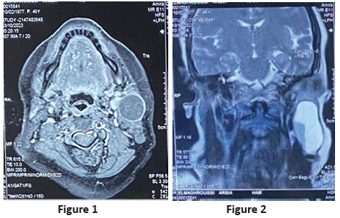
On radiological examination, cervical ultrasound revealed a left parotid cyst with an abscess appearance and MRI revealed a uniloculated cystic lesion of the left parotid gland.

Figure 1 & 2: Parotal MRI in axial and coronal section showing a uninoculated cystic lesion of the left parotid gland.

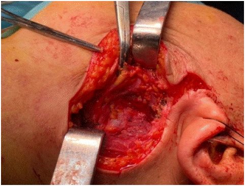
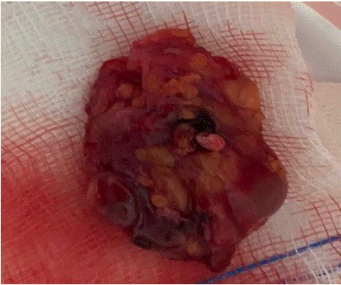
Figure 3: Intraoperative image of the tumor.

Figure 4: Image showing the operating room.
The patient benefited from surgical treatment which consisted of excision of the intraparotid cyst with respect to the intraparotid vascular and nerve elements and the piece was sent extemporaneously and came back in favor of an intraparotid lymphoepithelial cyst and the definitive anapathology confirmed the histological type of the tumor. The operative consequences are unremarkable as is the postoperative follow-up
Discussion
BLEC is a rare cystic tumor associated with benign embryonal dysplasia. The first case of BLEC was reported by Hildebrandt in 1895, but only 23 cases were published by researchers until 1981. With the emergence of the HIV epidemic, the incidence of BLEC in the parotid gland gradually increased, and researchers found that BLECs were closely linked to HIV infection in most cases, with BLEC as one of the first clinical manifestations. Approximately 3 to 6% of HIV-positive adults and 1 to 10% of HIV-positive children show symptoms of BLEC. In contrast, BLECs are rarely found in non-HIV-infected patients, and the exact prevalence of BLEC in this population has not been reported [1-4]. In this case, HIV test results include antibody and nucleic acid tests as well as CD4+ T lymphocyte examination was negative. In previous reports, the disease was also known as gill disease.
Split cyst and usually seen either in the mandibular angle at the bottom of the external regions or in in the anterior part of the sternocleidomastoid. This was found to occur less frequently in the mouth or in the parotid gland. In addition, its distribution among men and women is uniform. Although the catheter jam appears to be a cause, the source of the blockage is often not obvious. Furthermore, its pathogenesis remains unclear. Currently, the majority of researchers believe that extraglandular lymphoids, intraglandular lymphoid infiltration and/or hyperplasia with ductal and/or epithelial obstruction integration is the main cause. BLEC is a slow growing tumor, not associated with recurrence or metastases [5-8]. The main symptom of this disease is a slow-growing, painless, swollen mass, often associated with local symptoms such as dysphagia, dysphagia, dyspnea and stridor, and secondary Infection may present with acute inflammatory symptoms, such as redness, swelling and fever [3,8]. The diagnosis of BLEC mainly depends on clinical manifestations and preoperative auxiliaries examine. Preoperative auxiliary diagnostic procedures include CT, MRI, ultrasound and Fine Needle Aspiration (FNA) [9]. To date, the imaging features of this disease have been rarely reported. By reviewing the relevant literature [1,9], we found that the MRI manifestations of BLEC are typical. A single thin-walled round or oval cystic focus in the parotid gland, with clear boundaries and T2 hypersignal, hyposignal T1 in the cyst. On close examination, only the cyst wall shows mild signs. moderate enhancement and no enhancement is observed in the cyst. In this case, the MRI showed that small plaques with T2 hypersignal, T1 hyposignal without diffusion restriction and thin walls, discreetly accentuated T2 hypointensity in the parotid gland were caused by a viscous fluid in the capsule or lesions complicated by infection. MRI mainly shows a single thin-walled cyst lesion of the parotid gland, round or oval in shape, with a clear border. The wall shows a weak signal. Intracystic T1-weighted imaging showed low signal, while T2-weighted imaging showed high signal. In addition, the cyst wall was significantly improved upon enhancement scan, but no improvement was found in the cyst [2]. Ultrasonic features of the lesions included a circular shape with a complete capsule and smooth lining, and blood flow signals through cyst wall were not rich. The epithelial cells in a BLEC may contain keratin, and shed inflammatory cells can form a viscous gel-like yellowish-white liquid [10,11]. The internal echo of the cyst can generally appear as one of three types, including coarse granular hyperecho and cloud weak echo mixed signal, clear cystic echo, and cluster weak echo and cystic echo mixed. In this case, ultrasound showed typical mixed signals including coarse granular hyperechoic signal and cloud weak echo signal. FNA can be used as an important auxiliary method for the clinical diagnosis of lateral cervical lesions. The criteria for FNA cytological diagnosis of BLEC include thick, yellow, pustular fluid; non- nucleated cells; keratinocytes; and squamous epithelial cells of varying maturity [1]. Histologically, the walls of BLEC cysts are usually covered with squamous epithelium and/or, in some cases, columnar or cuboidal cells. Lymphoid tissue with or without germinal centers in subepithelial connective tissue is the most prominent morphological feature [12]. Histological examination of the mass case showed that the cyst wall was laminated squamous epithelium without epithelial nail process and the surface layer was mostly incomplete keratosis, surrounded by a large number of lymphoid stromata with lymphoid follicular formation and a center of occurrence. Differential diagnoses include: (1) Warthin tumor [2], which is more common in middle-aged and elderly patients and more likely to occur in the posterior lower pole of the parotid gland, with uneven internal density; (2) Intramuscular benign hemangioma [13], which is a fast-flowing intramuscular mass with uneven density and visible fat density in children that requires pathological examination for diagnosis; (3) Lymphoma [14], which is more common in men over 50 years old and appears as irregular soft tissue masses with a large range and uniform density on CT, without obvious calcification, cystic degeneration or necrosis, with diffuse growth to the surrounding area and mostly without adjacent bone destruction, and mild to moderate enhancement on enhanced scanning; (4) Thyroglossal duct cysts [15], which present as a painless mass in the front of the neck and are usually dumbbell shaped and movable when the tongue is extended or swallowed; CT shows a low-density, usually monocular parenchyma lesion during embryonic thyroid migration, mostly located in the midline and associated with the hyoid bone; (5) Lymphocele [16], which typically manifest as a cystic density focus with uniform density, clear boundary, thin cyst wall, no obvious exudation and calcification, and after enhancement, the cyst wall can present slightly uniform enhancement, with no enhancement in the cyst; (6) Metastatic lymph nodes [17]. which are accompanied by a history of primary tumor, and on CT exhibit uneven density, calcification, cystic or necrotizing changes, uneven edges, adhesion to surrounding tissues, and obvious annular or peripheral enhancement; and (7) Lymphadenitis [18], which is more common in children and present with local redness, swelling, heat and pain while appearing mostly oval with a thick wall and ring and uniform enhancement without obvious wall nodules and calcification, but with a blurred surrounding fat space.
The current treatment principles for this disease include early diagnosis, infection control, and cyst removal, and the treatment options include observation, repeated aspiration, sclerotherapy, radiotherapy, and surgical treatment [4]. Observation in asymptomatic patients show that repeated aspiration therapy is generally ineffective, and cysts will recur within weeks to months. Sclerotherapy is limited in scope and is generally only used for cases in which the cystic fluid can be aspirated, while radiotherapy is mostly used for HIV-infected BLEC [3]. For our patient, surgery was the best treatment and particular attention was given to the identification and avoiding injury to adjacent blood vessels and nerves during surgery. In this case, surgical treatment was chosen by the patient, and the surgical procedure consisted of an excision of the intraparotid cyst with preservation of the facial nerve branches and intraparotid vessels.
The Potential complications of surgical treatment include recurrence, persistent fistula formation, and cranial nerve injuries, which require close monitoring. In our case, post-operative follow-up was carried out weekly, monthly, quarterly and did not reveal any complications or recurrence.
Conclusion
BLEC in the absence of HIV infection is a rare benign cystic tumor of embryonic dysplasia, known as a branchial cleft cyst. The disease progresses slowly. Because the imaging, histopathology, and diagnosis and treatment characteristics of non-HIV-infected BLEC are poorly understood, the present case is reported to increase readers’ awareness of this disease.
References
- Joshi J, Shah S, Agarwal D, Khasgiwal A. Benign lymphoepithelial cyst of parotid gland: Review and case report. J OralMaxillofac Pathol. 2018; 22: S91-S97.
- Pillai S, Agarwal AC, Mangalore AB, Ramaswamy B, Shetty S. Benign Lymphoepithelial Cyst of the Parotid in HIV Negative Patient. J Clin Diagn Res. 2016; 10: MD05-MD06.
- Dave SP, Pernas FG, Roy S. The benign lymphoepithelial cyst and a classification system for lymphocytic parotid glandenlargement in the pediatric HIV population. Laryngoscope. 2007; 117: 106-113.
- Nahata V. Branchial Cleft Cyst. Indian J Dermatol. 2016; 61: 701.
- Zimmerman KO, Hupp SR, Bourguet-Vincent A, Bressler EA, Raynor EM, Turner DA, et al. Acute upper-airway obstruction by a lingual thyroglossal duct cyst and implications for advanced airway management. Respir Care. 2014; 59: e98-e102.
- Fujibayashi T, Itoh H. Lymphoepithelial (so-called branchial) cyst within the parotid gland. Report of a case and review ofthe literature. Int J Oral Surg. 1981; 10: 283-292.
- Chavan S, Deshmukh R, Karande P, Ingale Y. Branchial cleft cyst: A case report and review of literature. J Oral Maxillofac Pathol. 2014; 18: 150.
- Glosser JW, Pires CA, Feinberg SE. Branchial cleft or cervical lymphoepithelial cysts: etiology and management. J Am Dent Assoc. 2003; 134: 81-86.
- da Silva KD, Coelho LV, do Couto AM, de Aguiar MCF, Tarquínio SBC, Gomes APN, Mendonça EF, Batista AC, Nonaka CFW, de Sena LSB, Alves PM, Libório-Kimura TN, Louredo BVR, Câmara J, Caldeira PC. Clinicopathological and immunohistochemical features of the oral lymphoepithelial cyst: A multicenter study. J Oral Pathol Med. 2020; 49: 219-226.
- Reynolds JH, Wolinski AP. Sonographic appearance of branchial cysts. Clin Radiol. 1993; 48: 109-110.
- Hirota J, Maeda Y, Ueta E, Osaki T. Immunohistochemical and histologic study of cervical lymphoepithelial cysts. J Oral Pathol Med. 1989; 18: 202-205.
- Wee SJ, Park MC, Chung CM, Tak SW. Intramuscular hemangioma in the zygomaticus minor muscle: a case report and literature review. Arch Craniofac Surg. 2021; 22: 115-118.
- Thanarajasingam G, Bennani-Baiti N, Thompson CA. PET-CT in Staging, Response Evaluation, and Surveillance of Lymphoma. Curr Treat Options Oncol. 2016; 17: 24.
- Corvino A, Pignata S, Campanino MR, Corvino F, Giurazza F, Tafuri D, Pinto F, Catalano O.Thyroglossal duct cysts and site-specific differential diagnoses: imaging findings with emphasis on ultrasound assessment. J Ultrasound. 2020; 23: 139-149.
- Hekiert A, Newman J, Sargent R, Weinstein G. Spontaneous cervical lymphocele. Head Neck. 2007; 29: 77-80.
- Suh CH, Baek JH, Choi YJ, Lee JH. Performance of CT in the Preoperative Diagnosis of Cervical Lymph Node Metastasisin Patients with Papillary Thyroid Cancer: A Systematic Review and Meta-Analysis. AJNR Am J Neuroradiol. 2017; 38:154-161.
- Desai S, Shah SS, Hall M, Richardson TE, Thomson JE; Pediatric Research in Inpatient Settings (PRIS) Network. ImagingStrategies and Outcomes in Children Hospitalized with Cervical Lymphadenitis. J Hosp Med. 2020; 15: 197-203.