
Case Report
Austin J Otolaryngol. 2015;2(1): 1024.
Seeing the Face of Foreign Body Impaction
Justin Noroozian1*, Sharon Ramos2 and Harold S Pine2
1School of Medicine, University of Texas Medical Branch, USA
2Department of Otolaryngology, University of Texas Medical Branch, USA
*Corresponding author: Justin Noroozian, School of Medicine, University of Texas Medical Branch, Galveston, Texas 77550, USA
Received: November 15, 2014; Accepted: January 14, 2015; Published: January 19, 2015
Abstract
Foreign bodies are a common cause of morbidity and mortality in children up to 3-4 years of age. Foreign body impaction (FBI) in the upper aerodigestive tract can easily be identified by plain film radiography and treated with rigid endoscopy. With increasingly advanced radiographic imaging, we can more easily distinguish between disc batteries and coins to decrease the morbidity and mortality rates associated with ingestion.
Keywords: Foreign Body; Impaction; Ingestion; Airway
Case Presentation
A 4-year-old male presented to an urgent care center with a 7-day history of fever, mucopurulent rhinorrhea, cough, and decreased activity level. Pneumonia was suspected and the patient underwent full body x-ray. A circular, radio-paque foreign body was seen in the esophagus and the patient was referred to the local emergency department (ED). Repeat X-ray in the ED confirmed the presence of an approximately 20mm coin in the upper esophagus. Upon close inspection of the radiograph, the anterior surface of an American dime was clearly identified (Figure 1). Rigid esophagoscopy was performed and the coin was identified and removed. A scope was passed a second time demonstrating irritated but intact mucosa. The patient was discharged home the same day.
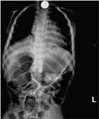
Figure 1: Full Body X-Ray (AP view).
Discussion/Conclusion
From the x-ray images we can clearly see the Roosevelt face of the dime. Running down the center of the coin we can also make out the torch from the tails side of the coin. Another distinguishing feature that allowed us to determine that the object was a dime is that we can see the single layer of ridging a disc battery would have a layer of ridging as well, but it would be more central to the coin (Figure 3). If you were to look at a regular dime, the face would be pointing left. What is interesting about this case is that upon looking at the x-ray, we noticed that the face of the dime was pointing in the opposite direction (to the right). This caught us by surprise, but we quickly came to the conclusion that this signified the dime would have had to be positioned in the patient’s esophagus with the face of the dime facing posteriorly, and the tail side of the dime facing anteriorly. The clarity of the dime characteristics that we were able to see on the x-ray was another interesting quality. According to Dr. Swischuk of The University of Texas Medical Branch in Galveston,“the face side of the dime has more tomography than the tails side, which is why we see more of the head and only a fraction of the torch.” Increased tomography, more density, and a little luck lead to the face being showcased more on x-ray (Figure 2). It is clear that with a disc battery the ridging is much more defined and located more interiorly than the ridging of a dime, as well as giving a double density shadow due to the bilaminar structure of batteries [1].

Figure 2: Magnified detail from Figure 1.
