Case Report
Austin J Otolaryngol. 2015;2(1): 1025.
Spontaneous Tongue and Pharynx Hematoma during Oral Anticoagulant Therapy
Hüseyin Günizi*
Department of Otolaryngology, University of Baskent Alanya Application and Research Center, Turkey
*Corresponding author: Hüseyin Günizi, Department of Otolaryngology, University of Baskent Alanya Application and Research Center / ANTALYA, Alanya, Turkey
Received: November 17, 2014; Accepted: January 16, 2015; Published: January 19, 2015
Introduction
Oral anticoagulant therapy is considerably important to prevent thromboembolic complications. Oral anticoagulant use has become more common in medical conditions such as deep vein thrombosis and pulmonary embolism and in patients with prosthetic cardiac valve and atrial fibrillation. During anticoagulant therapy, bleeding complication rate is 2-5.2%. Intracranial, genitourinary, skin and gastrointestinal hemorrhage are most frequently observed. Most of the cases with upper respiratory tract obstruction are retropharyngeal, sublingual and rarely laryngeal hematomas [1-3].
These complications can be controlled mostly with conservative methods. However, some cases may require endotracheal intubation and emergency tracheotomy. Pharyngeal hematomas may cause various clinical manifestations according to hematoma size, location and formation rate.
Case Presentation
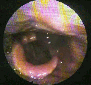
Seventy five-year old male patient presented to our emergency department with tongue swelling and dyspnea. The patient had mitral valve replacement and coronary bypass surgery 2 years ago and thus, he was using 10 mg warfarin sodium per day. He didn’t use another drug. However, the patient reported that his international normalized ratio (INR) level had not been checked for the last 2 months. In physical examination, the tongue was swelling and purple and the patient had dyspnea (Figure 1). Oxygen saturation was determined as 96-97%. In laboratory analysis, prothrombin time (PT) was 54.3 seconds, active partial thromboplastin time (APTT) was 43 seconds and INR was 6.18. Hemoglobin was 12.7, leukocyte count was 9330, thrombocyte count was 217.000, BUN was 22 mg/dL, creatinine was 0.90 mg/dL, Na was 141mEq/L and K was 4.5 mEq/L. In fiberoptic laryngoscopy, 2x3 cm hematoma that did not affect the rima glottis was observed in hypopahrynx, behind the left arythenoid (Figure 2). There were no reason to explain high INR level and sign of gastrointestinal bleeding or skin hematoma. The patient was transferred to the intensive care unit for close monitorization without laryngeal intubation and tracheotomy, because rima glottis was not obstructed and oxygen saturation was high enough, and existing high INR could cause development of a new source of bleeding. 4 lt/min was given to the patient. Oral anticoagulant was discontinued. Two units fresh frozen plasma transfusion was performed following cardiology consultation and injected subcutaneous enoxaparin sodium (120 mg per day). It was planned to reduce INR gradually to prevent additional cardiologic complications. Second day INR level was 3.53, and one unit fresh frozen plasma was transfusioned again. Fourth day INR level was 1.75 and the patient was transferred to the ward and started to take 5 mg warfarin sodium. Because of the patient’s hematuria, urine analysis was performed and it revealed erythrocyturia. INR value was reduced to therapeutical levels at follow-ups. Hematomas on tongue and pharynx were resolved and patient started taking oral foods. Physical examination and laboratory findings recovered expeditiously. INR level was 1.47 and the patient was discharged home after 6 days in the hospital with complete resolution of symptoms. After discharged, he came back for policlinic control and INR level was 1.82 one week later.

Figure 1: Tongue Hematoma.

Figure 2: Pharynx Submucosal Hematoma.
Discussion
Oral anticoagulants are classified into two groups according to their chemical structures: coumarin ve indandion derivatives. Among coumarin derivatives, the most tested and most preferred is Sodium Warfarin (coumadin). Such medications basically prevent the final step of synthesis of prothrombin, factor 7,9,10 which are vitamin K-dependent coagulation factors produced in the liver. Their effect appear at least 24 hours after the therapy initiation. The effect starts late, and ends after a latent period – a few days after the therapy discontinuation. The effect of oral anticoagulants is dosedependent. INR is the most valuable follow-up parameter to monitor oral anticoagulan use. Target INR value range is 2-3 to prevent thromboembolic events and avoid bleeding. If the value of INR is higher than 3, the likelihood of bleeding complication increases significantly [1].
The common adverse effect observed during oral anticoagulant therapy is spontaneous bleeding when high doses are given, and bleeding complications are seen in 2-5.2% of the patients. Thus, this medication should not be used in patients with diseases that predispose hemorrhage. Intracranial, genitourinary, skin and gastrointestinal hemorrhage are most frequently observed. Submandibular, sublingual, peritonsillar and retropharyngeal involvements are rare, but hematoma formation that obstructs airway is a serious complication [1]. Intraoral hematomas generally develop due to motor vehicle accidents, grand mal seizure or trauma following intubation in patients receiving oral anticoagulant [4-6].
Spontaneous tongue and pharynx hematomas due to warfarin sodium use are rare complications but may obstruct the airway and become life-threatening. Pharynx is one of the most important components of the respiratory tract. Thus, space-occupying lesions in this region may compromise airway quickly and may require urgent interventions. In patients with mild or moderate obstruction findings, the treatment approach includes evaluation of airway with flexible laryngoscopy, close monitoring of respiratory status, oxygen therapy, and vitamin K and fresh frozen plasma transfusion to correct the coagulation disorder. Some researchers have reported that in case of submucosal hematoma, awake fiberoptic intubation is safe [7]. The priority in the management of this complication is to control the airway safely. If intubation is impossible due to severe obstruction of oral cavity or the risk of new bleeding source, the patient may require emergency tracheostomy. Additionally, according to the clinical condition of the patient, correction of anticoagulation level alone may be enough to control the bleeding wihout surgical drainage. Withdrawal of oral anticoagulant therapy and rapid reduction of INR level may cause additional cardiologic problems, thus a gradual reduction is planned.
On the other hand, some researchers state that routine tracheotomy/cricothyrotomy should be performed in patients [2,3,8]. However, it is important to consider that prophylactic intubation or tracheotomy increases the risk of hematoma rupture and bleeding, thus it may compromise the airway. It would be more appropriate to prefer cricotomy over tracheotomy in such condition to minimize the risk of bleeding.
In many series, it has been reported that early surgical drainage is not safe and increases the airway obstruction even more and causes complications such as rehemorrhage. In a study, it has been reported that after early surgical drainage, the case was complicated with systemic infection [9]. It has been observed that non-operative approach is more useful in such patients. It may be perfomed at a later date in the absence of spontaneous drainage of hematoma. Hematomas are generally resolved spontaneuosly short time after withdrawal of anticoagulant therapy.
Conclusion
In patients who receive oral anticoagulants, the therapeutic level of the medication should be monitored closely. We do not recommend performing surgical drainage in upper respiratory tract hematomas that develop during oral anticoagulant therapy due to risk of rehemorrhage or serious and fatal complication such as sepsis. We believe that the optimal management of such patients includes the evaluation of airway and withdrawal of anticoagulant therapy and initiation of medical support therapy to decrease INR level, and that in case of airway compromise; tracheotomy/cricotomy should be performed to maintain the airway.
References
- Oake N, Jennings A, Forster AJ, Fergusson D, Doucette S, van Walraven C. Anticoagulation intensity and outcomes among patients prescribed oral anticoagulant therapy: a systematic review and meta-analysis. CMAJ. 2008; 179: 235-244.
- Bloom DC, Haegen T, Keefe MA. Anticoagulation and spontaneous retropharyngeal hematoma. J Emerg Med. 2003; 24: 389-394.
- Rosenbaum L, Thurman P, Krantz SB. Upper airway obstruction as a complication of oral anticoagulation therapy. Report of three cases. Arch Intern Med. 1979; 139: 1151-1153.
- Duong TC, Burtch GD, Shatney CH. Upper-airway obstruction as a complication of oral anticoagulation therapy. Crit Care Med. 1986; 14: 830-831.
- McGoldrick KE, Donlon JV. Sublingual hematoma following difficult laryngoscopy. Anesth Analg. 1979; 58: 343-344.
- Saah D, Braverman I, Elidan J, Nageris B. Traumatic macroglossia. Ann Otol Rhinol Laryngol. 1993; 102: 729-730.
- Keeling D, Baglin T, Tait C, Watson H, Perry D, Baglin C, et al. Guidelines on oral anticoagulation with warfarin - fourth edition. Br J Haematol. 2011; 154: 311-324.
- Uppal HS, Ayshford CA, Syed MA. Spontaneous supraglottic haemorrhage in a patient receiving warfarin sodium treatment. Emerg Med J. 2001; 18: 406-407.
- Evgeni B, Leonid K, Schwartz A, Efim R, Amit F, Alexander Z, et al. Spontaneous sublingual hematoma: Surgical or non-surgical management? International Journal of Case Reports and Images. 2012; 3: 1-4.